. 2017 Jan 13;292(2):748-759.
doi: 10.1074/jbc.M116.754960. Epub 2016 Nov 30.

Distinct Receptor Tyrosine Kinase Subsets Mediate Anti-HER2 Drug Resistance in Breast Cancer

Affiliations

Distinct Receptor Tyrosine Kinase Subsets Mediate Anti-HER2 Drug Resistance in Breast Cancer

Peter B Alexander et al. J Biol Chem. .

Abstract

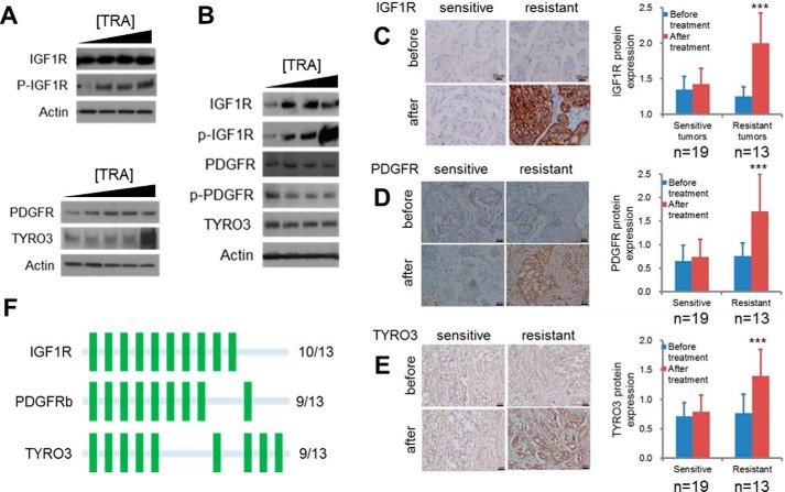
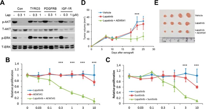
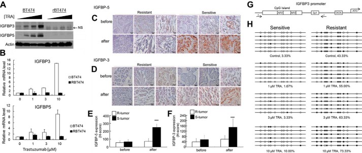
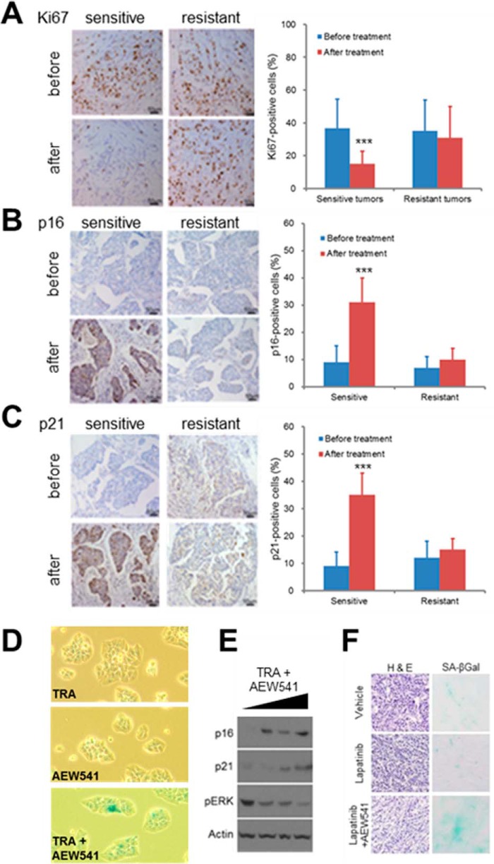
Targeted inhibitors of the human epidermal growth factor receptor 2 (HER2), such as trastuzumab and lapatinib, are among the first examples of molecularly targeted cancer therapy and have proven largely effective for the treatment of HER2-positive breast cancers. However, approximately half of those patients either do not respond to these therapies or develop secondary resistance. Although a few signaling pathways have been implicated, a comprehensive understanding of mechanisms underlying HER2 inhibitor drug resistance is still lacking. To address this critical question, we undertook a concerted approach using patient expression data sets, HER2-positive cell lines, and tumor samples biopsied both before and after trastuzumab treatment. Together, these methods revealed that high expression and activation of a specific subset of receptor tyrosine kinases (RTKs) was strongly associated with poor clinical prognosis and the development of resistance. Mechanistically, these RTKs are capable of maintaining downstream signal transduction to promote tumor growth via the suppression of cellular senescence. Consequently, these findings provide the rationale for the design of therapeutic strategies for overcoming drug resistance in breast cancer via combinational inhibition of the limited number of targets from this specific subset of RTKs.

Keywords: breast cancer; cellular senescence; drug resistance; human epidermal growth factor receptor 2 (HER2); insulin-like growth factor (IGF); receptor tyrosine kinase; targeted therapy.


Figures

FIGURE 1.
FIGURE 1.
Bioinformatic identification and experimental validation of RTKs mediating trastuzumab resistance. A, forest plot of the concordance index estimates of survival risk for each RTK in HER2 patients. Data are ordered and presented as the mean ± S.E. Higher concordance index numbers predict worse outcome. The most significant p values are also shown; *, p < 0.05; **, p < 0.01; ***, p < 0.001. B–D, SKBR3, BT474, or AU565 breast cancer cells engineered to stably express the indicated RTKs were cultured for 2 days in increasing amounts of lapatinib, and then cell viability was measured and plotted as a function of drug concentration. Error bars, S.E. Results are summarized in E. NR, no rescue; P, partial rescue; R, rescue. F–H, Kaplan-Meier curves of IGF1R, TYRO3, and PDGFRb in each of the HER2 molecular subtype. Gene expression is split on the median into low and high classifications, and data are presented as the probability of relapse-free survival or distant metastasis-free survival versus time in months.
FIGURE 2.
FIGURE 2.
RTKs mediating resistance are up-regulated in drug resistant tumors and cell lines as a result of HER2 inhibition. A and B, IGF1R, PDGFRb, and TYRO3 expression levels increase as a result of trastuzumab (TRA) treatment in drug-resistant breast cancer cells. rBT474 (A) or MDA-MB-361 (B) cells were cultured for 72 h in the presence of 1, 3, or 10 μm trastuzumab, after which cells were harvested and protein lysates were immunoblotted using the indicated antibodies. C–E, representative images and quantification of IGF1R, PDGFRb, and TYRO3 immunohistochemistry in resistant and sensitive tumors before and after neoadjuvant trastuzumab therapy. Samples were obtained from HER2-positive breast cancers by mammotome biopsy before therapy and from surgery after treatment. n = 19 sensitive tumors; 13 resistant tumors. Error bars, S.D.; ***, p < 0.01. See supplemental Table S3 for exact p values. F, frequency of RTK overexpression after HER2 inhibition based on immunohistochemistry analysis of unresponsive patient tumors. A green bar represents up-regulation of the indicated RTK in an individual tumor.
FIGURE 3.
FIGURE 3.
IGF1R, TYRO3, and PDGFRb activity is necessary to sustain signal transduction and tumor cell proliferation in response to HER2 inhibition. A, SKBR3 cells expressing TYRO3, IGF1R, or PDGFRb were treated with lapatinib (Lap) for 24 h, and then proteins were resolved by SDS-PAGE and immunoblotted using antibodies specific for the indicated signaling proteins. B, synergistic inhibition of trastuzumab-resistant cell growth by HER2 and IGF1R chemical inhibitors. rBT474 cells were cultured for 72 h in the presence of increasing concentrations of lapatinib, AEW541, or lapatinib plus AEW541 and then metabolically labeled with 2 μCi of [3H]thymidine for 16 h, after which DNA was precipitated and thymidine incorporation was measured by scintillation counting. C, rBT474 cells were cultured as in B but treated with the PDGFRb inhibitor sunitinib instead of AEW541. Error bars, S.E.; ***, p < 0.05. D and E, combined HER2 and IGF1R inhibition delays mammary tumor formation in mice. Immunodeficient BALB/c nude mice were orthotopically injected with rBT474 breast cancer cells, and, after allowing 1 week for tumor formation, were treated daily with vehicle, lapatinib, or lapatinib plus AEW541. Tumor sizes were measured twice weekly using vernier calipers until day 25, at which point mice were sacrificed, and dissected tumors were photographed (E). Mean tumor volumes ± S.E. are shown in D; ***, p < 0.05 (n = 4 mice/group).
FIGURE 4.
FIGURE 4.
Increased expression of IGFBP3 and IGFBP5 is associated with responsiveness to HER2 inhibition. A and B, expression of IGFBP3 and IGFBP5 in sensitive and resistant breast cancer cells after HER2 inhibition. BT474 or resistant BT474 (rBT474) breast cancer cells were cultured in the presence of 1, 3, or 10 μm trastuzumab for 72 h. IGFBP3 and IGFBP5 protein and mRNA levels were detected by immunoblotting (A) and qPCR (B), respectively. NS, nonspecific band. C–F, representative images (C and D) and quantification (E and F) of IGFBP immunohistochemistry in resistant (R) and sensitive (S) tumors before and after neoadjuvant trastuzumab therapy. The expression of both IGFBPs selectively increased only in responsive tumors. Samples were obtained from HER2-positive breast cancers by mammotome biopsy before therapy and from surgery after treatment. Error bars, S.D.; ***, p < 0.01. p values are provided in supplemental Table S3. G, structure of the IGFBP3 promoter. Locations of the primers used for bisulfite sequencing, TATA box, start codon, and transcription factor binding sites are indicated. H, methylation analysis of the IGFBP3 promoter in BT474 and rBT474 cells. BT474 (sensitive) or rBT474 (resistant) cells were cultured for 72 h in the presence of increasing concentrations of trastuzumab, after which genomic DNA was purified for bisulfite sequencing.
FIGURE 5.
FIGURE 5.
Effective anti-HER2 therapy results in tumor cell senescence. A–C, representative images and quantification of Ki67 (A), p16 (B), and p21 (C) immunohistochemistry in resistant and sensitive tumors before and after neoadjuvant trastuzumab therapy. Samples were obtained from HER2-positive breast cancers by mammotome biopsy before therapy and from surgery after treatment. Error bars, S.D.; ***, p < 0.01. Exact p values can be found in supplemental Table S3. D–F, combined pharmacological inhibition of HER2 and IGF1R leads to cellular senescence in drug-resistant breast cancer cells. MDA-MB-361 cells were treated for 72 h with trastuzumab (TRA) and AEW541 as indicated, and cellular senescence was assayed by SA-βgal staining (D) and immunoblotting cell lysates with antibodies specific for the senescence-associated proteins p16 and p21 (E). F, rBT474 breast cancer cells were grown as tumor xenografts in BALB/c nude mice (n = 4/group). Following 1 week of tumor establishment, mice were treated with either vehicle, lapatinib (50 mg/kg/day), or lapatinib combined with AEW541 (both at 50 mg/kg/day) for 18 days. Frozen tumor sections were stained with hematoxylin and eosin to examine tumor histology or for SA-βgal activity to detect cellular senescence.

References

    1. Jemal A., Center M. M., DeSantis C., and Ward E. M. (2010) Global patterns of cancer incidence and mortality rates and trends. Cancer Epidemiol. Biomarkers Prev. 19, 1893–1907 - PubMed
    1. Citri A., and Yarden Y. (2006) EGF-ERBB signalling: towards the systems level. Nat. Rev. Mol. Cell Biol. 7, 505–516 - PubMed
    1. Moasser M. M. (2007) The oncogene HER2: its signaling and transforming functions and its role in human cancer pathogenesis. Oncogene 26, 6469–6487 - PMC - PubMed
    1. Slamon D. J., Clark G. M., Wong S. G., Levin W. J., Ullrich A., and McGuire W. L. (1987) Human breast cancer: correlation of relapse and survival with amplification of the HER-2/neu oncogene. Science 235, 177–182 - PubMed
    1. Hynes N. E., and Lane H. A. (2005) ERBB receptors and cancer: the complexity of targeted inhibitors. Nat. Rev. Cancer 5, 341–354 - PubMed

MeSH terms