. 2017 Jan 20;292(3):872-883.
doi: 10.1074/jbc.M116.758102. Epub 2016 Nov 30.

Structural Analysis of Single Domain Antibodies Bound to a Second Neutralizing Hot Spot on Ricin Toxin's Enzymatic Subunit

Affiliations

Structural Analysis of Single Domain Antibodies Bound to a Second Neutralizing Hot Spot on Ricin Toxin's Enzymatic Subunit

Michael J Rudolph et al. J Biol Chem. .

Abstract

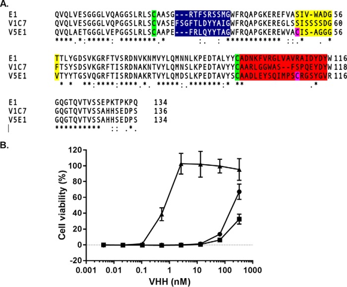
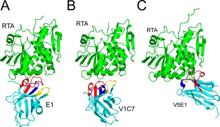
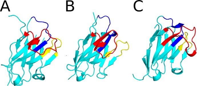
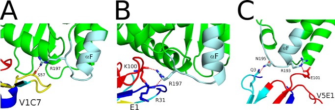
Ricin toxin is a heterodimer consisting of RTA, a ribosome-inactivating protein, and RTB, a lectin that facilitates receptor-mediated uptake into mammalian cells. In previous studies, we demonstrated that toxin-neutralizing antibodies target four spatially distinct hot spots on RTA, which we refer to as epitope clusters I-IV. In this report, we identified and characterized three single domain camelid antibodies (VHH) against cluster II. One of these VHHs, V5E1, ranks as one of the most potent ricin-neutralizing antibodies described to date. We solved the X-ray crystal structures of each of the three VHHs (E1, V1C7, and V5E1) in complex with RTA. V5E1 buries a total of 1,133 Å2 of surface area on RTA and makes primary contacts with α-helix A (residues 18-32), α-helix F (182-194), as well as the F-G loop. V5E1, by virtue of complementarity determining region 3 (CDR3), may also engage with RTB and potentially interfere with the high affinity galactose-recognition element that plays a critical role in toxin attachment to cell surfaces and intracellular trafficking. The two other VHHs, E1 and V1C7, bind epitopes adjacent to V5E1 but display only weak toxin neutralizing activity, thereby providing structural insights into specific residues within cluster II that may be critical contact points for toxin inactivation.

Keywords: antibody; antigen; crystal structure; epitope mapping; neutralizing; ricin; toxin.


Figures

FIGURE 1.
FIGURE 1.
Amino acid sequences and toxin neutralizing activities associated with E1, V1C7, and V5E1. A, primary amino acid sequence alignments of VHHs E1, V1C7, and V5E1. Highlighted are the conserved cysteine residues (green), two additional cysteine residues in V5E1 (pink), CDR1 (blue), CDR2 (yellow), and CDR3 (red). B, ricin toxin neutralizing activities associated with E1 (circles), V1C7 (squares), and V5E1 (triangles), as determined in a Vero cell cytotoxicity assay (see “Experimental Procedures”). The values shown are the average (with standard deviation) of two separate experiments each done in triplicate.
FIGURE 2.
FIGURE 2.
Competition ELISAs demonstrating that E1, V1C7, and V5E1 recognize epitopes within cluster II. A, ricin was captured on microtiter plates via ASF (top row) or mAbs representing epitope clusters I–IV, as indicated by colored vertical bars (left) and then probed with E1 (black bars), V1C7 (gray bars), and V5E1 (white bars), as described under the “Experimental Procedures.” The values shown are the average (with standard deviation) of a single representative experiment done in triplicate. B, competition ELISAs in which biotinylated ricin was mixed with indicated concentrations (x axis) of V5E1 in solution and then applied to microtiter plates coated with the four cluster II mAbs as follows: SyH7 (circles); PA1 (squares); PH12 (triangles); TB12 (inverted triangles). The plate was then washed and probed with avidin-HRP to detect bound ricin. The values shown are the % inhibition (y axis) as compared with mAb capture of biotinylated ricin without addition of VHH.
FIGURE 3.
FIGURE 3.
Characterization of epitope cluster II mAbs. A, ribbon diagram of RTA (PDB 1RTC) with relevant secondary structures labeled. The linear epitope recognized by SyH7 is shown in blue. B, competition ELISA in which SyH7 (at indicated concentrations) was mixed with biotinylated ricin in solution and then applied to microtiter plates coated with mAbs SyH7 (circles), PA1 (squares), PH12 (triangles), and TB12 (inverted triangles). The plates were then probed with avidin-HRP, and the amount that SyH7 that blocked biotin-ricin capture by the plate-bound mAbs is indicated on y axis (% inhibition). C, ricin toxin neutralizing activity of SyH7 (circles), PA1 (squares), PH12 (triangles), and TB12 (inverted triangles), as described under the “Experimental Procedures.”
FIGURE 4.
FIGURE 4.
V5E1 recognizes an epitope on RTA that is spatially distinct from E1 and V1C7's epitopes. Competition ELISA in which E1 (A), V1C7 (B), or V5E1 (C), at indicated concentrations, was mixed with biotinylated ricin in solution and then applied to microtiter plates coated with E1 (circles), V1C7 (squares), or V5E1 (triangles). The plates were then probed with avidin-HRP, and the amount that the soluble VHH blocked biotin-ricin capture by the plate bound antibody is indicated on y axis (% inhibition). D and E, structure of RTA-V5E1 complex was superpositioned onto the RTA-E1 or RTA-V1C7 structures to demonstrate the distinct binding profiles. RTA is colored green; V5E1 is magenta, and E1 and V1C7 are in cyan.
FIGURE 5.
FIGURE 5.
VHHs E1 and V1C7 interfere with neutralizing activity of cluster II mAbs. VHH E1 (squares) and V1C7 (triangles) mixed with indicated concentrations (x axis) of SyH7 (A), PA1 (B), PH12 (C), or TB12 (D) were incubated with ricin toxin and then applied to Vero cells, as described under the “Experimental Procedures.” For each panel, the circles represent mAb without the addition of E1 or V1C7. The VHHs were at a constant concentration of 133 nm (2 μg/ml). Cell viability was determined 48 h later. The values shown are the average (with standard deviation) of a single representative experiment done in triplicate.
FIGURE 6.
FIGURE 6.
X-ray crystal structures of RTA-VHH complexes. Structures of RTA in complex with VHHs E1 (A), V1C7 (B), and V5E1 (C). RTA (green) is presented in a similar orientation for each panel with α-helices D–G indicated, as necessary. VHHs are shaded in cyan with CDR1–3 colored blue, yellow, and red, respectively.
FIGURE 7.
FIGURE 7.
X-ray crystal structures of VHHs E1, V1C7, and V5E1. The structures of VHHs E1 (A), V1C7 (B), and V5E1 (C) are drawn as ribbon diagrams colored cyan with CDR1–3 colored blue, yellow, and red, respectively. The VHHs are oriented with CDR3 projecting out front.
FIGURE 8.
FIGURE 8.
Interfaces of E1, V1C7, and V5E1 with RTA. A–C, RTA (green) is drawn as a ribbon diagram with E1- (A), V1C7- (B), and V5E1 (C)-interacting regions colored red. RTA secondary structural elements are labeled as follows: α-helices A and C–G, β-strands a, d, e, and the 310-helix between α-helices C-D. D–F, surface representations of RTA (gray) with E1 (D), V1C7 (E), and V5E1 (F) contact points highlighted in red.
FIGURE 9.
FIGURE 9.
Close-up of the VHH interactions with the SyH7 epitope on RTA. RTA (green), the SyH7 epitope (colored pale cyan), and VHHs (cyan), V1C7 (A), E1 (B), and V5E1 (C) are drawn as ribbon diagrams. CDR1–3 are colored blue, yellow, and red, respectively. Key side chains are drawn as sticks and color-coordinated to the main chain. Hydrogen bonds are represented as red dashes.
FIGURE 10.
FIGURE 10.
Potential interactions of V5E1 with ricin holotoxin. RTB from ricin holotoxin (PDB code 2AAI) was superpositioned onto RTA-V5E1. RTA is colored green, V5E1 in cyan, and RTB in magenta. Lactose bound to RTB is shown as red sticks. The inset illustrates the proximity of V5E1 residue Arg-110 with RTB residues Asn-255 and Ser-238 involved in lactose recognition. Key residues forming interactions are drawn as sticks and color-coordinated to their respective main chain color. Hydrogen bonds are represented as red dashes.
FIGURE 11.
FIGURE 11.
V5E1 partially interferes with ricin attachment to terminal galactosides. A, dilutions of V5E1 (triangles) and V1C7 (squares) were mixed with biotinylated ricin and then applied to microtiter plates coated with ASF, as described under the “Experimental Procedures.” The % inhibition (y axis) values are based on ricin binding in the absence of antibodies. B, V5E1 at indicated concentrations was mixed with FITC-ricin and then applied to THP-1 cells at 4 °C. After 30 min, the THP-1 cells were washed, and the amount of bound ricin was determined by flow cytometry. The % inhibition (y axis) values are based on ricin binding in the absence of antibodies. Each panel is a representative experiment conducted in triplicate.

References

    1. Endo Y., Mitsui K., Motizuki M., and Tsurugi K. (1987) The mechanism of action of ricin and related toxins on eukaryotic ribosomes. J. Biol. Chem. 262, 5908–5912 - PubMed
    1. Endo Y., and Tsurugi K. (1987) RNA N-glycosidase activity of ricin A-chain. Mechanism of action of the toxic lectin ricin on eukaryotic ribosomes. J. Biol. Chem. 262, 8128–8130 - PubMed
    1. Sandvig K., Olsnes S., and Pihl A. (1976) Kinetics of binding of the toxic lectins abrin and ricin to surface receptors of human cells. J. Biol. Chem. 251, 3977–3984 - PubMed
    1. Spooner R. A., and Lord J. M. (2015) Ricin trafficking in cells. Toxins 7, 49–65 - PMC - PubMed
    1. Jandhyala D. M., Thorpe C. M., and Magun B. (2012) Ricin and Shiga toxins: effects on host cell signal transduction. Curr. Top. Microbiol. Immunol. 357, 41–65 - PMC - PubMed

LinkOut - more resources