Review
. 2016 Nov 30;81(1):e00037-16.
doi: 10.1128/MMBR.00037-16. Print 2017 Mar.

Yeast HMO1: Linker Histone Reinvented

Affiliations
Review

Yeast HMO1: Linker Histone Reinvented

Arvind Panday et al. Microbiol Mol Biol Rev. .

Abstract

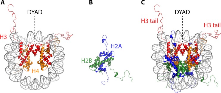
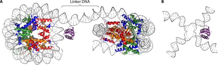
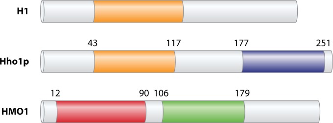
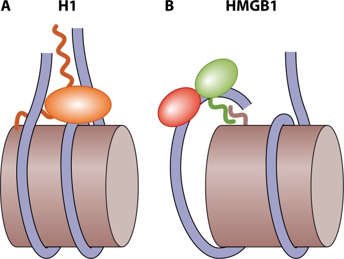
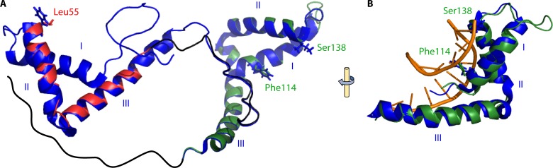
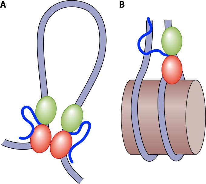
Eukaryotic genomes are packaged in chromatin. The higher-order organization of nucleosome core particles is controlled by the association of the intervening linker DNA with either the linker histone H1 or high mobility group box (HMGB) proteins. While H1 is thought to stabilize the nucleosome by preventing DNA unwrapping, the DNA bending imposed by HMGB may propagate to the nucleosome to destabilize chromatin. For metazoan H1, chromatin compaction requires its lysine-rich C-terminal domain, a domain that is buried between globular domains in the previously characterized yeast Saccharomyces cerevisiae linker histone Hho1p. Here, we discuss the functions of S. cerevisiae HMO1, an HMGB family protein unique in containing a terminal lysine-rich domain and in stabilizing genomic DNA. On ribosomal DNA (rDNA) and genes encoding ribosomal proteins, HMO1 appears to exert its role primarily by stabilizing nucleosome-free regions or "fragile" nucleosomes. During replication, HMO1 likewise appears to ensure low nucleosome density at DNA junctions associated with the DNA damage response or the need for topoisomerases to resolve catenanes. Notably, HMO1 shares with the mammalian linker histone H1 the ability to stabilize chromatin, as evidenced by the absence of HMO1 creating a more dynamic chromatin environment that is more sensitive to nuclease digestion and in which chromatin-remodeling events associated with DNA double-strand break repair occur faster; such chromatin stabilization requires the lysine-rich extension of HMO1. Thus, HMO1 appears to have evolved a unique linker histone-like function involving the ability to stabilize both conventional nucleosome arrays as well as DNA regions characterized by low nucleosome density or the presence of noncanonical nucleosomes.

Keywords: HMGB proteins; Saccharomyces cerevisiae; chromatin; linker histone; nucleosome.


Figures

FIG 1
FIG 1
Assembly of the nucleosome core particle. (A) Association of the (H3/H4)2 tetramer with DNA nucleates nucleosome assembly and defines the dyad axis. (B) H2A/H2B dimer. (C) Two H2A/H2B dimers are deposited to generate the nucleosome core particle. H3 N-terminal tails emerge near the DNA entry/exit points (based on data reported under PDB accession number 1KX5).
FIG 2
FIG 2
Histone H1 associates with linker DNA. (A) The globular domain of histone H1 (purple) binds the nucleosome at the dyad. The structure of a dinucleosome is depicted; the color coding for core histones is the same as that in Fig. 1. (B) Four-way junction DNA mimics the DNA configuration at the nucleosome dyad, perhaps explaining the preferred binding of H1 to such junctions. The dinucleosome represents the asymmetric unit in the structure of a tetranucleosome with one linker DNA trimmed for clarity (PDB accession number 1ZBB) (2). The H1 globular domain and its localization relative to the dyad are based on the structure of the chicken H5 globular domain in complex with a nucleosome (PDB accession number 4QLC) (33). The representation of the four-way junction is based on data reported under PDB accession number 3CRX.
FIG 3
FIG 3
Domain organization of H1, Hho1p, and HMO1. Metazoan H1 typically contains an ∼40-amino-acid N-terminal extension, followed by a globular domain of ∼80 amino acids (orange) and a long CTD characterized by S/TPXK-like repeats. Hho1p contains a lysine-rich N-terminal segment followed by a globular domain with similarity to that of H1 (orange). Another lysine-rich segment connects this globular domain to the second globular domain (purple). HMO1 contains box A (red), which has little similarity to consensus HMG domains, followed by a lysine-rich linker, the box B domain (green), and a lysine-rich CTD. Mammalian HMGB proteins have a domain organization similar to that of HMO1, except that the CTD is acidic.
FIG 4
FIG 4
Proposed interaction of H1 and HMGB proteins with nucleosomes. (A) H1 binds near the dyad such that the CTD mainly contacts one linker segment; this creates a stem-like structure that stabilizes the nucleosome core (32, 38). (B) For HMGB, interactions between the acidic CTD and the H3 N-terminal tail that exits near the dyad promote binding of HMGB to DNA; the DNA bending and underwinding induced may propagate to the nucleosome core to promote unwrapping or access to other factors (118, 119).
FIG 5
FIG 5
Model of HMO1 and its interaction with DNA. (A) HMO1 was modeled by using SwissModel in the automated mode, using human HMGB1 (PDB accession number 2YRQ) as the template. HMGB1 is shown in blue, and the HMO1 model is overlaid with box A and box B domains in red and green, respectively. Predicted HMO1-intercalating residues Leu55, from box A, and Phe114, from box B, are shown in a stick representation. Ser138 is located in the position occupied by the DNA-intercalating Ile residue in HMGB1 box B. Helices are identified with roman numerals. The HMO1 C-terminal extension (black) is inferred to interact with box A. (B) HMGB1 box B (blue) overlaid with HMO1 box B (green), showing the interaction of helix III in the DNA minor groove and the intercalation of Phe between DNA bases. HMGB1 box B DNA is based on data reported under PDB accession number 2GZK.
FIG 6
FIG 6
HMO1-mediated stabilization of genomic DNA. (A) On nucleosome-free DNA, HMO1 promotes the formation of loops and bridges that depend on the dimerization of the box A domains (151). Such topological domains may also be mediated by the concerted action of HMO1 and Top2 (195). (B) Both HMO1 and HMO1 deleted for its C-terminal domain can compete with human H1 for binding to nucleosomes, suggesting that HMO1 and H1 binding sites at least partially overlap (198). A possible nucleosome-stabilizing binding mode for HMO1 is illustrated, in which the structure-specific box A domain binds near the dyad and DNA bending by HMO1 is prevented due to the lysine-rich CTD contacting linker DNA.

References

    1. Luger K, Mäder AW, Richmond RK, Sargent DF, Richmond TJ. 1997. Crystal structure of the nucleosome core particle at 2.8 Å resolution. Nature 389:251–260. doi:10.1038/38444. - DOI - PubMed
    1. Schalch T, Duda S, Sargent DF, Richmond TJ. 2005. X-ray structure of a tetranucleosome and its implications for the chromatin fibre. Nature 436:138–141. doi:10.1038/nature03686. - DOI - PubMed
    1. Thastrom A, Bingham LM, Widom J. 2004. Nucleosomal locations of dominant DNA sequence motifs for histone-DNA interactions and nucleosome positioning. J Mol Biol 338:695–709. doi:10.1016/j.jmb.2004.03.032. - DOI - PubMed
    1. Wu B, Mohideen K, Vasudevan D, Davey CA. 2010. Structural insight into the sequence dependence of nucleosome positioning. Structure 18:528–536. doi:10.1016/j.str.2010.01.015. - DOI - PubMed
    1. Smith S, Stillman B. 1991. Stepwise assembly of chromatin during DNA replication in vitro. EMBO J 10:971–980. - PMC - PubMed

MeSH terms

LinkOut - more resources