. 2017 Jun 1;23(11):2831-2841.
doi: 10.1158/1078-0432.CCR-16-0463. Epub 2016 Nov 30.

The Bruton Tyrosine Kinase (BTK) Inhibitor Acalabrutinib Demonstrates Potent On-Target Effects and Efficacy in Two Mouse Models of Chronic Lymphocytic Leukemia

Affiliations

The Bruton Tyrosine Kinase (BTK) Inhibitor Acalabrutinib Demonstrates Potent On-Target Effects and Efficacy in Two Mouse Models of Chronic Lymphocytic Leukemia

Sarah E M Herman et al. Clin Cancer Res. .

Abstract

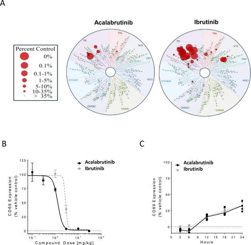
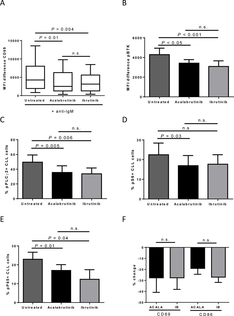
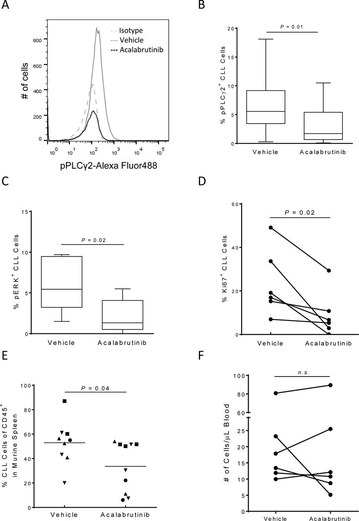
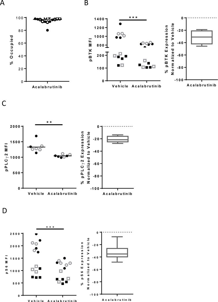
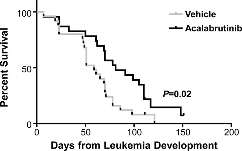
Purpose: Acalabrutinib (ACP-196) is a novel, potent, and highly selective Bruton tyrosine kinase (BTK) inhibitor, which binds covalently to Cys481 in the ATP-binding pocket of BTK. We sought to evaluate the antitumor effects of acalabrutinib treatment in two established mouse models of chronic lymphocytic leukemia (CLL).Experimental Design: Two distinct mouse models were used, the TCL1 adoptive transfer model where leukemic cells from Eμ-TCL1 transgenic mice are transplanted into C57BL/6 mice, and the human NSG primary CLL xenograft model. Mice received either vehicle or acalabrutinib formulated into the drinking water.Results: Utilizing biochemical assays, we demonstrate that acalabrutinib is a highly selective BTK inhibitor as compared with ibrutinib. In the human CLL NSG xenograft model, treatment with acalabrutinib demonstrated on-target effects, including decreased phosphorylation of PLCγ2, ERK, and significant inhibition of CLL cell proliferation. Furthermore, tumor burden in the spleen of the mice treated with acalabrutinib was significantly decreased compared with vehicle-treated mice. Similarly, in the TCL1 adoptive transfer model, decreased phosphorylation of BTK, PLCγ2, and S6 was observed. Most notably, treatment with acalabrutinib resulted in a significant increase in survival compared with mice receiving vehicle.Conclusions: Treatment with acalabrutinib potently inhibits BTK in vivo, leading to on-target decreases in the activation of key signaling molecules (including BTK, PLCγ2, S6, and ERK). In two complementary mouse models of CLL, acalabrutinib significantly reduced tumor burden and increased survival compared with vehicle treatment. Overall, acalabrutinib showed increased BTK selectivity compared with ibrutinib while demonstrating significant antitumor efficacy in vivo on par with ibrutinib. Clin Cancer Res; 23(11); 2831-41. ©2016 AACR.


Conflict of interest statement

Conflicts of Interest: M.G., F.K., R.I., T.C., A.K, R.U and B.L. are employees and equity holders of Acerta Pharma. J.B. A.J. and J.W. received research funding from Acerta Pharma. R.U. is an officer, equity holder and member of the board of directors of Acerta Pharma. A.W. received research funding from Pharmacylics and Acerta Pharma. C.U.N received funding from the Danish Cancer Society and has received travel grants/consultancy fees from Abbvie, Janssen, Gilead, Roche and Novartis. All other authors declare no competing financial interests.

Figures

Figure 1
Figure 1. Acalabrutinib is a potent and selective inhibitor of BTK
(A) Acalabrutinib and ibrutinib were profiled at 1 μM over a panel 395 wild-type human kinases, including mutants, at DiscoveRx kinase assays. The size of the red circles represents the extent of inhibition with larger circles meaning stronger inhibition compared to control signal as defined in the scale. (B) Mice (5/group/dose) were orally given vehicle, acalabrutinib or ibrutinib. After 3 hours, spleens were extracted and splenocytes stimulated with anti-IgM for 18 h, followed by CD69 expression analysis by flow cytometry. (C) Mice (5/group) received 25 mg/kg of vehicle, acalabrutinib or ibrutinib at time zero. Spleens were extracted at various time points and splenocytes stimulated with anti-IgM for 18 h, followed by CD69 expression analysis by flow cytometry. Black lines and symbols represent acalabrutinib, grey lines and symbols represent ibrutinib.
Figure 2
Figure 2. Acalabrutinib demonstrates equal in vitro on-target effects as ibrutinib
CLL patient MNCs from the peripheral blood or lymph node were treated in vitro with vehicle (untreated), or 1μM acalabrutinib or ibrutinib. (A) CLL patient MNCs (n=7) were pretreated with BCR-inhibitors for 3 hours and then stimulated with anti-IgM for 18 h, followed by CD69 expression analysis by flow cytometry. Results shown are for the CLL population. Shown is a min to max box and whisker plot. (B) CLL patient MNCs (n=5) were treated in vitro for 1 hour, fixed, permeabilized, and stained for phospho-BTK (pBTK). Results shown are for the CLL population. Shown is the mean (± SEM) MFI difference (pBTK-isotype control). (C-E) CLL patient MNCs (n=5) were treated in vitro for 1 hour, fixed, permeabilized, and stained for phospho-PLCγ2 (C), phospho-S6 (D) and phospho-NF-κB (E). Results shown are for the CLL population. The mean (± SEM) percent of CLL cells expressing the indicated readout is shown. (F) CLL patient MNCs (n=7) were treated for 24 hours followed by CD69 and CD86 expression analysis by flow cytometry. Results shown are for the CLL population. The mean (± SEM) percent change in CLL cells expressing the indicated readout is shown compared to untreated. ACALA: acalabrutinib, IB: ibrutinib. All Statistics were determined by paired student t-test.
Figure 3
Figure 3. Acalabrutinib demonstrates on target effects and reduced proliferation and tumor burden in the CLL xenograft mouse model
CLL MNCs (n=6) harvested from NSG mouse spleens (n=2–5 per patient) after 3 weeks of treatment were fixed, permeabilized, and stained with the indicated antibodies. Results shown are for the CLL population. (A) A representative histogram showing pPLCγ2 expression in a mouse treated with vehicle (solid grey line) compared to a mouse treated with acalabrutinib (solid black line). The dashed grey line represents the isotype control. (B-C) The median (± IQR) percent of pPLCγ2 (B) and pERK (C) is shown in a min to max box and whisker plot. (D) Percentage of CLL cells expressing KI67 is shown per patient for each treatment group. (E) Percentage of CLL cells among human CD45+ cells in the spleen. Each data point represents one mouse; symbols identify patients. (F) The absolute human CLL cell (CD45+/CD19+/CD5+) count in vehicle and acalabrutinib-treated mice per μl blood is shown. Data points represent the average measurements of 2–5 mice injected with MNCs from the same patient. All statistics were determined by paired student t-test.
Figure 4
Figure 4. Acalabrutinib demonstrates significant and sustained inhibition of BCR signaling in the TCL1 adoptive transfer model
Mice were engrafted with leukemic TCL1 cells and treated with vehicle or acalabrutinib. (A) % occupancy of Btk (n=14 per group) was determined after acalabrutinib treatment. Filled circles represent splenocytes harvested after one week of treatment and open circles represent splenocytes harvested after four weeks of treatment. (B-D) MFI expression (left) or % change in expression compared to vehicle (right) of pBtk (n=15 per group) (B), pPLCγ2 (n=7 per group) (C) and pS6 (n=14 per group) (D) evaluated in B-cell population by flow cytometry is shown. Filled symbols represent splenocytes harvested after one week of treatment and open symbols represent splenocytes harvested after four weeks of treatment. Circles and Squares differentiate independent experiments. Box and whisker plots show to min to max values. Asterisks indicate statistical significance as determined by normalized unpaired t-test. **P<0.01 and ***P<0.001.
Figure 5
Figure 5. Acalabrutinib increases survival in the TCL1 adoptive transfer model compared to vehicle
Mice engrafted with leukemic TCL1 cells were treated with vehicle (n=25) or acalabrutinib (n=23). Acalabrutinib increases survival compared to vehicle treatment. Median overall survival was 81 day vs 59 days, respectively, P=0.02.

References

    1. Buggy JJ, Elias L. Bruton Tyrosine Kinase (BTK) and Its Role in B-cell Malignancy. Int Rev Immunol. 2012;31:119–32. - PubMed
    1. Hendriks RW, Bredius RG, Pike-Overzet K, Staal FJ. Biology and novel treatment options for XLA, the most common monogenetic immunodeficiency in man. Expert Opin Ther Targets. 2011;15:1003–21. - PubMed
    1. Herman SE, Gordon AL, Hertlein E, Ramanunni A, Zhang X, Jaglowski S, et al. Bruton tyrosine kinase represents a promising therapeutic target for treatment of chronic lymphocytic leukemia and is effectively targeted by PCI-32765. Blood. 2011;117:6287–96. - PMC - PubMed
    1. Hendriks RW, Yuvaraj S, Kil LP. Targeting Bruton’s tyrosine kinase in B cell malignancies. Nat Rev Cancer. 2014;14:219–32. - PubMed
    1. Wiestner A. Targeting B-Cell receptor signaling for anticancer therapy: the Bruton’s tyrosine kinase inhibitor ibrutinib induces impressive responses in B-cell malignancies. J Clin Oncol. 2013;31:128–30. - PubMed

MeSH terms