. 2016 Nov 30;36(48):12117-12128.
doi: 10.1523/JNEUROSCI.2146-16.2016.

Adenosine Kinase Deficiency in the Brain Results in Maladaptive Synaptic Plasticity

Affiliations

Adenosine Kinase Deficiency in the Brain Results in Maladaptive Synaptic Plasticity

Ursula S Sandau et al. J Neurosci. .

Abstract

Adenosine kinase (ADK) deficiency in human patients (OMIM:614300) disrupts the methionine cycle and triggers hypermethioninemia, hepatic encephalopathy, cognitive impairment, and seizures. To identify whether this neurological phenotype is intrinsically based on ADK deficiency in the brain or if it is secondary to liver dysfunction, we generated a mouse model with a brain-wide deletion of ADK by introducing a Nestin-Cre transgene into a line of conditional ADK deficient Adkfl/fl mice. These AdkΔbrain mice developed a progressive stress-induced seizure phenotype associated with spontaneous convulsive seizures and profound deficits in hippocampus-dependent learning and memory. Pharmacological, biochemical, and electrophysiological studies suggest enhanced adenosine levels around synapses resulting in an enhanced adenosine A1 receptor (A1R)-dependent protective tone despite lower expression levels of the receptor. Theta-burst-induced LTP was enhanced in the mutants and this was dependent on adenosine A2A receptor (A2AR) and tropomyosin-related kinase B signaling, suggesting increased activation of these receptors in synaptic plasticity phenomena. Accordingly, reducing adenosine A2A receptor activity in AdkΔbrain mice restored normal associative learning and contextual memory and attenuated seizure risk. We conclude that ADK deficiency in the brain triggers neuronal adaptation processes that lead to dysregulated synaptic plasticity, cognitive deficits, and increased seizure risk. Therefore, ADK mutations have an intrinsic effect on brain physiology and may present a genetic risk factor for the development of seizures and learning impairments. Furthermore, our data show that blocking A2AR activity therapeutically can attenuate neurological symptoms in ADK deficiency.

Significance statement: A novel human genetic condition (OMIM #614300) that is based on mutations in the adenosine kinase (Adk) gene has been discovered recently. Affected patients develop hepatic encephalopathy, seizures, and severe cognitive impairment. To model and understand the neurological phenotype of the human mutation, we generated a new conditional knock-out mouse with a brain-specific deletion of Adk (AdkΔbrain). Similar to ADK-deficient patients, AdkΔbrain mice develop seizures and cognitive deficits. We identified increased basal synaptic transmission and enhanced adenosine A2A receptor (A2AR)-dependent synaptic plasticity as the underlying mechanisms that govern these phenotypes. Our data show that neurological phenotypes in ADK-deficient patients are intrinsic to ADK deficiency in the brain and that blocking A2AR activity therapeutically can attenuate neurological symptoms in ADK deficiency.

Keywords: adenosine kinase; epilepsy; gene mutation; human genetic disorder; learning and memory; mouse model.


Figures

Figure 1.
Figure 1.
Brain-specific deletion of ADK in AdkΔbrain mice. A, Transgenic strategy: exon 7 of the Adk allele is flanked with loxP sites in Adkfl/fl mice. B, Adk PCR on genomic DNA extracts from the cortex (C), striatum (S), hippocampus (Hp), cerebellum (Cb), heart (Ht), lung (L), liver (Lv), and spleen (Sp) from Adkfl/fl and AdkΔbrain mice. Tail DNA from an Adkfl/fl (T) and wild-type mouse (WT) were included as positive controls. Water (−) was included as a no-template control. Adk forward and reverse primer (P1 and P2) sites are depicted in A. C, ADK (40 kDa) Western blots on cortical, striatal, and hippocampal protein extracts from Adkfl/fl and AdkΔbrain mice; n = 2/genotype are used as representatives. ADK Western blots were reprobed with GAPDH (37 kDa) as a loading control. D, E, ADK immunohistochemistry of cortical brain tissue from Adkfl/fl (D) and AdkΔbrain (E) mice. F, G, Nissl stain of hippocampal formation from Adkfl/fl (F) and AdkΔbrain (G) mice.
Figure 2.
Figure 2.
Loss of brain ADK results in increased susceptibility to seizure induction. A, Representative cortical EEG trace that includes a complete DPCPX- induced seizure (110 s) in an AdkΔbrain mouse that received a total cumulative dose of 2.0 mg/kg DPCPX intraperitoneally. High-resolution portions (15 s) of the DPCPX-induced seizure are depicted in cortical/hippocampal EEG1 and EEG2. These EEG traces correspond to boxes demarcated with 1 and 2 in upper cortical trace. See Movie 2 for a matching behavioral seizure. B, DPCPX does not trigger seizures in Adkfl/fl control mice. Shown is a representative section of a cortical EEG trace (110 s) recorded after a cumulative dose of 3.5 mg/kg DPCPX intraperitoneally. C, DPCPX dose response (0.5 mg/kg, i.p., every 10 min) in AdkΔbrain mice (n = 6) and Adkfl/fl mice (n = 5). D, E, DPCPX (3 mg/kg, i.p.) administered to AdkΔbrain mice (n = 10) causes status epilepticus (D, ****p < 0.0001) and increased mortality (E, ****p < 0.0001) compared with DPCPX-treated Adkfl/fl mice (n = 10), vehicle-treated Adkfl/fl mice (n = 6), and vehicle-treated AdkΔbrain mice (n = 6). Statistical analysis: log–rank (Mantel–Cox) test.
Figure 3.
Figure 3.
Loss of brain ADK results in spontaneous seizures. A, Thirty seconds of representative hippocampal EEG traces from AdkΔbrain mice and C57BL6 (WT), Nestin-Cre+/−:Adk+/+ (Nes-Cre+/−), and Adkfl/fl control mice demonstrate comparable baseline seizure activity. B, Representative cortical EEG recording of a complete spontaneous convulsive seizure (55 s) from an AdkΔbrain mouse (top trace). Bottom traces are high-resolution cortical and hippocampal EEG recordings of a 20 s portion of seizure demarcated by the box in top trace. See Movie 3 for a matching behavioral seizure.
Figure 4.
Figure 4.
ADK deficiency in the brain results in cognitive impairment. A, Associative learning in the conditioned freezing paradigm is significantly decreased during conditioned stimulus 2 (CS2) in AdkΔbrain (n = 13) mice, compared with WT (n = 15), Nestin-Cre+/− (n = 10), and Adkfl/fl (n = 14) controls. Within AdkΔbrain mice, the percentage time freezing during CS3 and CS4 is significantly increased compared with baseline CS1 freezing. B, Contextual freezing is significantly impaired in AdkΔbrain mice (n = 13), compared with WT (n = 15), Nestin-Cre+/− (n = 10), and Adkfl/fl (n = 14) controls. Data are represented as the mean ± SEM; *p < 0.05, **p < 0.01, ***p < 0.001, ****p < 0.0001, ##p < 0.01.
Figure 5.
Figure 5.
AdkΔbrain mice have increased synaptic adenosine. A, Disinhibition of synaptic transmission by the A1R antagonist DPCPX (50 nm) is facilitated in AdkΔbrain vs Adkfl/fl slices (n = 5 mice each). DPCPX increases the fEPSP slope in AdkΔbrain slices versus baseline. B, Percentage change in fEPSP slope during the last 10 min of DPCPX. C, Decreased A1R mRNA levels of AdkΔbrain (n = 6) versus Adkfl/fl (n = 4 mice) cortex. D, Binding curve for [3H]DPCPX is reduced the AdkΔbrain versus Adkfl/fl cortex (n = 4 mice each). E, Average Bmax values from the binding curves of AdkΔbrain versus Adkfl/fl mice (Bmax: 73 ± 6.5 vs 239 ± 13.4 fmol/mg protein). Data are presented as the mean ± SEM. 100% corresponds to the averaged fEPSP recorded 10 min before drug perfusion (A, B, F, G). F, Input–output curves were obtained from hippocampal slices to address changes in synaptic transmission level. The input–output curves correspond to responses generated by increasing stimulation intensities (60–340 mA) in Adkfl/fl (n = 5) and AdkΔbrain (n = 5) mice. Results are shown as the mean ± SEM and statistical analysis was performed using an F test (*p < 0.05). *p < 0.05, **p < 0.01, ***p < 0.001, ##p < 0.01, ####p < 0.0001.
Figure 6.
Figure 6.
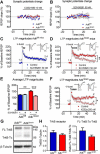
AdkΔbrain mice show enhanced A2A and TrkB receptor- dependent LTP. A, B, The A2AR-selective agonist CGS21680 (10 nm) and antagonist SCH58261 (50 nm) does not affect the fEPSP slope in Adkfl/fl and AdkΔbrain slices (n = 4 mice/genotype/drug). CE, SCH58261 restores normal theta-burst (3 × 3)-induced LTP in AdkΔbrain mice. C, D, Average change in fEPSP slopes in the absence versus presence of SCH58261 in Adkfl/fl (C) and AdkΔbrain (D) slices (n = 5 mice/genotype/drug). E, Percentage of change in the fEPSP slopes recorded at 52–60 min after LTP induction. F, The TrkB inhibitor K252a (200 nm) restores normal theta-burst (3 × 3)-induced LTP in AdkΔbrain slices (absence: 150 ± 6.7% vs presence: 101 ± 7.0%, n = 5 mice each,). Insets in C, D, and F are representative traces of six consecutive responses composed of the stimulus artifact, presynaptic volley, and fEPSP obtained before (1, 3) and after (2, 4) the theta-burst stimuli in the absence (1, 2) and presence (3, 4) of drug. LTP was significant (####) after theta-burst stimuli versus baseline (C, D, F). G, Representative blot of full-length TrkB receptor (FL-TrkB, 145 kDa), truncated TrkB receptor (TrkB-Tc, 95 kDa), and β-tubulin (55 kDa, loading control) is shown. H, Averaged immunodensities of TrkB receptor normalized to β-tubulin and the ratio of FL-TrkB to TrkB-Tc (n = 8 mice/genotype). Results are presented as mean ± SEM. 100% corresponds to the averaged fEPSP slope recorded 10 min before LTP induction. *p < 0.05, **p < 0.01, ####p < 0.0001.
Figure 7.
Figure 7.
Blockade of A2AR activity ameliorates the inducible seizure phenotype and cognitive impairment in AdkΔbrain mice. A, Increased survival time of AdkΔbrain mice treated with SCH 58261 (0.5 mg/kg, i.p., 30 min in advance, n = 6) or Ana-12 (0.5 mg/kg, i.p., 4 h in advance, n = 6) before the DPCPX (3 mg/kg, i.p.) challenge compared with DPCPX controls (n = 10). B, Contingency analysis indicates a significant decrease in the percentage of AdkΔbrain:A2AR−/− mice (3 of 10) that develop the inducible seizure phenotype compared with AdkΔbrain (12 of 12) and AdkΔbrain:A1R−/− (8 of 8) mice. C, Kaplan–Meier survival curve indicating increased AdkΔbrain:A1R−/− (n = 18) mortality compared with AdkΔbrain (n = 18) and AdkΔbrain:A2AR−/− (n = 10). D, Associative learning indexed as total percentage time freezing during the CS 2–4 is restored to Adkfl/fl (n = 13) control levels in AdkΔbrain:A2AR−/− (n = 12) mice and is significantly increased compared with AdkΔbrain (n = 13) mice. E, Contextual memory indexed as total percentage time freezing during the 8 min contextual freezing trial is significantly increased in AdkΔbrain:A2AR−/− (n = 12) and Adkfl/fl (n = 13) mice, compared with AdkΔbrain (n = 13) mice. Data are represented as the mean ± SEM; **p < 0.01, ***p < 0.001, ****p < 0.0001.

Similar articles

Cited by

References

    1. Batalha VL, Pego JM, Fontinha BM, Costenla AR, Valadas JS, Baqi Y, Radjainia H, Müller CE, Sebastião AM, Lopes LV. Adenosine A(2A) receptor blockade reverts hippocampal stress-induced deficits and restores corticosterone circadian oscillation. Mol Psychiatry. 2013;18:320–331. doi: 10.1038/mp.2012.8. - DOI - PubMed
    1. Bjursell MK, Blom HJ, Cayuela JA, Engvall ML, Lesko N, Balasubramaniam S, Brandberg G, Halldin M, Falkenberg M, Jakobs C, Smith D, Struys E, von Döbeln U, Gustafsson CM, Lundeberg J, Wedell A. Adenosine kinase deficiency disrupts the methionine cycle and causes hypermethioninemia, encephalopathy, and abnormal liver function. Am J Hum Genet. 2011;89:507–515. doi: 10.1016/j.ajhg.2011.09.004. - DOI - PMC - PubMed
    1. Boison D. The adenosine kinase hypothesis of epileptogenesis. Prog Neurobiol. 2008;84:249–262. doi: 10.1016/j.pneurobio.2007.12.002. - DOI - PMC - PubMed
    1. Boison D. Adenosine kinase: exploitation for therapeutic gain. Pharmacol Rev. 2013;65:906–943. doi: 10.1124/pr.112.006361. - DOI - PMC - PubMed
    1. Boison D. Adenosinergic signaling in epilepsy. Neuropharmacology. 2016a;104:131–139. - PMC - PubMed

Publication types

LinkOut - more resources