. 2017 Jan 25;45(2):547-555.
doi: 10.1093/nar/gkw1173. Epub 2016 Nov 29.

Elasticity of the transition state for oligonucleotide hybridization

Affiliations

Elasticity of the transition state for oligonucleotide hybridization

Kevin D Whitley et al. Nucleic Acids Res. .

Abstract

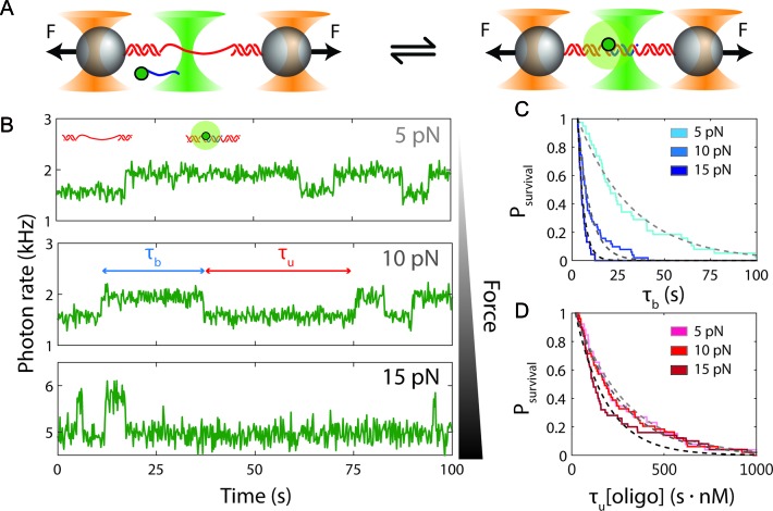
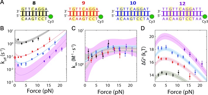
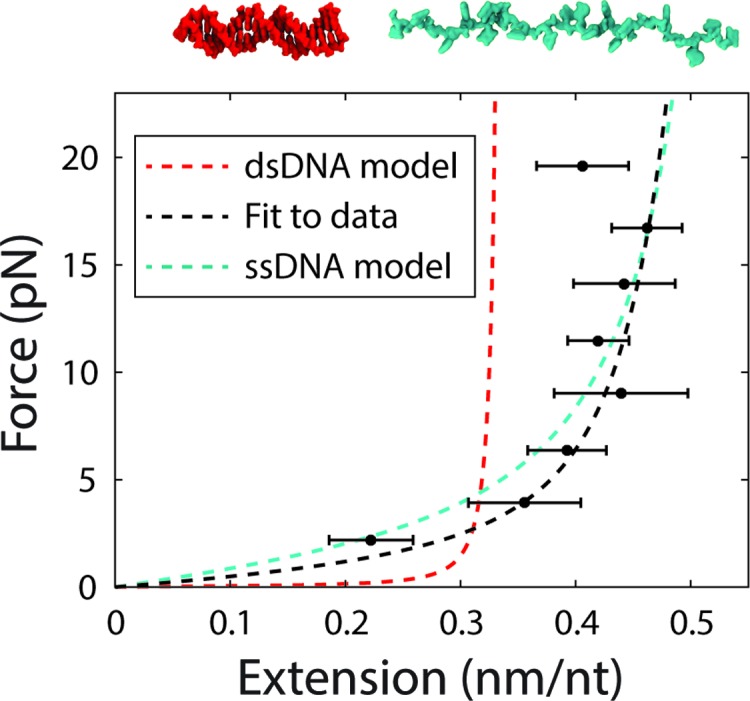
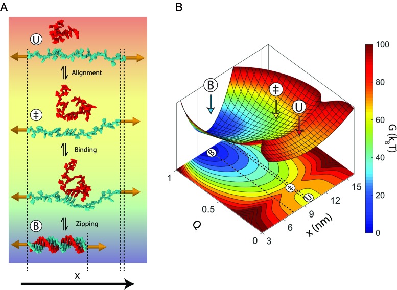
Despite its fundamental importance in cellular processes and abundant use in biotechnology, we lack a detailed understanding of the kinetics of nucleic acid hybridization. In particular, the identity of the transition state, which determines the kinetics of the two-state reaction, remains poorly characterized. Here, we used optical tweezers with single-molecule fluorescence to observe directly the binding and unbinding of short oligonucleotides (7-12 nt) to a complementary strand held under constant force. Binding and unbinding rate constants measured across a wide range of forces (1.5-20 pN) deviate from the exponential force dependence expected from Bell's equation. Using a generalized force dependence model, we determined the elastic behavior of the transition state, which we find to be similar to that of the pure single-stranded state. Our results indicate that the transition state for hybridization is visited before the strands form any significant amount of native base pairs. Such a transition state supports a model in which the rate-limiting step of the hybridization reaction is the alignment of the two strands prior to base pairing.


Figures

Figure 1.
Figure 1.
Measurement of single-oligonucleotide hybridization kinetics under force. (A) Schematic of the hybridization assay (not to scale). An engineered DNA molecule (red) containing a short, central ssDNA region flanked by long double-stranded DNA (dsDNA) handles is held under constant force by polystyrene beads (grey spheres) held in optical traps (orange cones). A fluorescence excitation laser (green cone) is focused on the central ssDNA region. Short oligonucleotides (blue) labeled with a Cy3 fluorophore at the 3′ end (green disk) bind and unbind to the complementary ssDNA sequence in the center of the tethered DNA. The binding and unbinding is observed by the fluorescence emitted from the attached fluorophores. (B) Representative time traces showing 10-nt probes binding and unbinding a DNA construct held under three constant forces (5, 10 and 15 pN, ± 0.02 pN each). The lifetimes of the oligonucleotide bound states, formula image and the unbound states, formula image, are measured from the increase and decrease of the fluorescence intensity. (C and D) The survival probabilities of the bound and unbound states over time for the three forces are shown. The probabilities are fitted to a single exponential function (dotted lines) for each force. Because the binding reaction displays second-order kinetics, the survival probabilities of the unbound states is plotted versus formula image.
Figure 2.
Figure 2.
Force-dependence of oligonucleotide hybridization kinetics and thermodynamics. (A) The four oligonucleotides used in this study, bound to their complementary sequences on the DNA construct (GC pairs highlighted). (B and C) Force-dependence of the unbinding (koff) and binding (kon) rate constants for each probe length (error bars: s.e.m.). The dotted lines show the force-dependent model (Equations 3–5) using parameters obtained from the globally fitted data (Table 1). Open circle: measured zero-force unbinding rate constant from (11). Shaded regions represent 95% confidence intervals. (D) Force-dependence of the standard-state equilibrium free energy Δ(F) between bound and unbound states for each probe length. The dotted lines show the force-dependent model (Equation 6) using parameters from the literature and those determined empirically. Shaded regions represent 95% confidence intervals.
Figure 3.
Figure 3.
Force-extension curve of the transition state for hybridization. The end-to-end extension of the transition state was calculated from Equation (7). The model for the transition state using the fitted parameters for P and h (black dotted line) is plotted alongside the models for dsDNA (red dotted line) and ssDNA (cyan dotted line) for comparison.
Figure 4.
Figure 4.
Model for nucleic acid hybridization. (A) Schematic depicting nucleic acid hybridization of a 22-nt oligonucleotide (red) to a complementary strand (cyan) under a force of 15 pN. In the unbound state (U), the strand under tension encounters a random-coil oligonucleotide. Most encounters between these two strands do not result in duplex formation because they are not aligned properly with respect to one another. When both strands transiently form a short stretch of aligned nucleotides (‡), they are prepared to bind to one another. The two strands then bind and rapidly zip together to form the bound-state duplex (B). The dsDNA in this schematic was created using Visual Molecular Dynamics (VMD) (59) using Protein Data Bank (PDB) entry 1BNA. (B) Model energy landscape corresponding to the schematic in A at 15 pN. Two reaction coordinates are shown: the end-to-end extension of the strand held under tension, x, and the fraction of native duplex contacts formed, Q. Dotted lines on the x-Q projection are shown to clarify the end-to-end extension of each state.

References

    1. Erlich H.A., Gelfand D., Sninsky J.J. Recent advances in the polymerase chain reaction. Science. 1991;252:1643–1651. - PubMed
    1. Raj A., van den Bogaard P., Rifkin S.A., van Oudenaarden A., Tyagi S. Imaging individual mRNA molecules using multiple singly labeled probes. Nat. Methods. 2008;5:877–879. - PMC - PubMed
    1. Dorsett Y., Tuschl T. siRNAs: applications in functional genomics and potential as therapeutics. Nat. Rev. Drug Discov. 2004;3:318–329. - PubMed
    1. Yurke B., Turber A.J., Mills A.P., Jr, Simmel F.C., Neumann J.L. A DNA-fuelled molecular machine made of DNA. Nature. 2000;406:605–608. - PubMed
    1. Sherman W.B., Seeman N.C. A precisely controlled DNA biped walking device. Nano Lett. 2004;4:1203–1207.

Publication types