Repression of YAP by NCTD disrupts NSCLC progression
- PMID: 27903989
- PMCID: PMC5356801
- DOI: 10.18632/oncotarget.13668
Repression of YAP by NCTD disrupts NSCLC progression
Abstract
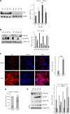
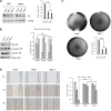
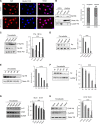
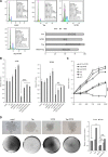
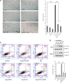
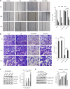
The efficacy of available lung cancer therapeutic interference is significantly limited by various resistance mechanisms to those drugs. Activation of the oncogene YAP underlying the initiation, progression, and metastasis of lung cancer associates with poor prognosis and confers drug resistance against targeted therapy. In this study, we evaluated the specificity of norcantharidin (NCTD) in repressing YAP to inhibit non-small cell lung carcinoma (NSCLC) progression. Our study revealed that YAP signal pathways were aberrantly activated in lung cancer tissues and cells which rendered more proliferative and invasive phenotypes to human lung cancer cells. We confirmed that NCTD specifically repressed YAP signaling pathway to interfere the YAP-mediated non-small cell lung carcinoma progression and metastasis via arresting cell cycle, enhancing apoptosis and inducing senescence. We also found NCTD-mediated repression of YAP decreased epithelial-to-mesenchymal transition (EMT) and reduced the motile and invasive cellular phenotype in vitro via enhancing E-cadherin and decreasing fibronectin/vimentin. Mechanistic investigations revealed that NCTD transcriptionally downregulated YAP and post-translationally modulated the subcellular redistribution of YAP between nucleus and cytoplasm. Collectively, our results indicated that NCTD is a novel therapeutic drug candidate for NSCLC which specifically and sensitively target YAP signal pathway.
Keywords: EMT; NCTD; YAP; cell cycle arrest; lung cancer.
Conflict of interest statement
The authors declare no competing financial interests.
Figures
Similar articles
-
Norcantharidin reverses cisplatin resistance and inhibits the epithelial mesenchymal transition of human non‑small lung cancer cells by regulating the YAP pathway.Oncol Rep. 2018 Aug;40(2):609-620. doi: 10.3892/or.2018.6486. Epub 2018 Jun 12. Oncol Rep. 2018. Retraction in: Oncol Rep. 2021 Jan;45(1):408. doi: 10.3892/or.2020.7830. PMID: 29901163 Free PMC article. Retracted.
-
Suppression of YAP by DDP disrupts colon tumor progression.Oncol Rep. 2018 May;39(5):2114-2126. doi: 10.3892/or.2018.6297. Epub 2018 Mar 6. Oncol Rep. 2018. Retraction in: Oncol Rep. 2021 Jan;45(1):406. doi: 10.3892/or.2020.7843. PMID: 29512779 Free PMC article. Retracted.
-
Norcantharidin inhibits Wnt signal pathway via promoter demethylation of WIF-1 in human non-small cell lung cancer.Med Oncol. 2015 May;32(5):145. doi: 10.1007/s12032-015-0592-0. Epub 2015 Mar 27. Med Oncol. 2015. PMID: 25814287
-
A brief review: some compounds targeting YAP against malignancies.Future Oncol. 2019 May;15(13):1535-1543. doi: 10.2217/fon-2019-0035. Epub 2019 May 8. Future Oncol. 2019. PMID: 31066301 Review.
-
Angiomotin Family Members: Oncogenes or Tumor Suppressors?Int J Biol Sci. 2017 Jun 1;13(6):772-781. doi: 10.7150/ijbs.19603. eCollection 2017. Int J Biol Sci. 2017. PMID: 28656002 Free PMC article. Review.
Cited by
-
Knockdown of YAP inhibits growth in Hep-2 laryngeal cancer cells via epithelial-mesenchymal transition and the Wnt/β-catenin pathway.BMC Cancer. 2019 Jul 3;19(1):654. doi: 10.1186/s12885-019-5832-9. BMC Cancer. 2019. PMID: 31269911 Free PMC article.
-
A Yes-Associated Protein (YAP) and Insulin-Like Growth Factor 1 Receptor (IGF-1R) Signaling Loop Is Involved in Sorafenib Resistance in Hepatocellular Carcinoma.Cancers (Basel). 2021 Jul 29;13(15):3812. doi: 10.3390/cancers13153812. Cancers (Basel). 2021. PMID: 34359714 Free PMC article.
-
Context-dependent roles of YAP/TAZ in stem cell fates and cancer.Cell Mol Life Sci. 2021 May;78(9):4201-4219. doi: 10.1007/s00018-021-03781-2. Epub 2021 Feb 13. Cell Mol Life Sci. 2021. PMID: 33582842 Free PMC article. Review.
-
Hippo/YAP signaling's multifaceted crosstalk in cancer.Front Cell Dev Biol. 2025 Jul 2;13:1595362. doi: 10.3389/fcell.2025.1595362. eCollection 2025. Front Cell Dev Biol. 2025. PMID: 40673277 Free PMC article. Review.
-
The miR 495-UBE2C-ABCG2/ERCC1 axis reverses cisplatin resistance by downregulating drug resistance genes in cisplatin-resistant non-small cell lung cancer cells.EBioMedicine. 2018 Sep;35:204-221. doi: 10.1016/j.ebiom.2018.08.001. Epub 2018 Aug 23. EBioMedicine. 2018. Retraction in: EBioMedicine. 2021 Jan;63:103168. doi: 10.1016/j.ebiom.2020.103168. PMID: 30146342 Free PMC article. Retracted.
References
-
- Jemal A, Bray F, Center MM, Ferlay J, Ward E, Forman D. Global cancer statistics. CA Cancer J Clin. 2011;61:69–90. - PubMed
-
- Smith RA, Cokkinides V, Brawley OW. Cancer screening in the United States, 2009: a review of current American Cancer Society guidelines and issues in cancer screening. CA Cancer J Clin. 2009;59:27–41. - PubMed
-
- Navab R, Strumpf D, Bandarchi B, Zhu CQ, Pintilie M, Ramnarine VR, Ibrahimov E, Radulovich N, Leung L, Barczyk M, Panchal D, To C, Yun JJ, et al. Prognostic gene-expression signature of carcinoma-associated fibroblasts in non-small cell lung cancer. Proc Natl Acad Sci USA. 2011;108:7160–7165. - PMC - PubMed
-
- Siegel R, Naishadham D, Jemal A. Cancer statistics, 2013. CA Cancer J Clin. 2013;63:11–30. - PubMed
MeSH terms
Substances
LinkOut - more resources
Full Text Sources
Other Literature Sources
Medical

