. 2016 Nov 15;8(11):4682-4693.
eCollection 2016.

Melatonin promotes diabetic wound healing in vitro by regulating keratinocyte activity

Affiliations

Melatonin promotes diabetic wound healing in vitro by regulating keratinocyte activity

Ruipeng Song et al. Am J Transl Res. .

Abstract

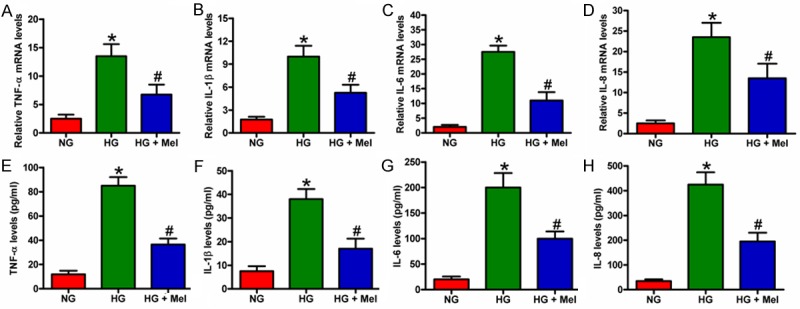
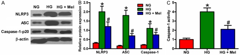
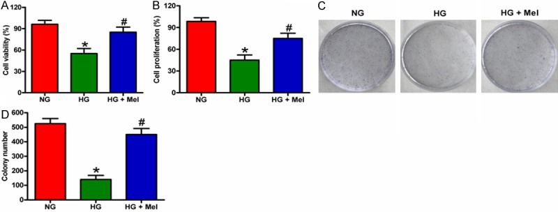
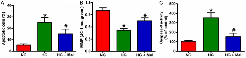
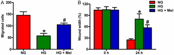
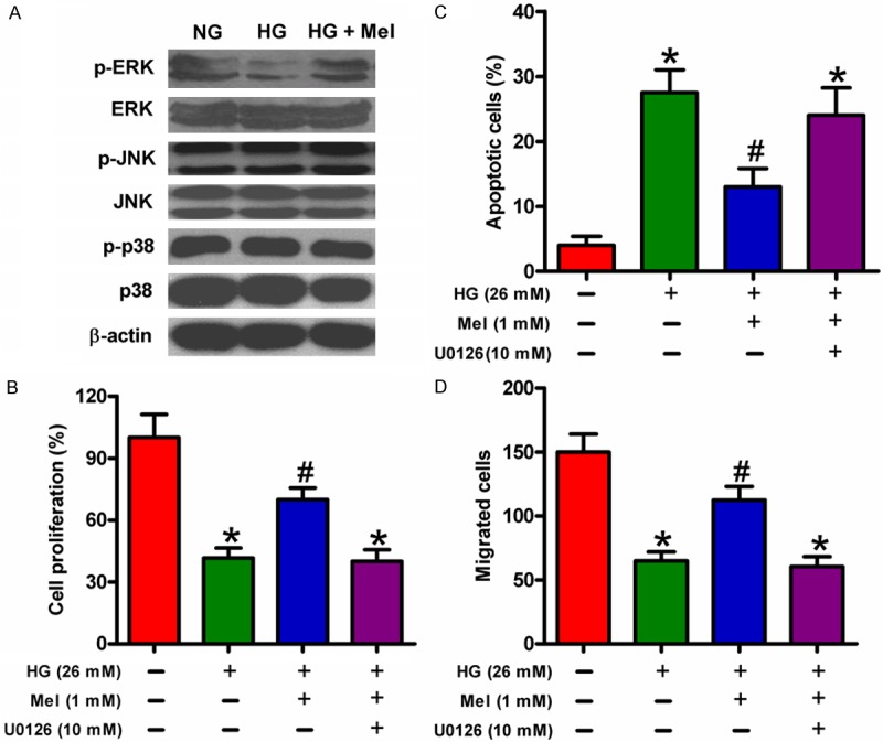
Diabetic patients are at high risk of developing delayed cutaneous wound healing. Proper keratinocyte proliferation and migration are crucial steps during re-epithelialization. Melatonin (Mel) accelerates wound repair in full-thickness incisional wounds; however, its role in diabetic wound healing is unknown. This study explored the role of Mel in diabetic wound healing in vitro by using high glucose (HG)-cultured keratinocytes. Mel reduced the HG-induced mRNA expression and release of pro-inflammatory cytokines, including tumor necrosis factor-α, interleukin (IL)-1β, IL-6, and IL-8, in keratinocytes. Mel inhibited oxidative stress, as evidenced by reduced production of reactive oxygen species and malondialdehyde and increased activity of superoxide dismutase in HG-stimulated keratinocytes. Mel also inhibited HG-induced nucleotide binding oligomerization domain-like receptor family pyrin domain-containing 3 inflammasome activation in keratinocytes. HG-induced reduced migration and proliferation and increased apoptosis of keratinocytes were counteracted by Mel treatment. The pro-proliferative, pro-migratory, and anti-apoptotic effects of Mel on HG-treated keratinocytes were mediated by extracellular signal-regulated kinase signaling pathway. Results collectively suggested that Mel is an alternative therapeutic strategy to ameliorate poor condition for diabetic wound healing by regulating keratinocyte activity.

Keywords: Diabetic wound healing; chronic inflammation; high glucose; keratinocyte; melatonin; oxidative stress.


Figures

Figure 1
Figure 1
Mel reduced the mRNA expression and release of pro-inflammatory cytokines in HG-cultured keratinocytes. Keratinocytes were treated with or without 1 mM Mel 24 h prior to treatment with NG (6 mM) or HG (26 mM) for 72 h. (A-D) mRNA expression of (A) TNF-α, (B) IL-1β, (C) IL-6, and (D) IL-8 was measured by qPCR assays. GAPDH was used as endogenous control. (E-H) Release of (E) TNF-α, (F) IL-1β, (G) IL-6, and (H) IL-8 in the supernatants of the cells were detected by ELISAs. Data are means ± SD of three independent experiments. *P < 0.05 vs. NG group; #P < 0.05 vs. HG group. NG: normal glucose; HG: high glucose; Mel: melatonin.
Figure 2
Figure 2
Mel inhibited ROS and MDA generation and enhanced SOD activity in HG-stimulated keratinocytes. Keratinocytes were treated with or without 1 mM Mel 24 h prior to treatment with NG (6 mM) or HG (26 mM) for 72 h. (A) After treatment, the cells were labeled with DCFH-DA to detect ROS generation. (B and C) (B) MDA level and (C) SOD activity were measured by specific commercial kits. Data are means ± SD of three independent experiments. *P < 0.05 vs. NG group; #P < 0.05 vs. HG group. NG: normal glucose; HG: high glucose; Mel: melatonin.
Figure 3
Figure 3
Mel inhibited HG-induced activation of NLRP3 inflammasome in keratinocytes. Keratinocytes were treated with or without 1 mM Mel 24 h prior to treatment with NG (6 mM) or HG (26 mM) for 72 h. (A) Western blot analyses of NLRP3, ASC, and caspase-1-p20 in keratinocytes. β-actin was used as endogenous control. (B) Quantification of NLRP3, ASC, and caspase-1-p20 in (A) was normalized to that of β-actin. (C) Caspase-1 activity in keratinocytes was measured by a colorimetric assay kit. Data are means ± SD of three independent experiments. *P < 0.05 vs. NG group; #P < 0.05 vs. HG group.
Figure 4
Figure 4
Mel increased the proliferation of HG-treated keratinocytes. (A and B) Keratinocytes were treated with or without 1 mM Mel 24 h prior to treatment with NG (6 mM) or HG (26 mM) for 72 h. (A) Cell viability was assessed by MTT assay. (B) Cell proliferation was measured by BrdU incorporation assay. (C and D) Keratinocytes were treated with or without 1 mM Mel 24 h prior to treatment with NG (6 mM) or HG (26 mM). The cells were subsequently cultured for 14 days, and the medium was replaced every 3 days. The cell colonies were stained and photographed. (D) The number of colonies in (C) was counted. Data are means ± SD of three independent experiments. *P < 0.05 vs. NG group; #P < 0.05 vs. HG group. NG: normal glucose; HG: high glucose; Mel: melatonin.
Figure 5
Figure 5
Mel reduced the apoptosis of HG-treated keratinocytes. Keratinocytes were treated with or without 1 mM Mel 24 h prior to treatment with NG (6 mM) or HG (26 mM) for 72 h. A. Apoptosis of keratinocytes was analyzed by flow cytometry and the apoptotic rate was calculated. B. JC-1 staining was performed to evaluate the cell apoptosis. C. Quantification of caspase-3 activity in keratinocytes. Data are means ± SD of three independent experiments. *P < 0.05 vs. NG group; #P < 0.05 vs. HG group. NG: normal glucose; HG: high glucose; Mel: melatonin.
Figure 6
Figure 6
Mel promoted the migration of HG-treated keratinocytes. Keratinocytes were treated with or without 1 mM Mel 24 h prior to treatment with NG (6 mM) or HG (26 mM) for 72 h. A. Keratinocyte migration was assayed quantitatively using the Transwell migration assay. After adding keratinocytes into the upper wells and incubating overnight, the migrated cells were counted. B. Scratch wound healing assay was performed to evaluate the effect of Mel on keratinocyte migration. The percentage of wound closures at the indicated time points was measured and calculated. Data are means ± SD of three independent experiments. *P < 0.05 vs. NG group; #P < 0.05 vs. HG group. NG: normal glucose; HG: high glucose; Mel: melatonin.
Figure 7
Figure 7
Pro-proliferative, pro-migratory, and anti-apoptotic effects of Mel on HG-treated keratinocytes were mediated by ERK signaling. A. Keratinocytes were treated with or without 1 mM Mel 24 h prior to treatment with NG (6 mM) or HG (26 mM) for 72 h. Representative Western blot results of ERK1/2, p-ERK1/2, JNK, p-JNK, p38 MAPK, and p-p38 MAPK are shown. β-actin was used as endogenous control. B-D. Keratinocytes were preincubated with or without U0126 (10 μM) for 30 min. The cells were treated with or without 1 mM Mel 24 h prior to treatment with NG (6 mM) or HG (26 mM) for 72 h. B. Keratinocyte proliferation was analyzed using the BrdU assay. C. Flow cytometry was performed to measure apoptotic cells. D. Migration of keratinocyte was measured by Transwell migration assay. Data are means ± SD of three independent experiments. *P < 0.05 vs. NG group; #P < 0.05 vs. HG group. HG: high glucose; Mel: melatonin.

References

    1. Boulton AJ, Vileikyte L, Ragnarson-Tennvall G, Apelqvist J. The global burden of diabetic foot disease. Lancet. 2005;366:1719–1724. - PubMed
    1. Bartus CL, Margolis DJ. Reducing the incidence of foot ulceration and amputation in diabetes. Curr Diab Rep. 2004;4:413–418. - PubMed
    1. Singer AJ, Clark RA. Cutaneous wound healing. N Engl J Med. 1999;341:738–746. - PubMed
    1. Falanga V. Wound healing and its impairment in the diabetic foot. Lancet. 2005;366:1736–1743. - PubMed
    1. Pierce GF. Inflammation in nonhealing diabetic wounds: the space-time continuum does matter. Am J Pathol. 2001;159:399–403. - PMC - PubMed

LinkOut - more resources