EXAMINATION OF THE PATELLOFEMORAL JOINT
Abstract
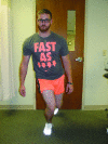
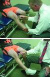
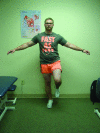
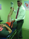
Patellofemoral pain is one of the leading causes of knee pain in athletes. The many causes of patellofemoral pain make diagnosis unpredictable and examination and treatment difficult. This clinical commentary discusses a detailed physical examination routine for the patient with patellofemoral pain. Critically listening and obtaining a detailed medical history followed by a clearly structured physical examination will allow the physical therapist to diagnose most forms of patellofemoral pain. This clinical commentary goes one step further by suggesting an examination scheme and order in which it should be performed during the examination process. This step-by-step guide will be helpful for the student or novice therapist and serve as review for those that are already well versed in patellofemoral examination.
Keywords: Patellofemoral assessment and Clinical reasoning; evaluation.
Figures

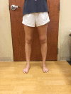
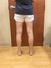
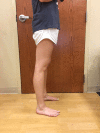


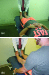




Similar articles
-
Validity of Combining History Elements and Physical Examination Tests to Diagnose Patellofemoral Pain.Arch Phys Med Rehabil. 2018 Apr;99(4):607-614.e1. doi: 10.1016/j.apmr.2017.10.014. Epub 2017 Nov 9. Arch Phys Med Rehabil. 2018. PMID: 29128344
-
Physical examination of the patellofemoral joint.Clin Sports Med. 2014 Jul;33(3):403-12. doi: 10.1016/j.csm.2014.03.002. Epub 2014 Apr 18. Clin Sports Med. 2014. PMID: 24993407 Review.
-
Anterior knee pain in the athlete.Clin Sports Med. 2014 Jul;33(3):437-59. doi: 10.1016/j.csm.2014.03.010. Epub 2014 May 24. Clin Sports Med. 2014. PMID: 24993409 Review.
-
Patellofemoral joint abnormalities in athletes: evaluation by kinematic magnetic resonance imaging.Top Magn Reson Imaging. 1991 Sep;3(4):71-95. doi: 10.1097/00002142-199109000-00007. Top Magn Reson Imaging. 1991. PMID: 1910829
-
Physical and arthroscopic examination techniques of the patellofemoral joint.J Orthop Sports Phys Ther. 1998 Nov;28(5):277-85. doi: 10.2519/jospt.1998.28.5.277. J Orthop Sports Phys Ther. 1998. PMID: 9809276 Review.
Cited by
-
Comparing hip and knee focused exercises versus hip and knee focused exercises with the use of blood flow restriction training in adults with patellofemoral pain.Eur J Phys Rehabil Med. 2022 Apr;58(2):225-235. doi: 10.23736/S1973-9087.22.06691-6. Epub 2022 Jan 5. Eur J Phys Rehabil Med. 2022. PMID: 34985237 Free PMC article. Clinical Trial.
-
Distal femoral osteotomy for the treatment of chronic patellofemoral instability improves gait patterns.Arch Orthop Trauma Surg. 2025 Mar 8;145(1):176. doi: 10.1007/s00402-025-05788-x. Arch Orthop Trauma Surg. 2025. PMID: 40056196 Free PMC article.
-
Botulinum toxin injections as salvage therapy is beneficial for management of patellofemoral pain syndrome.Knee Surg Relat Res. 2021 Oct 29;33(1):39. doi: 10.1186/s43019-021-00121-3. Knee Surg Relat Res. 2021. PMID: 34715941 Free PMC article.
-
Fulkerson Osteotomy Effect on Knee Joint Position Sense, Gait Kinematics, and Functional Level.Orthop J Sports Med. 2025 Apr 30;13(4):23259671251331975. doi: 10.1177/23259671251331975. eCollection 2025 Apr. Orthop J Sports Med. 2025. PMID: 40322747 Free PMC article.
-
Telemedicine Examination of the Knee.Cureus. 2023 Apr 1;15(4):e37009. doi: 10.7759/cureus.37009. eCollection 2023 Apr. Cureus. 2023. PMID: 37139029 Free PMC article.
References
-
- Lankhorst NE Bierma-Zeinstra SM van Middelkoop M. Risk factors for patellofemoral pain syndrome: a systematic review. J Orthop Sports Phys Ther. 2012;42(2):81-94. - PubMed
-
- Nunes GS Stapait EL Kirsten MH de Noronha M Sanatos GM. Clinical test for diagnosis of patellofemoral pain syndrome: systematic review with meta-analysis. Phys Ther Sport. 2013;14(1):54-59. - PubMed
-
- Fredricson M Yoon K. Physical examination and patellofemoral pain syndrome. Am J Phys Med Rehabil. 2006;85(3):234-243. - PubMed
-
- Crossley KM Stefanik JJ Selfe J, et al. 2016 Patellofemoral pain consensus statement from the 4th International Patellofemoral Pain Research Retreat, Manchester. Part 1: Terminology, definitions, clinical examination, natural history, patellofemoral osteoarthritis and patient-reported outcome measures. Br J Sports Med. 2016;50:839-843. - PMC - PubMed
-
- Crossley KM van Middelkoop M Callaghan MJ Collins NJ Rathleff MS Barton CJ. 2016 Patellofemoral pain consensus statement from the 4th International Patellofemoral Pain Research Retreat, Manchester. Part 2: Recommended physical interventions (exercise, taping, bracing, foot orthoses and combined interventions. Br J Sports Med. 2016;50:844-852. - PMC - PubMed
LinkOut - more resources
Full Text Sources
