. 2016 Dec;11(6):903-925.

Current Concepts in Treatment of Patellofemoral Osteochondritis Dissecans

Affiliations

Current Concepts in Treatment of Patellofemoral Osteochondritis Dissecans

Chris Juneau et al. Int J Sports Phys Ther. 2016 Dec.

Abstract

Identification, protection, and management of patellofemoral articular cartilage lesions continue to remain on the forefront of sports medicine rehabilitation. Due to high-level compression forces that are applied through the patellofemoral (PF) joint, managing articular cartilage lesions is challenging for sports medicine specialists. Articular cartilage damage may exist in a wide spectrum of injuries ranging from small, single areas of focal damage to wide spread osteoarthritis involving large chondral regions. Management of these conditions has evolved over the last two centuries, most recently using biogenetic materials and cartilage replacement modalities. The purpose of this clinical commentary is to discuss PF articular cartilage injuries, etiological variables, and investigate the evolution in management of articular cartilage lesions. Rehabilitation of these lesions will also be discussed with a focus on current trends and return to function criteria.

Level of evidence: 5.

Keywords: Articular cartilage; anterior knee pain; osteochondral defect; osteochondritis dissecans; patellofemoral pain.


Figures
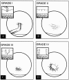
Figure 1.
Figure 1.
Outerbridge classification system. Used with permission of the Journal of Orthopedics.
Figure 2.
Figure 2.
Trochlear chondromalacia on radiographs. Lateral (A) and merchant (B) radiographs of the knee demonstrate undulation of the articular surface of the medial trochlea (white arrows), representing reactive proliferation of the subchondral bone indicating overlying chondromalacia.
Figure 3.
Figure 3.
Trochear microfracture. A) Preoperative sagittal proton density fat-saturated knee MR image shows a full-thickness cartilage defect at the superior aspect of the trochlea with mild subarticular marrow edema (white arrow). B) Post-operative sagittal proton density fat-saturated knee MR image performed 3 months after microfracture demonstrates partial fibrocartilage fill of the microfracture site with minimal residual subarticular marrow edema (white arrowhead). C) Follow-up sagittal proton density fat-saturated knee MR image performed almost 3 years after the microfracture shows near complete fill of the microfracture site with a combination of fibrocartilage and reactive subarticular bone proliferation (black arrowhead). There is complete resolution of the subarticular marrow edema.
Figure 4.
Figure 4.
Trochlear osteochondral allograph. A) Pre-operative sagittal proton density fat-saturated knee MR image shows a large full-thickness cartilage defect in the inferior central trochlea with minimal reactive proliferation of the subchondral cortical bone (white arrow). B & C) Post-operative sagittal proton density fat-saturated (B) and T1 non-fat saturated (C) images of the same region demonstrates interval placement of osteochondral allograph in region of previous full-thickness cartilage loss. The osteochondral graph is flush to the native cartilage with small fluid clefts at the interface between the native and transplanted cartilage (white arrowheads). There is complete osseous incorporation of the graft with normal bone marrow signal within the graft and absence of linear fluid-like signal or cystic change at the interface between the graft and native bone (black arrowheads).

References

    1. Frisbie DD Oxford JT Southwood L, et al. Early events in cartilage repair after subchondral bone microfracture. Clin Orthop Relat Res. 2003(407):215-227. - PubMed
    1. Haggart G. Surgical treatment of degenerative arthritis of the knee joint. J Bone Joint Surg Am. 1940;22-B:717-729.
    1. Magnuson P. Joint debridement: surgical treatment of degenerative arthritis. Surgical Gynecology and Obstetrics. 1941;73:1-9. - PubMed
    1. Kruger T Wohlrab D Birke A Hein W. Results of arthroscopic joint debridement in different stages of chondromalacia of the knee joint. Arch Orthop Trauma Surg. 2000;120(5-6):338-342. - PubMed
    1. Pridie KH. A new method of treatment for severe fractures of the os calcis; a preliminary report. Surg Gynecol Obstet. 1946;82:671-675. - PubMed

LinkOut - more resources