. 2017 Jul;222(5):2309-2328.
doi: 10.1007/s00429-016-1343-5. Epub 2016 Nov 30.

Distinct cortical and sub-cortical neurogenic domains for GABAergic interneuron precursor transcription factors NKX2.1, OLIG2 and COUP-TFII in early fetal human telencephalon

Affiliations

Distinct cortical and sub-cortical neurogenic domains for GABAergic interneuron precursor transcription factors NKX2.1, OLIG2 and COUP-TFII in early fetal human telencephalon

Ayman Alzu'bi et al. Brain Struct Funct. 2017 Jul.

Abstract

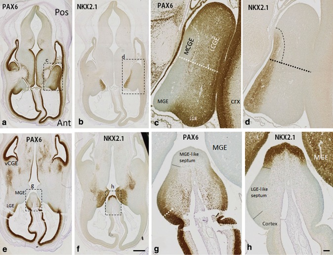
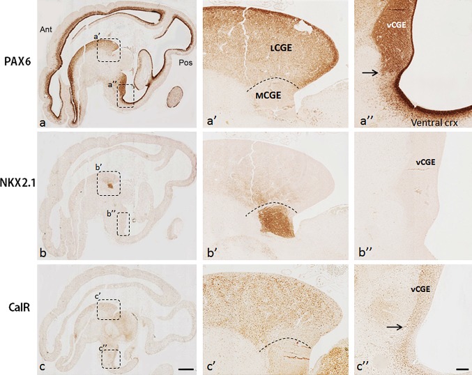
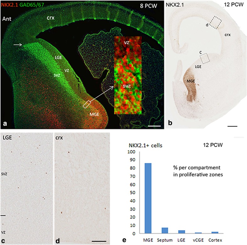
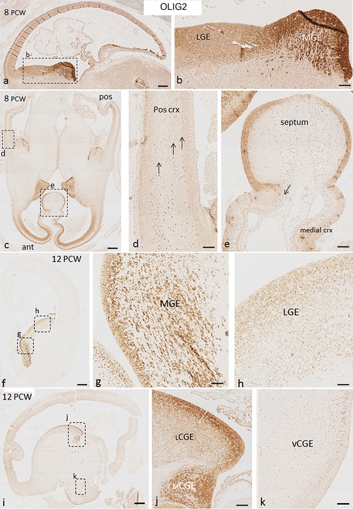
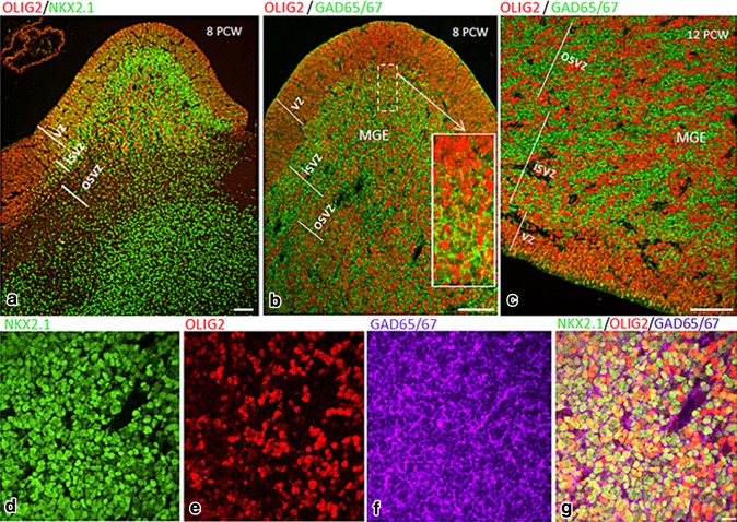
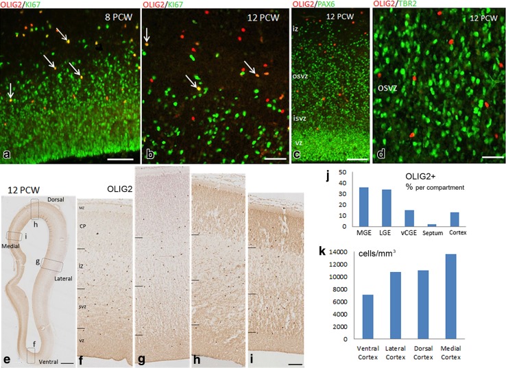
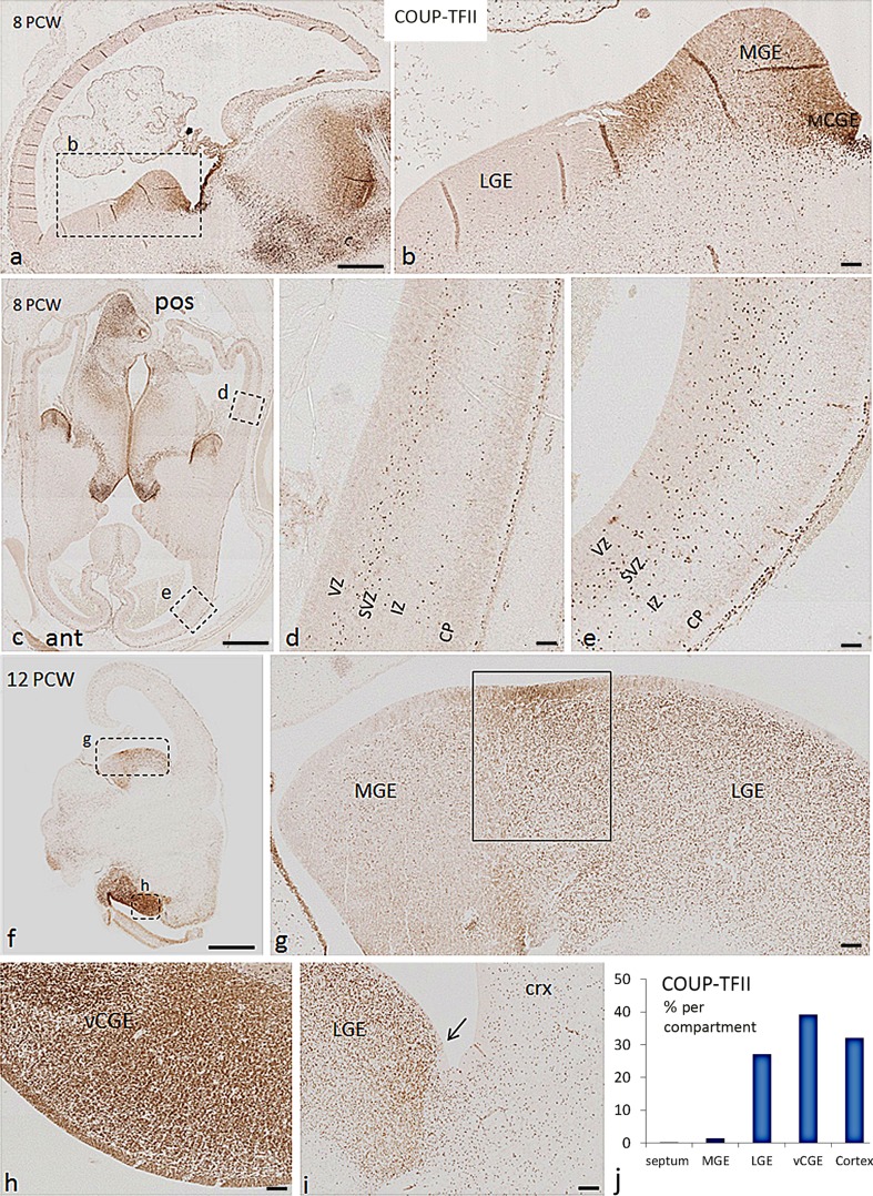
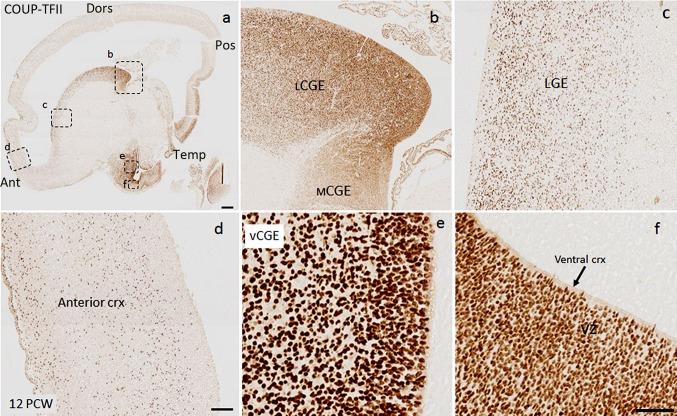
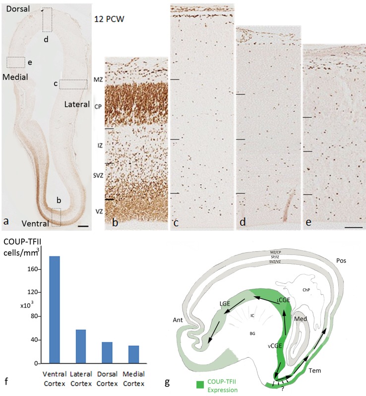
The extent of similarities and differences between cortical GABAergic interneuron generation in rodent and primate telencephalon remains contentious. We examined expression of three interneuron precursor transcription factors, alongside other markers, using immunohistochemistry on 8-12 post-conceptional weeks (PCW) human telencephalon sections. NKX2.1, OLIG2, and COUP-TFII expression occupied distinct (although overlapping) neurogenic domains which extended into the cortex and revealed three CGE compartments: lateral, medial, and ventral. NKX2.1 expression was very largely confined to the MGE, medial CGE, and ventral septum confirming that, at this developmental stage, interneuron generation from NKX2.1+ precursors closely resembles the process observed in rodents. OLIG2 immunoreactivity was observed in GABAergic cells of the proliferative zones of the MGE and septum, but not necessarily co-expressed with NKX2.1, and OLIG2 expression was also extensively seen in the LGE, CGE, and cortex. At 8 PCW, OLIG2+ cells were only present in the medial and anterior cortical wall suggesting a migratory pathway for interneuron precursors via the septum into the medial cortex. By 12 PCW, OLIG2+ cells were present throughout the cortex and many were actively dividing but without co-expressing cortical progenitor markers. Dividing COUP-TFII+ progenitor cells were localized to ventral CGE as previously described but were also numerous in adjacent ventral cortex; in both the cases, COUP-TFII was co-expressed with PAX6 in proliferative zones and TBR1 or calretinin in post-mitotic cortical neurons. Thus COUP-TFII+ progenitors gave rise to pyramidal cells, but also interneurons which not only migrated posteriorly into the cortex from ventral CGE but also anteriorly via the LGE.

Keywords: Ganglionic eminences; Inhibitory interneurons; Neurodevelopment; Neuronal fate specification; Pallium; Subpallium.


Figures

Fig. 1
Fig. 1
Complementary expression of PAX6 and NKX2.1 in the ganglionic eminences and septum of human fetal forebrain at 8 PCW. a PAX6 was expressed in a gradient with higher expression in the proliferative zone of the cortex to lower expression in the LGE and its caudal extension (lCGE). b NKX2.1 expression was mainly confined to the MGE and its caudal extension (mCGE). c, d Higher magnification of boxed areas in a and b. e, f Ventral sections cut at the level of the septum; PAX6 was densely expressed in the proliferative zone of vCGE, but no NKX2.1 expression was found in vCGE. g, h Higher magnification of boxed areas in e and f. Similar to the ganglionic eminences, PAX6 was expressed in a gradient from the cortex part to dorsal part of the septum (LGE-like septum) and NKX2.1 was exclusively expressed in the most ventral part of the septum (MGE-like septum). Scale bars 1 mm in f (and for a, b, and e); 100 µm in h (and for c, d, and g). Ant anterior, Pos posterior
Fig. 2
Fig. 2
Subdivisions of the CGE in sagittal sections at 12 PCW. a, a′, a″ PAX6 was expressed in the proliferative zone of lCGE but not mCGE; PAX6 was also expressed in the vCGE with a distinct cortical/subcortical boundary (arrow in a″). b, b′, b″ NKX2.1 was largely confined to the caudal extension of MGE (mCGE) but with some dispersion into the lCGE. c, c′, c″ Calretinin (CalR) was preferentially expressed in the VZ and SVZ of the lCGE and only scattered cells were observed in mCGE; CalR was also preferentially expressed in vCGE with distinct pallial/subpallial boundary (arrow in c″). Scale bars 1 mm in c (and for a, b) c 100 µm in c″ (and for a′, a″, b′, b″, c′). Ant anterior, Pos posterior
Fig. 3
Fig. 3
Expression of NKX2.1 in the human fetal forebrain. a Double labelling for GAD65/67 (green) and NKX2.1 (red) in coronal section at 8 PCW. GAD65/67 was expressed mainly in the subventricular zone (SVZ) of the MGE and LGE. In LGE, GAD65/67 immunoreactivity also showed a clear cortical/subcortical boundary anteriorly (Ant; arrow). NKX2.1 was expressed in the ventricular zone (VZ) and subventricular zone (SVZ) of the MGE, and in cells probably migrating through the non-proliferative mantle zone of LGE toward the cortex (crx). The high magnification inset in a shows NKX2.1/GAD65/67 co-localisation in the cells of the SVZ only (NKX2.1 in the nuclei, GAD65/67 in the cytoplasm). No NKX2.1+ cells were found in the cortex at 8 PCW. bd At 12 PCW, the majority of cells in the MGE were NKX2.1 immunoreactive; scattered NKX2.1+ cells were found in the proliferative zones of the LGE and cortex. e Distribution of NKX2.1+ cells in the proliferative zones of different regions of human fetal forebrain at 12 PCW (see Table 2 for more details). In a, green vertical stripes in the cortex are an artefact caused by section folding. Scale bars 1 mm in a and b, 200 µm in the inset for a; 100 µm in d (and for c)
Fig. 4
Fig. 4
Expression pattern of OLIG2 in human fetal forebrain at 8 and 12 PCW. a OLIG2 was strongly expressed in the proliferative zones of MGE, weaker expression was observed in LGE. b Higher magnification of boxed area in a. c Anterior (ant) cortex was heavily populated with OLIG2+ cells, whereas no OLIG2+ cells were found in the most posterior cortex. d Higher magnification of boxed area in c, OLIG2+ cells from the GE appeared to be only starting to invade the posterior cortex (arrows). e OLIG2 was expressed in the proliferative zone of septum, with a stream of OLIG2+ cells appearing to migrate (arrow) into the medial cortex (crx). f, g Similar to 8 PCW, OLIG2 was strongly expressed in the proliferative zone of MGE at 12 PCW, with aggregations of OLIG+ cells amongst OLIG2− cells. h Relatively weaker expression was observed in LGE. i, j Expression pattern of OLIG2 in the MGE and LGE extended to the mCGE and lCGE, respectively. k Scattered OLIG2+ cells were observed in the vCGE. In a dark vertical stripes in the cortex are an artefact caused by section folding. Scale bars 1 mm in a, c, f, and i; 100 µm in b, d, e, g, h, j, and k
Fig. 5
Fig. 5
a Double labelling for NKX2.1 (green) and OLIG2 (red) in the MGE at 8 PCW showed three populations of cells located in the MGE: the first expressed only OLIG2 (red), the second expressed only NKX2.1 (green), and a third population co-localized these two markers (yellow). b, c Double labelling for OLIG2 (red) and GAD65/67 (green) in the MGE at 8 and 12 PCW showed that most of the OLIG2+ cells (nuclear staining) were double labelled with GAD65/67. Magnification inset in b shows double labelling in the SVZ but not the VZ. dg Triple labelling for NKX2.1 (green), OLIG2 (red), and GAD65/67 (purple) in the MGE at 8 PCW showed that many cells co-expressed the transcription factors NKX2.1 and OLIG2 (nuclear staining, yellow) and GAD65/67 (cytoplasmic, purple). Scale bars 100 µm in a; 50 µm in b (15 µm in inset), c 20 µm in g (and for df)
Fig. 6
Fig. 6
a Significant number of OLIG2+ cells (red) in the anterior cortex co-expressed the cell division marker KI67 (green) at 8 PCW (arrows). b Many OLIG2+ cells co-expressed KI67 at 12 PCW (arrows). c OLIG2+ cells (red) did not co-express the radial glial progenitor cell marker PAX6 (green). d OLIG2+ cells (red) did not co-express the intermediate progenitor cell marker TBR2 (green). e OLIG2 expression in the cortex (coronal section) at 12 PCW. OLIG2 expression in ventral cortex (f), lateral cortex (g), dorsal cortex (h), and medial cortex (i). j Distribution of OLIG2+ cells in the proliferative zones of different regions of human fetal forebrain at 12 PCW. k Average density of OLIG2+ cells in the ventral cortex, lateral cortex, dorsal cortex, and medial cortex of 12 PCW human fetal brain. Scale bars 50 µm in a; 20 µm in b; 50 µm in c; 20 µm in d; 1 mm in e; and 100 µm in i (and for g and h)
Fig. 7
Fig. 7
Expression pattern of COUP-TFII in human fetal forebrain at 8 and 12 PCW. a, b At 8 PCW, COUP-TFII was mainly expressed in the caudal part of the ganglionic eminences and at the boundary between MGE and LGE. Scattered cells were also observed in MGE and LGE. c Expression pattern of COUP-TFII in the anterior (ant) and posterior (pos) cortex at 8 PCW. d Magnification of boxed area in the posterior cortex in c; COUP-TFII expression appeared to be restricted to two migratory streams, one in the subventricular zone (SVZ) and one at the border between the intermediate zone (IZ) and the cortical plate (CP). e Magnification of boxed area in the anterior cortex in c COUP-TFII+ cells was found in all layers of the cortex. fh At 12 PCW, COUP-TFII was highly expressed in vCGE, moderately expressed in LGE, with scattered cells found in the MGE. Strong expression was observed in the VZ/SVZ at the boundary between MGE and LGE (boxed area in g). i Distribution of COUP-TFII expression showed a distinct cortical/subcortical boundary (arrow) between LGE and cortex (crx). j The distribution of COUP-TFII+ cells in the proliferative zones of different regions of human fetal forebrain at 12 PCW. Scale bars 1 mm a, c, f; 100 µm in b, d, e, g, h, and i
Fig. 8
Fig. 8
COUP-TFII was predominantly expressed in vCGE but spanned sub-cortical/cortical domains in human fetal forebrain. a COUP-TFII expression in a sagittal section at 12 PCW. bd COUP-TFII was highly expressed in the proliferative zone of lCGE with lower expression in the LGE and anterior cortex (ant). e The strongest expression was observed in the proliferative zone of vCGE where COUP-TFII+ cells were not organized radially. f Strong expression of COUP-TFII in the VZ of ventral/temporal cortex with radial nuclear morphology of COUP-TFII+ cells. Scale bars 1 mm in a; 100 µm in d (and for b and c); 100 µm in f (and for e). Dors dorsal, Pos posterior, Temp temporal, Crx cortex
Fig. 9
Fig. 9
a COUP-TFII expression in frontal cortex (coronal section, 12 PCW). b COUP-TFII expression in ventral cortex, c lateral cortex, d dorsal cortex, and e the medial cortex. f Average density of COUP-TFII+ cells in the ventral, lateral, dorsal, and medial cortex. g Schematic diagram showing the distribution of COUP-TFII+ cells in the telencephalon and proposed migratory paths from the vCGE to the anterior and posterior cortex (large arrows). COUP-TFII progenitors also underwent division in the ventral cortex, but the migratory paths and phenotype of the cells remain unclear (small arrows). Scale bars 1 mm in a, 100 µm in e (and for bd). Ant anterior cortex, Pos posterior cortex, Tem temporal cortex, Med medial cortex, BG basal ganglia, ChP choroid plexus, MZ/CP marginal zone/cortical plate, SP/IZ presubplate/intermediate zone, SVZ/VZ subventricular zone/ventricular zone

References

    1. Al-Jaberi N, Lindsay S, Sarma S, Bayatti N, Clowry GJ. The early fetal development of human neocortical gabaergic interneurons. Cereb Cortex. 2015;25:631–645. doi: 10.1093/cercor/bht254. - DOI - PMC - PubMed
    1. Arshad A, Vose LR, Vinukonda G, Hu F, Yoshikawa K, Csiszar A, Brumberg JC, Ballabh P. Extended production of cortical interneurons into the third trimester of human gestation. Cereb Cortex. 2016;26:2242–2256. doi: 10.1093/cercor/bhv074. - DOI - PMC - PubMed
    1. Ballesteros-Yáñez I, Muñoz A, Contreras J, Gonzalez J, Rodriguez-Veiga E, DeFelipe J. Double bouquet cell in the human cerebral cortex and a comparison with other mammals. J Comp Neurol. 2005;486:344–360. doi: 10.1002/cne.20533. - DOI - PubMed
    1. Bayatti N, Moss JA, Sun L, Ambrose P, Ward JF, Lindsay S, Clowry GJ. A molecular neuroanatomical study of the developing human neocortex from 8 to 17 postconceptional weeks revealing the early differentiation of the subplate and subventricular zone. Cereb Cortex. 2008;18:1536–1548. doi: 10.1093/cercor/bhm184. - DOI - PMC - PubMed
    1. Bayatti N, Sarma S, Shaw C, Eyre JA, Vouyiouklis DA, Lindsay S, Clowry GJ. Progressive loss of PAX6, TBR2, NEUROD and TBR1 mRNA gradients correlates with translocation of EMX2 to the cortical plate during human cortical development. Eur J Neurosci. 2008;28:1449–1456. doi: 10.1111/j.1460-9568.2008.06475.x. - DOI - PMC - PubMed

MeSH terms

LinkOut - more resources