Review
. 2017 Feb;8(1):49-57.
doi: 10.1007/s13244-016-0535-0. Epub 2016 Nov 30.

Imaging of patellar fractures

Affiliations
Review

Imaging of patellar fractures

Mohamed Jarraya et al. Insights Imaging. 2017 Feb.

Abstract

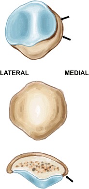
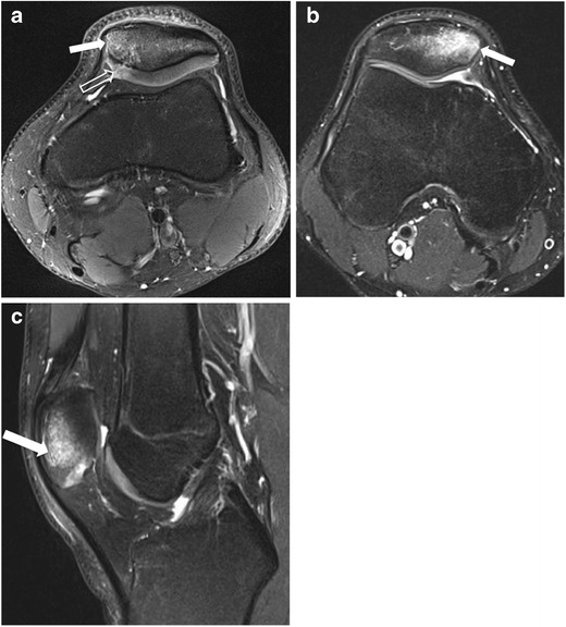
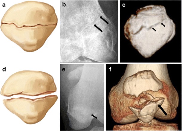
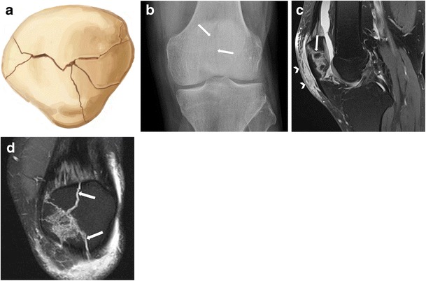
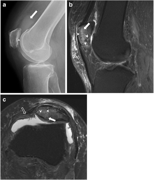
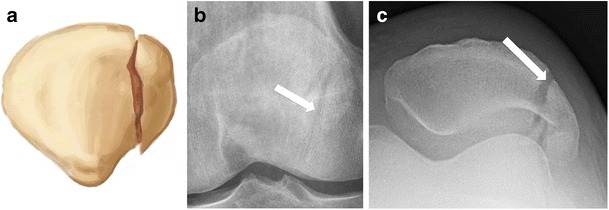
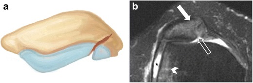
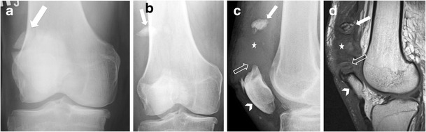
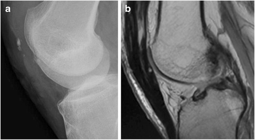
Patellar fractures account for approximately 1% of all skeletal fractures and may result from direct, indirect, or combined trauma. Because of the importance of patellar integrity for knee extension and the risk of associated injury to the extensor mechanism, accurate reporting and description of fracture type is paramount for appropriate management. This pictorial essay aims to review the normal anatomy of the patella, the mechanisms of injury and different types of patellar fractures, with a brief introduction to therapeutic management. Teaching Points • Patellar fractures are classified according to their morphology and degree of displacement.• Direct trauma results in stellate fractures.• Indirect trauma results in transverse fractures.• Displacement should raise suspicion for retinacular injury.

Keywords: Conventional radiograph; Extensor mechanism rupture; Fracture; MRI; Patella.


Figures

Fig. 1
Fig. 1
Drawing showing, from top to bottom, the anterior, posterior, and axial views of the patella. The proximal part is termed the basis, and the distal part, the apex. The distal pole is entirely devoid of articular cartilage. A longitudinal ridge divides the cartilaginous surface into medial and lateral facets. The odd facet (arrows) is located at the peripheral aspect of the medial facet, and is devoid of cartilage
Fig. 2
Fig. 2
Examples of patellar contusions. (a) Axial fat-suppressed proton density-weighted MR image in a 20-year-old man shows hyperintense signal of the medial patellar bone marrow (solid arrow) and a fracture line through the adjacent cartilage (open arrow). Conventional radiographs were negative for fracture (not shown). (b) Axial and (c) sagittal fat-suppressed proton density MR images in another 20-year-old man following direct trauma, revealing bone marrow oedema (arrow) without cartilage damage. Conventional radiographs were also negative for fracture (not shown)
Fig. 3
Fig. 3
Two examples of transverse fractures in young adults. (a, d) Drawing, (b, e) anteroposterior radiograph, and (c, f) 3D CT surface reconstruction. (ac) Non-displaced and (df) displaced transverse fractures through the body of the patella. Displaced fractures are associated with a higher risk of retinacular injury
Fig. 4
Fig. 4
Patellar fracture of the lower pole in a 39-year-old man. (a) Drawing and (b) anteroposterior and (c) lateral radiographs show a transverse fracture of the lower pole of the patella
Fig. 5
Fig. 5
Sleeve fracture of the lower patellar pole in an 11-year-old boy after falling on his flexed knee during a football game, with a popping sound. The patient presented to the ER 5 days later. (a) Lateral knee radiograph and (b) sagittal fat-suppressed T2-weighted MR image 3 days after knee x-ray show small bony fragment fracture from the lower pole (arrow), extending anteriorly, with detachment of a thin shell of cortical bone (arrowheads), consistent with a sleeve fracture. Note the injury of the distal attachment of the patellar tendon (open arrow). Images courtesy of Arnold Carlson Merrow, Jr., MD - Cincinnati Children’s Hospital Medical Center, OH, USA
Fig. 6
Fig. 6
(a) Drawing, (b) anteroposterior radiograph, and (c) sagittal and (d) coronal fat-suppressed proton density MR images showing non-displaced stellate fracture (solid arrows) in a 36-year-old man. Note the subcutaneous prepatellar oedema related to direct trauma mechanism (arrowheads)
Fig. 7
Fig. 7
CT reformatted image showing displaced comminuted fracture. (a) Drawing, (b) anteroposterior radiograph, and (c) sagittal computed tomography reformat in a middle-aged man following direct blow to the knee in a car accident, showing displaced comminuted fracture. Note that the degree of displacement is better appreciated on the reformatted CT image than on conventional radiograph
Fig. 8
Fig. 8
Stellate fracture with retinacular injury in a 40-year-old woman with persistent anterior knee pain for 2 weeks ago after a fall. (a) Lateral knee radiograph shows joint effusion (arrow) and a subtle linear lucency through the patellar undersurface (arrowhead), suggesting fracture. (b) Sagittal and (c) axial fat-suppressed proton density MR images reveal several fracture lines (arrowheads) consistent with stellate fracture, bone marrow oedema (solid arrow), and partial thickness tear of the medial retinaculum from its patellar attachment (open arrow)
Fig. 9
Fig. 9
Vertical fracture of the right patella in a 34-year-old man. (a) Drawing and (b) anteroposterior and (c) skyline views of the right knee show a vertical fracture of the lateral facet (red arrows). Note that the fracture line is more conspicuous on the tangential than the anteroposterior view
Fig. 10
Fig. 10
Osteochondral fracture from lateral patellar dislocation in a 27-year-old woman. (a) Drawing shows an incomplete osteochondral fracture. (b) Axial fat-suppressed proton density MR image shows an osteochondral injury to the articular surface (open arrow) with bone marrow oedema of the medial patella (solid arrow) and lateral aspect of the lateral femoral condyle (arrowhead), in keeping with transient lateral patellar dislocation. Note also a moderate amount of joint effusion (star)
Fig. 11
Fig. 11
Two examples of incidental bipartite patella. (a) Anteroposterior and (b) tangential patellar radiographs of the right knee in the first example show a patellar fragment at the superolateral aspect of the right patella consistent with bipartite patella. In the second, (c) axial and (d) coronal fat-suppressed proton density MR images of the right knee demonstrate a well-corticated osseous fragment (solid arrow) at the superolateral aspect of the patella. Note the high signal of the synchondrosis (open arrows), the well-preserved underlying cartilage of the patella, and the lack of bone marrow oedema at the adjacent bone that differentiate this variant from a patellar fracture
Fig. 12
Fig. 12
Separation of patella bipartite in a 70-year-old man. (a) Anteroposterior radiograph of the right knee demonstrates an incidental bipartite patella (arrow). Two years later, the patient presented with anterior knee pain after a fall. (b) Anteroposterior and (c) lateral radiographs and (d) sagittal T1-weighted MR image show separation of the superolateral fragment (solid arrow) within the anterolateral soft tissues of the knee, patella baja (arrowhead), obliteration of the suprapatellar fat (open arrow), and soft tissue swelling and haemorrhage from disruption of the quadriceps tendon (star)
Fig. 13
Fig. 13
Frontal knee radiographs showing examples of common patellar fracture fixation constructs using tension band wiring. (a) Modified anterior tension band (MATB) technique (also called K-wire technique) with vertical figure eight loop configuration and wire twist. (b) MATB with vertical figure eight and wire twist in addition to cerclage. (c) Cannulated screws with horizontal figure eight loop
Fig. 14
Fig. 14
Example of total patellectomy. (a) Lateral knee radiograph and (b) sagittal proton density MR image of the knee after total patellectomy as a result of surgery following a severely comminuted fracture

References

    1. Melvin JS, Karunakar MA. Patella fractures and extensor mechanism injuries. In: Court-Brown CB, Heckman JD, McQueen MM, Ricci WM, Tornetta P III, editors. Rockwood and Green’s Fractures in adults. Philadelphia: Wolters Kluwer; 2004. pp. 2269–2302.
    1. Gwinner C, Mardian S, Schwabe P, Schaser KD, Krapohl BD, Jung TM (2016) Current concepts review: Fractures of the patella. GMS Interdiscip Plast Reconstr Surg DGPW 5:Doc01 - PMC - PubMed
    1. Pengas IP, Assiotis A, Khan W, Spalding T. Adult native knee extensor mechanism ruptures. Injury. 2016 - PubMed
    1. Melvin JS, Mehta S. Patellar fractures in adults. J Am Acad Orthop Surg. 2011;19:198–207. doi: 10.5435/00124635-201104000-00004. - DOI - PubMed
    1. Scapinelli R. Blood supply of the human patella. Its relation to ischaemic necrosis after fracture. J Bone Joint Surg (Br) 1967;49:563–570. - PubMed

LinkOut - more resources