. 2016 Nov 30;540(7631):134-138.
doi: 10.1038/nature20169.

The SND proteins constitute an alternative targeting route to the endoplasmic reticulum

Affiliations

The SND proteins constitute an alternative targeting route to the endoplasmic reticulum

Naama Aviram et al. Nature. .

Abstract

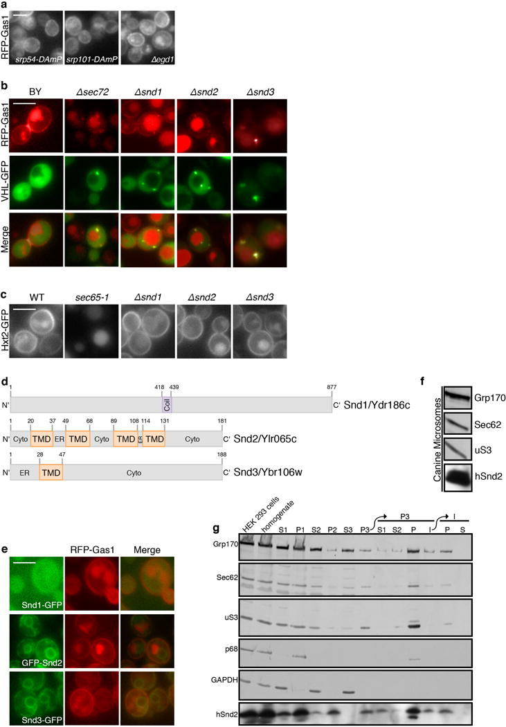
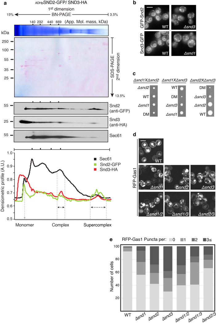
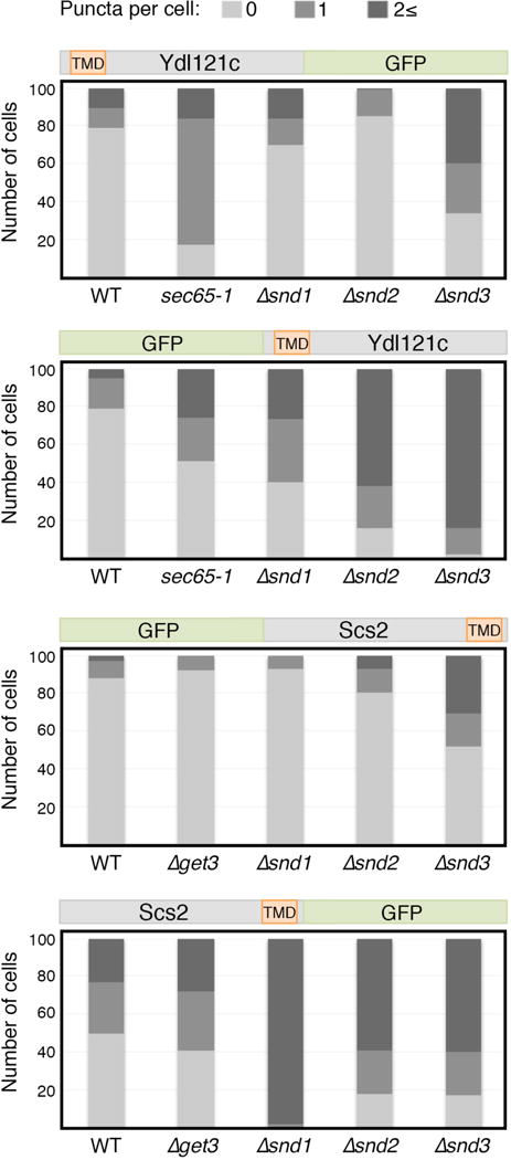
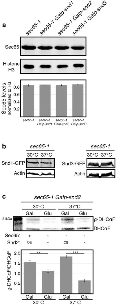
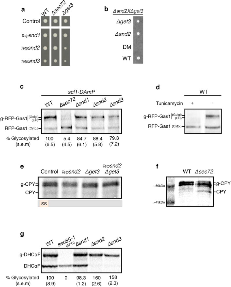
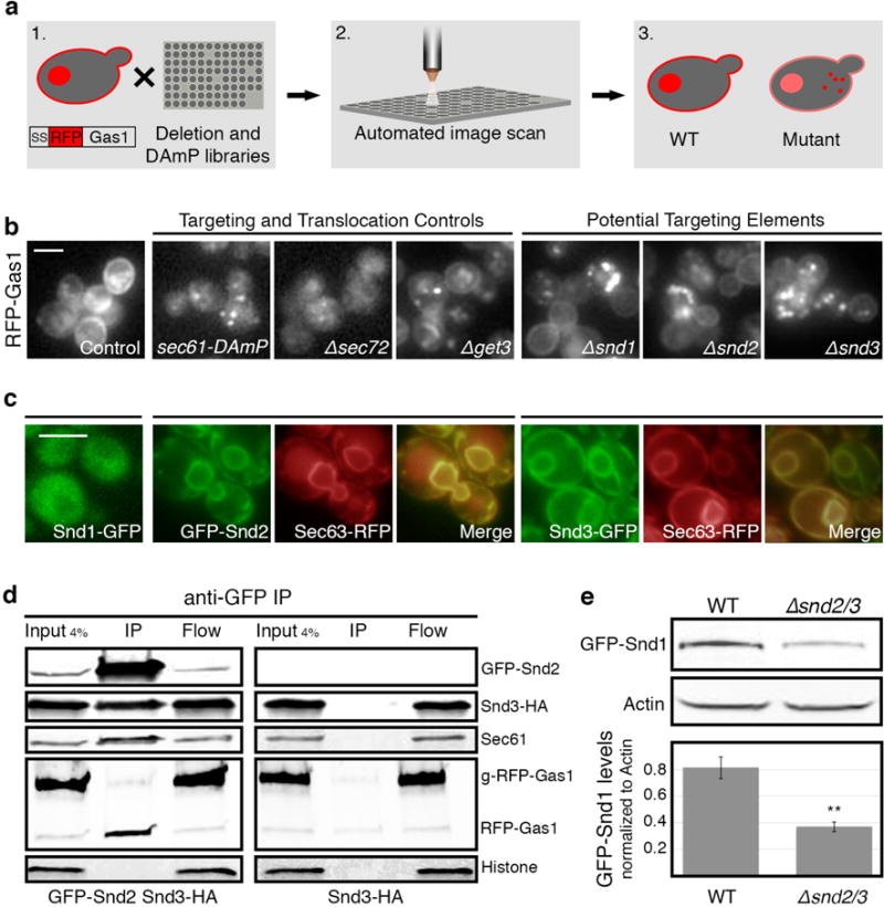
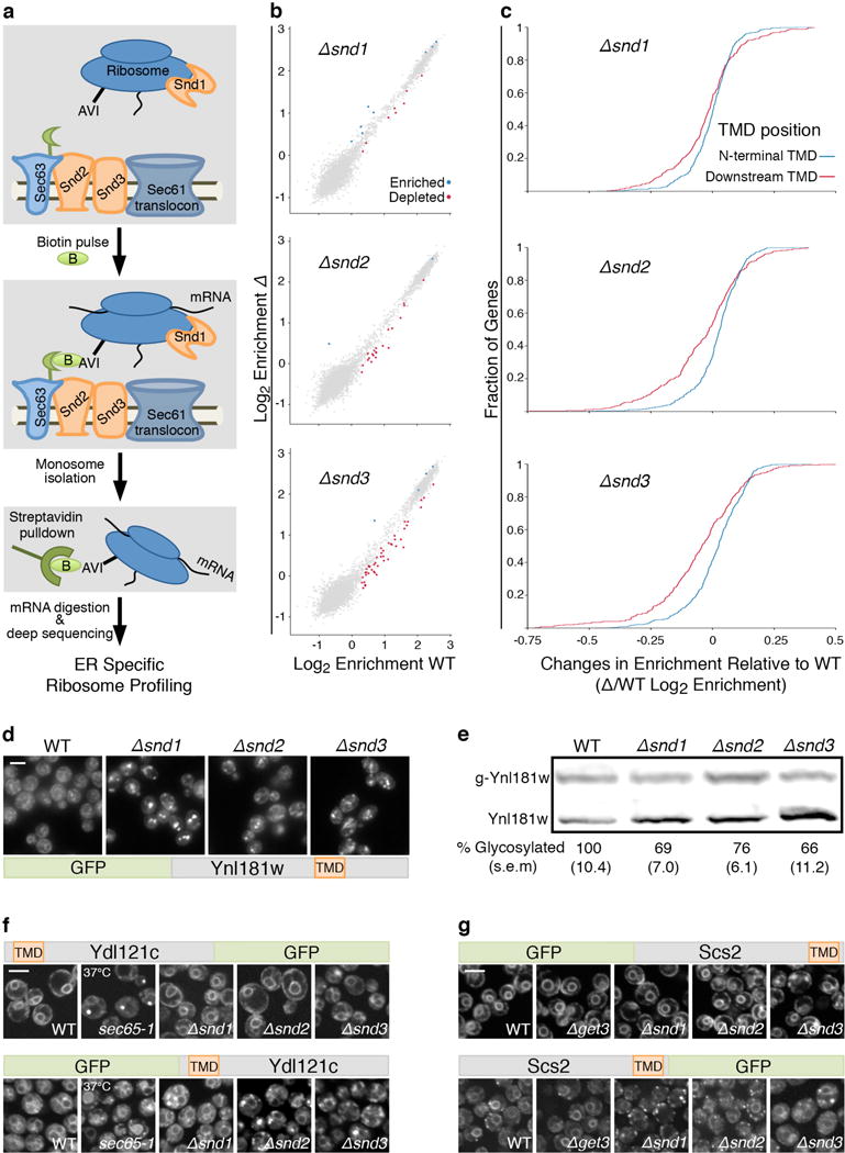
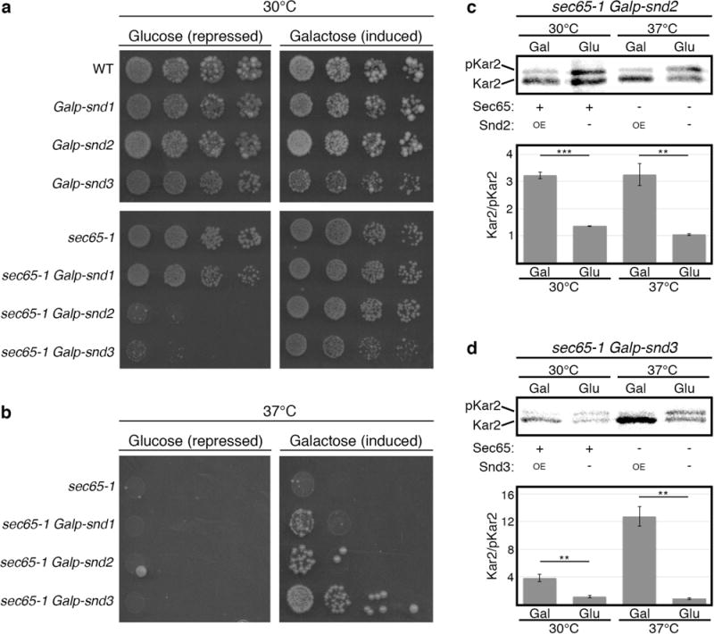
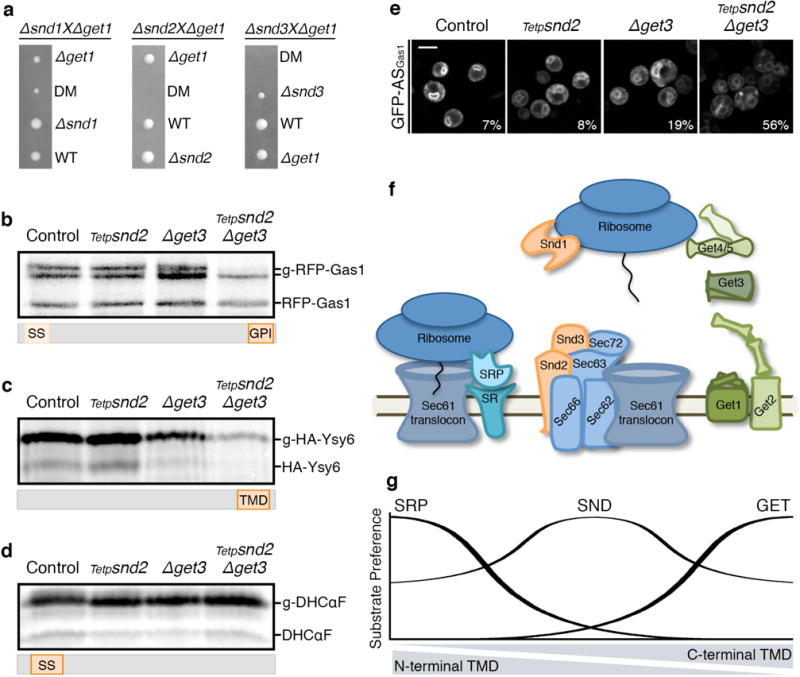
In eukaryotes, up to one-third of cellular proteins are targeted to the endoplasmic reticulum, where they undergo folding, processing, sorting and trafficking to subsequent endomembrane compartments. Targeting to the endoplasmic reticulum has been shown to occur co-translationally by the signal recognition particle (SRP) pathway or post-translationally by the mammalian transmembrane recognition complex of 40 kDa (TRC40) and homologous yeast guided entry of tail-anchored proteins (GET) pathways. Despite the range of proteins that can be catered for by these two pathways, many proteins are still known to be independent of both SRP and GET, so there seems to be a critical need for an additional dedicated pathway for endoplasmic reticulum relay. We set out to uncover additional targeting proteins using unbiased high-content screening approaches. To this end, we performed a systematic visual screen using the yeast Saccharomyces cerevisiae, and uncovered three uncharacterized proteins whose loss affected targeting. We suggest that these proteins work together and demonstrate that they function in parallel with SRP and GET to target a broad range of substrates to the endoplasmic reticulum. The three proteins, which we name Snd1, Snd2 and Snd3 (for SRP-independent targeting), can synthetically compensate for the loss of both the SRP and GET pathways, and act as a backup targeting system. This explains why it has previously been difficult to demonstrate complete loss of targeting for some substrates. Our discovery thus puts in place an essential piece of the endoplasmic reticulum targeting puzzle, highlighting how the targeting apparatus of the eukaryotic cell is robust, interlinked and flexible.


Conflict of interest statement

Ribosome-profiling data are deposited in Gene Expression Omnibus (GEO) under accession number GSE85686. The authors declare no competing financial interests.

Figures

Extended Data Figure 1
(a) RFP-Gas1 localization is not affected by mutants in SRP or NAC Fluorescent micrographs of RFP-Gas1 confirm that it is not mislocalized when components of SRP, SRP receptor or NAC are compromised (control image can be found in Fig.1b). Scale bars throughout figure, 5 μm. (b) SND mutants accumulate RFP-Gas1 in inclusions Fluorescent micrographs of RFP-Gas1 confirm that its accumulation in Δsnd strains colocalize with the cytosolic inclusion marker, VHL-GFP. (c) SND deletions do not have a non-specific effect on translation, targeting or translocation. A fluorescently tagged SRP substrate (Hxt2-GFP) was mislocalized only in the temperature sensitive strain, sec65-1, when grown in the restrictive temperature of 37°C (under these conditions the cells are depleted for functional SRP). SND deleted strains display normal cell surface localization of Hxt2. (d) Schemes of SND proteins Schematic representation of the structural elements and topology-predictions of Snd1 (top), Snd2 (middle) and Snd3 (bottom). Numbers indicate the number of amino acids in the proteins. (e) GFP-tagged SND proteins are functional RFP-Gas1 is correctly localized in all GFP-tagged SND proteins, indicating that the tag does not disrupt their function and endogenous localization. (f) An ortholog of Snd2 is present in canine microsomes A mammalian ortholog of Snd2 (hSnd2) is present in canine pancreatic rough microsomes, which are routinely used as a source of mammalian ER proteins, as seen by immunoblotting with an antibody against hSnd2 which was shown to be specific in siRNA mediated gene silencing experiments. (g) Endogenous hSnd2 is localized to the human rough ER HEK293 cells were homogenized and subfractionated into various pellet (P) and supernatant (S) fractions. Fractions were analyzed by SDS-PAGE and immunoblotting. hSnd2 co-fractionated with the rough ER markers, Grp170 and Sec62, and the ribosomal protein uS3 but not with the nuclear and cytosolic proteins p68 and GAPDH. The areas of interest of luminescence images from a single western blot are shown. For gel source data see Supplementary Figure 1.
Extended Data Figure 2
(a) Snd2 and Snd3 form a complex together with the Sec61 translocon BN-PAGE followed by 2nd dimension SDS-PAGE. Densitometry quantification revealed that Sec61 migrates in four distinct complexes, as well as a monomer. Interestingly, we found both Snd2 and Snd3 to reside together in two of these complexes, one of an approximate molecular mass of ~669 kDa, and a second supercomplex of a higher molecular mass. We postulate that the two Sec61/SND complexes may differ in size depending on the presence of additional auxiliary components. For gel source data see Supplementary Figure 1. (b) Loss of each SND protein affects the localization of the others Fluorescent micrographs showing that Snd2 is mislocalized upon deletion of SND3 and Snd3 is mislocalized upon deletion of SND1, suggesting a functional dependence between the three proteins. Scale bars throughout figure, 5 μm. (c) Growth rates reveal the genetic interactions between the SND genes Heterozygous diploids of Δsnd were sporulated and tetrad-dissected to retrieve haploids. Tetrads obtained demonstrate an epistatic interaction between SND1 and SND2 mutants, and a synthetic sick interaction between SND3 and the SND1/2 mutants. As SND3 is more than an order of magnitude more abundant than SND1/2, it is possible that this interaction is due to an independent cellular function. (d) RFP-Gas1 localization is comparable between SND single and double mutants Fluorescent micrographs of RFP-Gas1 in SND single and double mutants show that they are epistatic to each other in terms of their effect on targeting. (e) Quantification of RFP-Gas1 mis-localization in SND double mutants Quantification of the RFP-Gas1 mislocalization phenotype in SND single and double mutants (Extended Data Fig. 2d) reveals a buffering epistatic interaction between SND genes (100 cells were counted per strain).
Extended Data Figure 3
Extended Data Figure 3. Substrate affinity to a targeting pathway depends on the position of its transmembrane domain
Quantification of the mislocalization phenotype in Fig. 2F and Fig. 2G confirms that re-positioning of a substrate’s TMD can alter its dependence on the different targeting pathways.
Extended Data Figure 4
(a) Overexpression of SND genes does not affect SRP levels SND genes were over-expressed by growth on galactose in 30°C, and levels of Sec65 protein were measured by western-blot and normalized to Histone H3 loading control. No apparent change in sec65-1 levels was detected, implying that the rescue observed in Fig. 3b–d is not due to increased SRP levels (data shown are means +/− s.e.m., n=3, biological replicates). (b) Levels of SND proteins do not change in SRP-depleted cells SND proteins were C-terminally tagged on the sec65-1 background, and their levels were measured by western-blot when grown in either permissive or restrictive temperatures (30°C and 37°C respectively), and normalized to Actin loading control. No apparent change in Snd1 or Snd3 levels was observed. Snd2 levels were below detection threshold (data not shown). (c) SND2 overexpression increases the translocation of DHCαF Pulse radioactive metabolic labeling followed by DHCαF immunoprecipitation was used to measure the translocation rate of the DHCαF. SND2 overexpression showed significantly higher translocation when compared to its repression by glucose, regardless to the functional state of sec65-1 (data shown are means +/− s.e.m. **p<0.01, ***p<0.001, by two-tailed Student’s t-test, n=3, biological replicates). For all gel source data see Supplementary Figure 1.
Extended Data Figure 5
(a) Repression of SND genes is epistatic with SEC72 and synthetic sick with GET3 Growth rate of strains with the SND genes expressed under the regulation of a repressible Tet-promoter were measured when grown on Tetracycline. The growth rate of Δsec72 Tetp-SNDs conditional double mutants is identical to the control, indicating that they are epistatic to one another. The Δget3 Tetp-SNDs conditional double mutants are sick, yet viable. (b) Double deletion of SND2 and GET3 is lethal Heterozygous diploids of Δsnd2 and Δget3 were sporulated and tetrad-dissected to retrieve haploids. Tetrads obtained demonstrate a synthetic lethal interaction between SND2 and GET3. (c) RFP-Gas1 translocation is moderately affected by SND single deletions Pulse radioactive metabolic labeling followed by RFP-Gas1 immunoprecipitation was used to measure RFP-Gas1 translocation rates. Percentage of glycosylated ER and Golgi forms (indicated by 2 black lines) was reduced to 5% in Δsec72, while in Δsnd1, Δsnd2 and Δsnd3 it was reduced to 85%, 88% and 79% respectively (data shown are means (s.e.m.), n=3, biological replicates). All strains in this assay were attenuated for degradation with the scl1-DAmP proteasome hypomorphic allele. (d) Verification of the glycosylated forms of RFP-Gas1 Pulse radioactive metabolic labeling followed by RFP-Gas1 immunoprecipitation was performed in the presence and absence of the glycosylation inhibitor Tunicamycin, allowing the identification of three forms of RFP-Gas1: Cytosolic, ER and Golgi (mature). (e) CPY targeting is not affected by double mutants of the SND and GET pathways Same methodology as in (c) was used to follow the SS-containing protein CPY in the conditional double mutant for SND2/GET3. A mild decrease in the glycosylated forms was observed in the SND2 single mutant, however there was no translocation defect in the GET3 single mutant or in the conditional double mutant. This result repeated in three independent biological repeats. (f) MW of cytosolic CPY and translocated CPY (g-CPY) CPY was metabolically labeled in a control strain and a partially translocated pool was visualized with a ladder to provide a size reference to (e). (g) DHCαF translocation is not hampered by SND single deletions Same methodology as in (c) was used to measure the translocation rate of the SRP-dependent substrate, DHCαF. In the temperature sensitive strain, sec65-1, in the restrictive temperature (37°C), there was no translocated substrate. Δsnd1’s translocation efficiency was comparable to the WT control. Δsnd2 and Δsnd3 translocation efficiency was significantly higher: ~160% glycosylated protein compared to the WT control (data shown are means (s.e.m.), n=3, biological repeats). For all gel source data see Supplementary Figure 1.
Figure 1
Figure 1. A systematic screen uncovers uncharacterized ER targeting elements
(a) A systematic screen for localization of SS-RFP-Gas1 on background of yeast mutant libraries. (b) Mutants of SND1/2/3 (Srp iNDependent targeting), affect SS-RFP-Gas1 targeting similarly to known translocation/targeting mutants. Scale bars throughout figure, 5 μm. (c) Localization of GFP tagged Snd1/2/3. ER is marked by Sec63-RFP. (d) Anti-GFP immunoprecipitation of GFP-Snd2/Snd3-HA strain and the negative control Snd3-HA. GFP-Snd2 co-immunoprecipitated with Snd3-HA, Sec61, and the uninserted, cytosolic form of RFP-Gas1. (e) GFP-Snd1 levels decrease in Δsnd2/3 compared to WT. (Data shown are means +/− s.e.m. **p<0.01, by two-tailed Student’s t-test, n=3, biological replicates). For gel source data see Supplementary Figure 1.
Figure 2
Figure 2. SNDs affect the targeting of proteins with downstream transmembrane domains
(a) Schematic of proximity-specific ribosome-profiling. (b) Translational enrichment on the ER surface. Significantly enriched/depleted transcripts in Δsnds compared to WT: blue/red circles. (c) Cumulative distribution of ER-enrichments of proteins with downstream TMDs (after 95 amino-acids, red) or with an N-terminal TMD (in the 1st 95 amino-acids, blue). (d) Microscopy images of GFP-Ynl181w. Scale bars throughout figure, 5 μm. (e) Western blot of translocation efficiency of glycosylatable HA (HA-Gly) tagged Ynl181w (data shown are means (s.e.m.), n=3, biological replicates). (f) Microscopy of re-engineered Ydl121c or (g) Scs2, on the background of targeting mutants. For gel source data see Supplementary Figure 1.
Figure 3
Figure 3. SND proteins can compensate for loss of SRP
SND genes were expressed under the repressible (glucose) or inducible (galactose) Gal1 promoter. (a) Growth in permissive-temperature (30°C) (mild compromise of SRP). Repression of SNDs leads to a synthetic-sick phenotype. (b) Growth in restrictive temperature (37°C). Over-expression of SNDs rescues lethality. (c, d) Metabolic labeling of Kar2. When overexpressing either SND2 (c) or SND3 (d), Kar2 was translocated significantly better than when SND2/3 were repressed (data shown are means +/− s.e.m., **p<0.01 ***p<0.001, by two-tailed Student’s t-test, n=3, biological replicates). Stronger Snd3-dependent translocation may explain the stronger rescue of this strain (b). For gel source data see Supplementary Figure 1.
Figure 4
Figure 4. The GET and SND pathways act as backup for targeting in-vivo
(a) Tetrads from Δsnd/Δget diploids demonstrate a synthetic sick/lethal interaction. (b–d) Metabolic labeling of RFP-Gas1 (b), HA-Ysy6 (c) or DHCαF (d) showing decrease in translocated forms only for SRP-independent substrates in the conditional SND2/GET3 double-mutant. Accumulation of pre-inserted forms cannot be observed due to lack of proteasomal inhibition. Results repeated in three biological replicates. (e) GFP fused to Gas1 GPI-anchoring sequence (GFP-ASGas1). Percentage of cells (from 100) with mistargeting depicted on images. Scale bar, 5 μm. (f) Scheme of the eukaryotic ER-targeting apparatus. (g) Model of the ER-targeting pathways’ interplay. For gel source data see Supplementary Figure 1.

Comment in

References

    1. Rapoport Ta. Protein translocation across the eukaryotic endoplasmic reticulum and bacterial plasma membranes. Nature. 2007;450:663–9. - PubMed
    1. Walter P, Johnson AE. Signal sequence recognition and protein targeting to the endoplasmic reticulum membrane. Annu Rev Cell Biol. 1994;10:87–119. - PubMed
    1. Favaloro V, Spasic M, Schwappach B, Dobberstein B. Distinct targeting pathways for the membrane insertion of tail-anchored (TA) proteins. J Cell Sci. 2008;121:1832–40. - PMC - PubMed
    1. Stefanovic S, Hegde RS. Identification of a targeting factor for posttranslational membrane protein insertion into the ER. Cell. 2007;128:1147–59. - PubMed
    1. Jonikas MC, et al. Comprehensive characterization of genes required for protein folding in the endoplasmic reticulum. Science. 2009;323:1693–7. - PMC - PubMed

Publication types

MeSH terms