. 2016 Dec 1;15(1):161.
doi: 10.1186/s12933-016-0477-3.

Expression of insulin receptor (IR) A and B isoforms, IGF-IR, and IR/IGF-IR hybrid receptors in vascular smooth muscle cells and their role in cell migration in atherosclerosis

Affiliations

Expression of insulin receptor (IR) A and B isoforms, IGF-IR, and IR/IGF-IR hybrid receptors in vascular smooth muscle cells and their role in cell migration in atherosclerosis

N Beneit et al. Cardiovasc Diabetol. .

Abstract

Background: Abnormal proliferation and migration of vascular smooth muscle cells (VSMCs) is a major contributor to the development of atherosclerotic process. In a previous work, we demonstrated that the insulin receptor isoform A (IRA) and its association with the insulin-like growth factor-I receptor (IGF-IR) confer a proliferative advantage to VSMCs. However, the role of IR and IGF-IR in VSMC migration remains poorly understood.

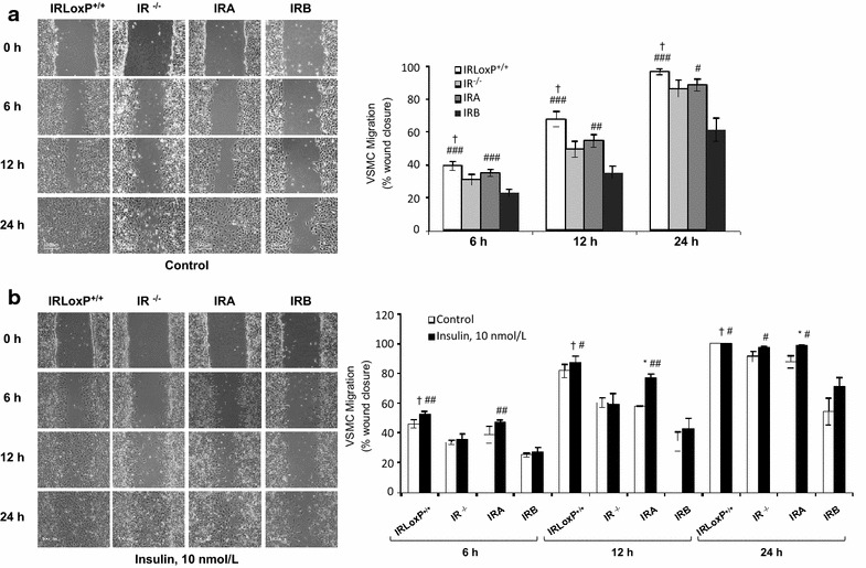
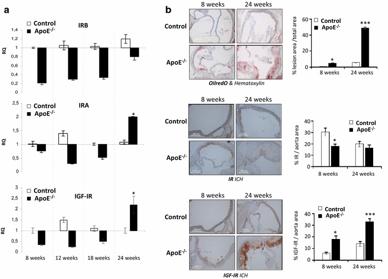
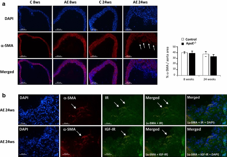
Methods: Wound healing assays were performed in VSMCs bearing IR (IRLoxP+/+ VSMCs), or not (IR-/- VSMCs), expressing IRA (IRA VSMCs) or expressing IRB (IRB VSMCs). To study the role of IR isoforms and IGF-IR in experimental atherosclerosis, we used ApoE-/- mice at 8, 12, 18 and 24 weeks of age. Finally, we analyzed the mRNA expression of total IR, IRB isoform, IGF-IR and IGFs by qRT-PCR in the medial layer of human aortas.

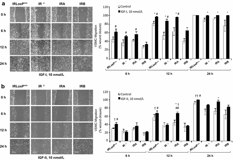
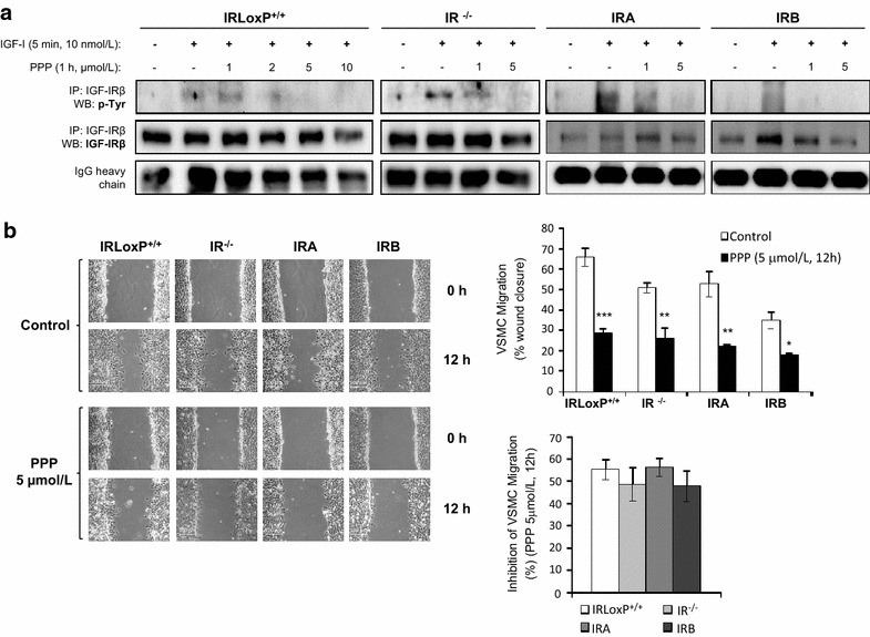
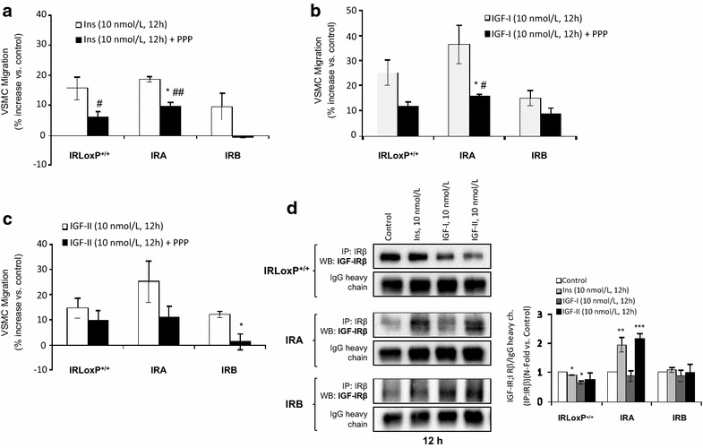
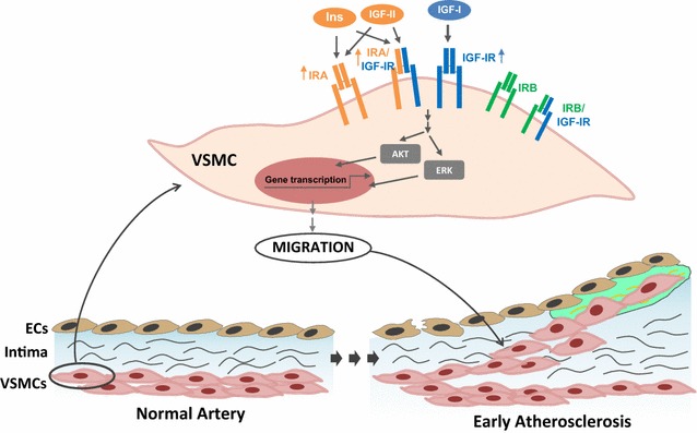
Results: IGF-I strongly induced migration of the four cell lines through IGF-IR. In contrast, insulin and IGF-II only caused a significant increase of IRA VSMC migration which might be favored by the formation of IRA/IGF-IR receptors. Additionally, a specific IGF-IR inhibitor, picropodophyllin, completely abolished insulin- and IGF-II-induced migration in IRB, but not in IRA VSMCs. A significant increase of IRA and IGF-IR, and VSMC migration were observed in fibrous plaques from 24-week-old ApoE-/- mice. Finally, we observed a marked increase of IGF-IR, IGF-I and IGF-II in media from fatty streaks as compared with both healthy aortas and fibrolipidic lesions, favoring the ability of medial VSMCs to migrate into the intima.

Conclusions: Our data suggest that overexpression of IGF-IR or IRA isoform, as homodimers or as part of IRA/IGF-IR hybrid receptors, confers a stronger migratory capability to VSMCs as might occur in early stages of atherosclerotic process.

Keywords: Atherosclerosis; Insulin receptor; Migration; Vascular smooth muscle cells.


Figures

Fig. 1
Fig. 1
Insulin induces migration in IRA VSMCs. a Analysis of basal migration of four VSMC lines at 6, 12 and 24 h. The experiments were performed by wound healing assays and the percentage of wound healing closure quantified (using TScratch program) at different time points. Experiments were performed 4–7 times. b Effect of insulin on wound healing closure in four VSMC lines at 6, 12 and 24 h. Photomicrographs (10x magnification) were selected the more representative. Experiments were performed at least 3 times. *p < 0.05 vs. each control; p < 0.05 vs. IR−/− VSMCs; #p < 0.05, ##p < 0.005, ###p < 0.001 vs. IRB VSMCs
Fig. 2
Fig. 2
IGF-I strongly induces VSMC migration and IGF-II has a lesser effect on IRA VSMC migration. Effect of IGF-I (a) or IGF-II (b) on wound healing closure in four lines of VSMCs at 6, 12 and 24 h. Photomicrographs (10x magnification) were selected the more representative. Experiments were performed at least 3 times. *p < 0.05 vs. each control; p < 0.05, ††p < 0.005 vs. IR−/− VSMCs; #p < 0.05, ##p < 0.005 vs. IRB VSMCs
Fig. 3
Fig. 3
IGF-IR contributes to basal VSMC migration. a Effect of PPP (IGF-IR inhibitor) at different doses on IGF-IR tyrosine phosphorylation of four VSMC lines. We performed the immunoprecipitation against IGF-IRβ followed by Western blot against p-tyrosine. b Effect of PPP on basal VSMC migration at 12 h by wound healing closure and the percentage of inhibition of VSMC migration induced by PPP. Experiments were performed 4–7 times. *p < 0.05, **p < 0.005, ***p < 0.001 vs. each control
Fig. 4
Fig. 4
Effect of IGF-IR inhibition on VSMC migration induced by insulin or IGFs. Quantification of PPP effect on VSMC migration stimulated with insulin (a), IGF-I (b) or IGF-II (c) at 12 h by wound healing closure. *p < 0.05 vs. each control; #p < 0.05, ##p < 0.005 vs. IRB VSMCs. d Formation of hybrid receptors (IRA/IGF-IR or IRB/IGF-IR) in IRLoxP+/+, IRA and IRB VSMCs stimulated with insulin, IGF-I or IGF-II. We performed the immunoprecipitation against IRβ followed by Western blot against IGF-IRβ. Experiments were performed at least 3 times. *p < 0.05, **p < 0.005, ***p < 0.001 vs. each control
Fig. 5
Fig. 5
IR and IGF-IR expression in aorta from an experimental model of atherosclerosis. a By qRT-PCR, we studied IRA, IRB and IGF-IR mRNA expression in aorta from ApoE−/− mice at 8, 12, 18 and 24 weeks of age and their respective controls. *p < 0.05 vs. each control. b Representative photomicrographs (10× magnification) and quantifications of OilRedO staining and immunohistochemistry of IR and IGF-IR in aortic roots from ApoE−/− at 8 and 24 weeks of age and their respective controls. *p < 0.05 vs. each control; ***p < 0.001 vs. each control. AE ApoE−/− mice, C control mice. C 8ws (n = 5); AE 8ws (n = 5); C12 ws (n = 5); AE12 ws (n = 8); C18 ws (n = 8); AE 18ws (n = 8); C 24ws (n = 8); AE 24ws (n = 8)
Fig. 6
Fig. 6
VSMCs migrated into the intima of atherosclerotic plaques express IR or IGF-IR. a Representative photomicrographs (10× magnification) and quantifications of immunofluorescence of α-SMA in aortic roots from ApoE−/− mice at 8 and 24 weeks of age and their respective controls. b Representative photomicrographs (20× magnification) of double immunofluorescence against IR or IGF-IR (green staining) and α-SMA (red staining) in aortic roots from ApoE−/− mice at 24 weeks of age. DAPI staining was performed to localize nuclei of cells presented in aortic roots (blue staining). White arrows indicate VSMCs in the intima of fibrous plaque from 24-week-old ApoE−/− mice. AE ApoE−/− mice, C control mice. C 8ws (n = 5); AE 8ws (n = 5); AE 18ws (n = 8); C 24ws (n = 8); AE 24ws (n = 8)
Fig. 7
Fig. 7
Proposed model of role of IR isoforms, IGF-IR and IR/IGF-IR hybrid receptors in VSMC migration. We propose that IGF-I mainly through IGF-IR, while insulin and IGF-II by binding IRA homodimers or IRA/IGF-IR hybrid receptors might favor VSMC migration and contribute to plaque growth in early states of atherosclerosis. ECs endothelial cells, VSMCs vascular smooth muscle cells

References

    1. Libby P, Ridker PM, Hansson GK. Progress and challenges in translating the biology of atherosclerosis. Nature. 2011;473:317–325. doi: 10.1038/nature10146. - DOI - PubMed
    1. Bennett MR, Sinha S, Owens GK. Vascular smooth muscle cells in atherosclerosis. Circ Res. 2016;118:692–702. doi: 10.1161/CIRCRESAHA.115.306361. - DOI - PMC - PubMed
    1. Ross R. The pathogenesis of atherosclerosis: a perspective for the 1990s. Nature. 1993;362:801–809. doi: 10.1038/362801a0. - DOI - PubMed
    1. Bornfeldt KE, Raines EW, Nakano T, Graves LM, Krebs EG, Ross R. Insulin-like growth factor-I and platelet-derived growth factor-BB induce directed migration of human arterial smooth muscle cells via signaling pathways that are distinct from those of proliferation. J Clin Invest. 1994;93:1266–1274. doi: 10.1172/JCI117081. - DOI - PMC - PubMed
    1. Bayes-Genis A, Conover CA, Schwartz RS. The insulin-like growth factor axis: a review of atherosclerosis and restenosis. Circ Res. 2000;86:125–130. doi: 10.1161/01.RES.86.2.125. - DOI - PubMed

Publication types

MeSH terms