. 2016 Dec 1;6(1):42.
doi: 10.1186/s13395-016-0113-7.

Nuclear bodies reorganize during myogenesis in vitro and are differentially disrupted by expression of FSHD-associated DUX4

Affiliations

Nuclear bodies reorganize during myogenesis in vitro and are differentially disrupted by expression of FSHD-associated DUX4

Sachiko Homma et al. Skelet Muscle. .

Abstract

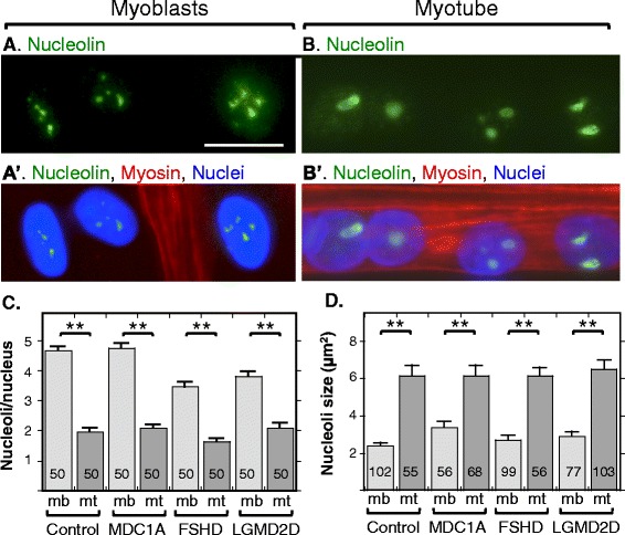
Background: Nuclear bodies, such as nucleoli, PML bodies, and SC35 speckles, are dynamic sub-nuclear structures that regulate multiple genetic and epigenetic processes. Additional regulation is provided by RNA/DNA handling proteins, notably TDP-43 and FUS, which have been linked to ALS pathology. Previous work showed that mouse cell line myotubes have fewer but larger nucleoli than myoblasts, and we had found that nuclear aggregation of TDP-43 in human myotubes was induced by expression of DUX4-FL, a transcription factor that is aberrantly expressed and causes pathology in facioscapulohumeral dystrophy (FSHD). However, questions remained about nuclear bodies in human myogenesis and in muscle disease.

Methods: We examined nucleoli, PML bodies, SC35 speckles, TDP-43, and FUS in myoblasts and myotubes derived from healthy donors and from patients with FSHD, laminin-alpha-2-deficiency (MDC1A), and alpha-sarcoglycan-deficiency (LGMD2D). We further examined how these nuclear bodies and proteins were affected by DUX4-FL expression.

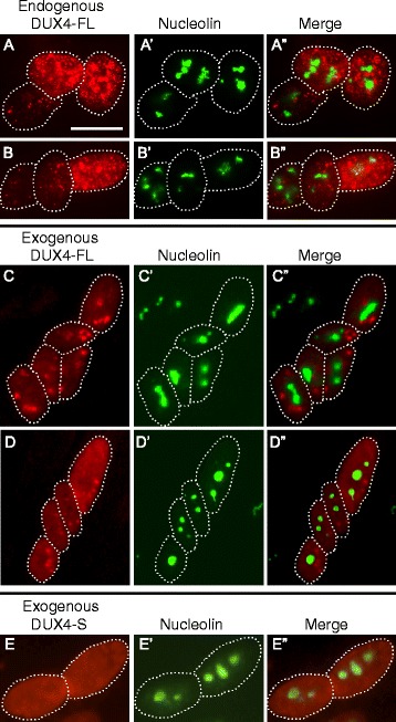
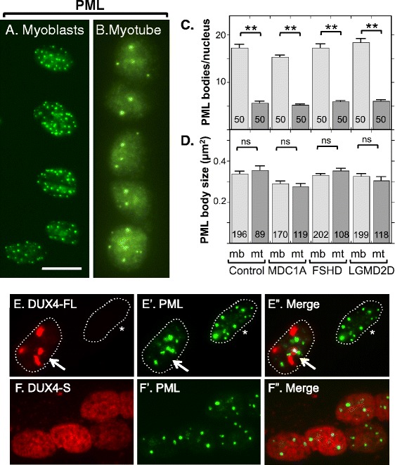
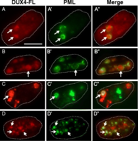
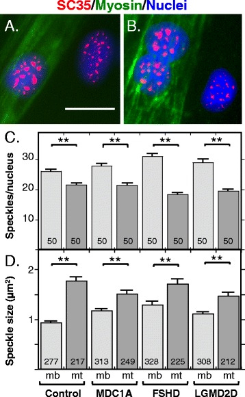
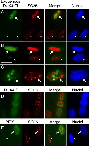
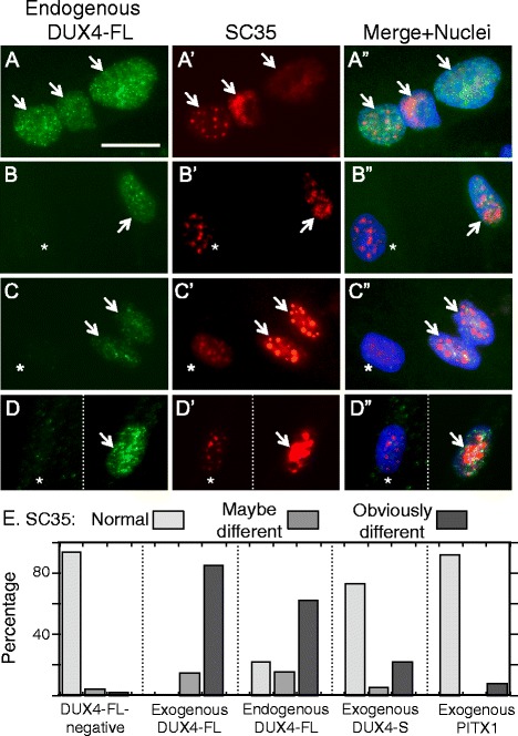
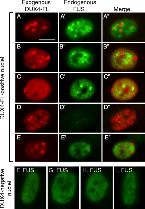
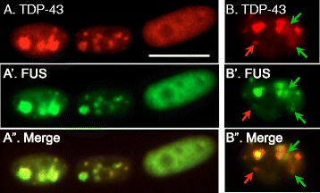
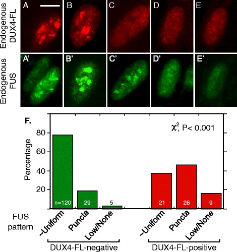
Results: We found that nucleoli, PML bodies, and SC35 speckles reorganized during differentiation in vitro, with all three becoming less abundant in myotube vs. myoblast nuclei. In addition, though PML bodies did not change in size, both nucleoli and SC35 speckles were larger in myotube than myoblast nuclei. Similar patterns of nuclear body reorganization occurred in healthy control, MDC1A, and LGMD2D cultures, as well as in the large fraction of nuclei that did not show DUX4-FL expression in FSHD cultures. In contrast, nuclei that expressed endogenous or exogenous DUX4-FL, though retaining normal nucleoli, showed disrupted morphology of some PML bodies and most SC35 speckles and also co-aggregation of FUS with TDP-43.

Conclusions: Nucleoli, PML bodies, and SC35 speckles reorganize during human myotube formation in vitro. These nuclear body reorganizations are likely needed to carry out the distinct gene transcription and splicing patterns that are induced upon myotube formation. DUX4-FL-induced disruption of some PML bodies and most SC35 speckles, along with co-aggregation of TDP-43 and FUS, could contribute to pathogenesis in FSHD, perhaps by locally interfering with genetic and epigenetic regulation of gene expression in the small subset of nuclei that express high levels of DUX4-FL at any one time.

Keywords: DUX4; FUS; Facioscapulohumeral muscular dystrophy; Myotube; Nucleoli; PML bodies; SC35 speckles; TDP-43.


Figures

Fig. 1
Fig. 1
Myotubes had fewer, but larger, nucleoli than myoblasts. Immunostaining for nucleolin (green) was used to identify nucleoli and staining for myosin heavy chain (red) was used to distinguish nuclei in myotubes from those in myoblasts. a, a’ Myoblast nuclei, three of which are shown, typically had four or five nucleoli. b, b’ Myotube nuclei, of which four are shown from a single myotube, usually had one to three nucleoli that were typically larger than those in the myoblast nuclei. c Quantitation of nucleoli in myoblasts (light gray bars) and myotubes (dark gray bars) in cultures of healthy control, MDC1A, DUX4-negative FSHD, and LGMD2D myogenic cells. All cultures showed similar decreases in nucleolar number in myotube vs. myoblast nuclei. Error bars = s.e.m. **P < 0.01 by t test. Nucleoli were counted in n = 50 nuclei. d Quantitation of the cross-sectional areas of nucleoli in myoblasts (light gray bars) and myotubes (dark gray bars) in cultures of healthy control, MDC1A, DUX4-negative FSHD, and LGMD2D myogenic cells. All cultures showed similar increases in nucleolar size in myotube vs. myoblast nuclei. Scale bar in A = 20 μm. Error bars = s.e.m. **P < 0.01 by t test. Number of nucleoli measured as indicated on each data bar
Fig. 2
Fig. 2
Nucleoli appeared to be unaffected by expression of DUX4-FL. ab” Staining for endogenously expressed DUX4-FL (red) and nucleolin (green) in three nuclei within a single myotube. In these nuclei, DUX4-FL expression did not appear to affect nucleolar structure, and there was little or no overlap of punctate staining for DUX4-FL with nucleolin. In all panels, dotted lines indicate approximate borders of individual nuclei. cd” Staining for endogenously expressed DUX4-FL (red) and nucleolin (green) in several nuclei within a single myotube. Exogenous DUX4-FL expression, whether predominantly punctate (c) or predominantly uniform (d) also did not appear to affect nucleolar structure, and there was also little or no overlap of exogenous DUX4-FL and nucleolin staining. e e” When expressed in myotubes, the non-cytotoxic, short DUX-S isoform was uniformly distributed in nuclei and did not appear to affect nucleolar structure. Bar in A = 20 μm for rows ad and 15 μm for row e
Fig. 3
Fig. 3
Myotubes had fewer PML bodies than myoblasts and the structure of most PML bodies was not affected by DUX4-FL expression. a Human myoblasts from a healthy donor typically had 10 to 20 or sometimes more PML-positive structures (green). b Nuclei in myotubes typically had four to eight PML bodies. c Quantitation of PML bodies in myoblasts (light gray bars) and myotubes (dark gray bars) in healthy control, MDC1A, DUX4-FL-negative, and LGMD2D myogenic cells. All cultures showed similar decreases in PML body number in myotube vs. myoblast nuclei. Error bars = s.e.m. **P < 0.01 by t test, with all PML bodies counted in n = 50 nuclei. d Quantitation of the cross-sectional areas of PML bodies in myoblasts (light gray bars) and myotubes (dark gray bars) in healthy control, MDC1A, DUX4-FL-negative FSHD, and LGMD2D myogenic cells. All cultures showed no significant changes in PML body size in myotube vs. myoblast nuclei. Error bars = s.e.m. n.s. not significant (P > 0.05) by t test. Number of PML bodies measured as indicated on each bar. e–e” In most, but not all (see Fig. 4), nuclei with punctate DUX4-FL fluorescence, PML bodies showed minor or no disruption, and there was little overlap between DUX4-FL and PML fluorescence. Nuclei in myotubes are shown. Arrow = DUX4-FL-positive nucleus, asterisk = DUX4-FL-negative nucleus. f–f” DUX4-S was typically uniformly distributed within the nuclei, and expression of DUX4-S did not affect PML body morphology. Nuclei in myotubes are shown. Bar in a = 20 μm for a, b, f, and 15 μm for e
Fig. 4
Fig. 4
The structures of PML bodies in a small fraction of nuclei were disrupted by DUX4-FL expression. Each row shows one myotube nucleus immunostained as indicated for DUX4-FL or PML along with a merged image. Dotted lines show the approximate outlines of each nucleus. In rows a–a” and b–b”, the arrows point to regions where PML staining appears to envelop DUX4-FL aggregates; and in rows cc” and dd”, the arrows point to PML staining that appears to be intertwined with small DUX4-FL aggregates. Bar in a = 10 μm
Fig. 5
Fig. 5
SC35-containing speckles in myotube nuclei were fewer in number but larger in size than those in myoblast nuclei. a, b Immunostaining for SC35 (red) was used to identify SC35 speckles and staining for myosin heavy chain (green) was used to identify myotubes and thus distinguish nuclei in myotubes from those in myoblasts. SC35 speckles in myotube nuclei typically appeared to be fewer in number and sometimes larger than those in myoblasts. c Quantitation of the number of SC35 speckles in myoblast nuclei (mb, light gray bars) and in myotube nuclei (mt, dark gray bars). As indicated, speckles were counted in healthy control, MDC1A, and LGMD2D myogenic cells, as well as in the DUX4-FL-negative nuclei of FSHD myogenic cells. In each type of cells, the average number of SC35 speckles was lower in myotube nuclei than in myoblast nuclei. Error bars = s.e.m. **P < 0.01 by t test. Speckles were counted in n = 50 nuclei. d Quantitation of the cross-sectional areas (μm2) of SC35 speckles in myoblasts (mb, light gray bars) and myotubes (mt, dark gray bars). As indicated, speckles were measured in healthy control, MDC1A, and LGMD2D myogenic cells, as well as in the DUX4-FL-negative nuclei of FSHD myogenic cells. In each type of cell, the average size of SC35 speckles was higher in myotube nuclei than in myoblast nuclei. Error bars = s.e.m. **P < 0.01 by t test. The number of speckles measured is indicated on each bar. Bar in a = 20 μm
Fig. 6
Fig. 6
SC35 speckles in most nuclei were disrupted by exogenous DUX4-FL expression. a–c BacMam-mediated expression of DUX4-FL (green) in healthy control myotubes caused SC35 speckles (red) to show an altered morphology. Arrows indicate nuclei that expressed DUX4-FL, and asterisks indicate nearby nuclei that were DUX4-FL-negative. SC35 speckles in the DUX4-FL-positive nuclei typically showed larger aggregates and/or more intense staining. SC35 speckles were disrupted in both nuclei with punctate DUX4-FL staining (rows a, c) and in nuclei with uniform DUX4-FL staining (row b). In nuclei with punctate DUX4-FL (rows a, c), there was little or no overlap between DUX4-FL and SC35 staining. d, e In contrast to expression of DUX4-FL, exogenous, BacMam-mediated expression of DUX4-S (row d) or PITX1 (row e) did not markedly affect SC35 speckles in most nuclei. See Fig. 7e for quantitation of the extent to which exogenous DUX4-FL, DUX4-S, and PITX1 affects SC35 speckles using blind assays. Bar in b = 20 μm for a, b, d, and e and 12 μm for c
Fig. 7
Fig. 7
SC35 speckles were disrupted in many nuclei by expression of DUX4-FL from its endogenous promoter. a–d” Endogenous expression of DUX4-FL (green) in FSHD myotubes caused SC35 speckles (red) to show an altered morphology. Arrows indicate nuclei that expressed DUX4-FL, and asterisks indicate nearby nuclei that were DUX4-FL-negative. In panel d, the dotted line indicates where empty space was cropped from the image so that two nearby neighboring nuclei could be juxtaposed for presentation. The most common changes to SC35 speckles in the DUX4-FL-positive nuclei were the appearance of larger aggregates and/or more intense staining. Less common changes included loss of most speckles (e.g., rightmost nucleus in row a) and disorganized speckles (e.g., row b). SC35 speckles were disrupted in both nuclei with punctate DUX4-FL staining (rows a, c) and in nuclei with uniform DUX4-FL staining (row b). In nuclei with punctate DUX4-FL (rows a and c), there was little or no overlap between DUX4-FL and SC35 staining. Some nuclei also showed little effect of DUX4-FL (e.g., leftmost nucleus in row a). e Quantitation of SC35 speckle morphology. As described in the text, a blind test was used to classify speckle patterns into three groups: (i) similar to the majority of controls (normal, light gray bars), (ii) maybe different from controls (medium gray bars), or (iii) obviously different from controls (dark gray bars). Nuclei that expressed either endogenous or exogenous DUX4-FL had much higher frequencies of obviously different SC35 speckle patterns, compared to nuclei in healthy control cultures or to nuclei that expressed exogenous DUX4-S or PITX1. Bar in a = 20 μm
Fig. 8
Fig. 8
Exogenous DUX4-FL expression induced abnormalities in FUS expression. At 24 h after addition of BacMam vector, healthy control myotubes were examined for expression of exogenous DUX4-FL (red) and FUS (green). a–c” About 40–50% of the DUX4-FL-positive myotube nuclei showed punctate immunostaining for FUS (panels ac). In addition, DUX4-FL itself showed punctate staining in some nuclei and merged images indicated that FUS and DUX4-FL puncta were not usually co-localized. d–d” About 10% of the DUX4-FL-positive myotube nuclei showed little or no staining for FUS (panel d). e–e” The remaining approximately half of the DUX4-FL-positive myotube nuclei showed a more uniform distribution of FUS staining in the nucleus (though excluded from nucleoli), even when DUX4-FL staining was itself punctate. fi For comparison, myotube nuclei that did not express DUX4-FL typically showed the more uniform pattern of FUS staining (green) without large puncta, which was similar to the nucleus in panel e. Bar in a = 10 μm for ae” and 8 μm for fi
Fig. 9
Fig. 9
Exogenous DUX4-FL expression induced nuclear co-aggregates of FUS and TDP-43. a-a” Double immunostaining of TDP-43 (red) and FUS (green) in myotubes which also expressed exogenous DUX4-FL showed that TDP-43 and FUS staining were almost completely co-localized. b–b” In rare myotube nuclei, some TDP-43 puncta (red arrows) and FUS puncta (green arrows) did not co-localize with the other protein, even though most of the FUS and TDP-43 in the same nucleus were co-localized. Bar in a = 10 μm
Fig. 10
Fig. 10
Induction of nuclear aggregates of FUS by endogenous expression of DUX4-FL in FSHD myotubes. a–e” Nuclei were double stained for endogenous DUX4-FL (red, top row) and endogenous FUS (green, lower row) in FSHD myotubes. The five nuclei illustrate a range of staining patterns ranging from mostly large, though non-overlapping, puncta for both proteins (e.g., a, b) to more uniform staining or very small puncta (e.g., d, e). f Quantitation of blind assays in which observers classified FUS staining patterns in DUX4-negative and DUX4-positive myotube nuclei as (i) mostly uniform (as illustrated in Fig. 7e–i), (ii) consisting largely of punctate staining (e.g., as in a, b of this figure), or (iii) showing a low intensity signal or no signal (e.g., as in Fig. 9d). Expression of DUX4-FL was associated with a significantly increased percentage of nuclei in which FUS showed a punctate staining indicative of aggregation or a loss of signal intensity. Bar in a = 10 μm

Similar articles

Cited by

References

    1. Laker RC, Ryall JG. DNA methylation in skeletal muscle stem cell specification, proliferation, and differentiation. Stem Cells Int. 2016;2016:5725927. doi: 10.1155/2016/5725927. - DOI - PMC - PubMed
    1. Sincennes MC, Brun CE, Rudnicki MA. Concise review: epigenetic regulation of myogenesis in health and disease. Stem Cells Transl Med. 2016;5:282–90. doi: 10.5966/sctm.2015-0266. - DOI - PMC - PubMed
    1. Horak M, Novak J, Bienertova-Vasku J. Muscle-specific microRNAs in skeletal muscle development. Dev Biol. 2016;410:1–13. doi: 10.1016/j.ydbio.2015.12.013. - DOI - PubMed
    1. Daxinger L, Tapscott SJ, van der Maarel SM. Genetic and epigenetic contributors to FSHD. Curr Opin Genet Dev. 2015;33:56–61. doi: 10.1016/j.gde.2015.08.007. - DOI - PMC - PubMed
    1. Musumeci G, Castrogiovanni P, Coleman R, Szychlinska MA, Salvatorelli L, Parenti R, Magro G, Imbesi R. Somitogenesis: from somite to skeletal muscle. Acta Histochem. 2015;117:313–28. doi: 10.1016/j.acthis.2015.02.011. - DOI - PubMed

MeSH terms