. 2016 Nov;6(11):160185.
doi: 10.1098/rsob.160185.

The distinct fate of smooth and rough Mycobacterium abscessus variants inside macrophages

Affiliations

The distinct fate of smooth and rough Mycobacterium abscessus variants inside macrophages

Anne-Laure Roux et al. Open Biol. 2016 Nov.

Abstract

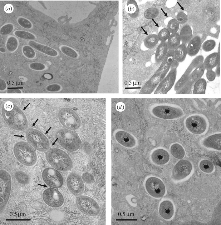
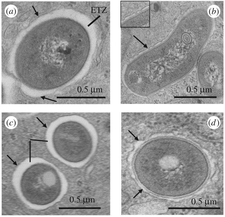
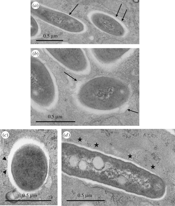
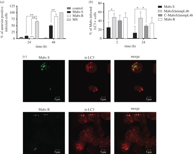
Mycobacterium abscessus is a pathogenic, rapidly growing mycobacterium responsible for pulmonary and cutaneous infections in immunocompetent patients and in patients with Mendelian disorders, such as cystic fibrosis (CF). Mycobacterium abscessus is known to transition from a smooth (S) morphotype with cell surface-associated glycopeptidolipids (GPL) to a rough (R) morphotype lacking GPL. Herein, we show that M. abscessus S and R variants are able to grow inside macrophages and are present in morphologically distinct phagosomes. The S forms are found mostly as single bacteria within phagosomes characterized by a tightly apposed phagosomal membrane and the presence of an electron translucent zone (ETZ) surrounding the bacilli. By contrast, infection with the R form leads to phagosomes often containing more than two bacilli, surrounded by a loose phagosomal membrane and lacking the ETZ. In contrast to the R variant, the S variant is capable of restricting intraphagosomal acidification and induces less apoptosis and autophagy. Importantly, the membrane of phagosomes enclosing the S forms showed signs of alteration, such as breaks or partial degradation. Although not frequently encountered, these events suggest that the S form is capable of provoking phagosome-cytosol communication. In conclusion, M. abscessus S exhibits traits inside macrophages that are reminiscent of slow-growing mycobacterial species.

Keywords: Mycobacterium abscessus; innate response; macrophages; phagosome; rapid-growing mycobacteria.


Figures

Figure 1.
Figure 1.
Ultrastructural differences of the M. abscessus S- or R-containing phagosomes. Bone marrow-derived murine Mϕ (BMDM) were infected with either the S (a,d) or R (b,c) variants of M. abscessus for 3 h. At 24 h p.i., cells were fixed and processed for TEM. Thin sections were analysed for phagocytic uptake of each variant (a,b) and the morphological appearance of R (c) or S (d)-containing phagosomes. S forms (a) reside within phagosomes and none are found in phagocytic cups whereas most of the R forms (b) are still clustered in phagocytic cups (arrows) at 24 h p.i. (c) Once formed, the R-containing phagosomes usually comprise several bacteria (social phagosomes, indicated with arrows). (d) The S-containing phagosomes typically comprise a single bacterium (loner phagosomes, indicated with stars).
Figure 2.
Figure 2.
Growth of M. abscessus S (Mabs S) and R (Mabs R) variants in different cells. Wild-type murine (a) or human (b) Mϕ were infected with S and R variants. Amikacin treatment was applied to avoid extracellular growth (see Experimental procedures). The number of CFU was determined at the indicated times p.i. (c,d) Wild-type and cftr−/− murine Mϕ were infected as mentioned above. Intracellular growth was similar in murine wild-type Mϕ and in murine Mϕ knock-out for cftr (cftr−/−). Error bars indicate the s.e.m., based on the results from three independent experiments.
Figure 3.
Figure 3.
Morphological appearance of the electron translucent layer (ETZ) of the cell wall of M. abscessus within phagosomes. Bone marrow-derived murine Mϕ (BMDM) were infected with various M. abscessus strains for 3 h. At selected time points p.i., cells were fixed and processed for TEM. The cell wall ultrastructure of the different strains was assessed on 100–200 different bacterial profiles. (a) S variant: the electron translucent outermost layer (ETZ) of the wall was thick and apposed to the smooth phagosome membrane (arrows) all around. (b) R variant: in the absence of GPL production, the ETZ was thin and the phagosome membrane had a wavy appearance (arrows); in some instances a close contact was observed at discrete sites (arrow and insert). (c) Mycobacterium abscessus ΔmmpL4b complemented strain: as for the S variant. (d) Mycobacterium abscessus ΔmmpL4b mutant: no GPL produced and thin ETZ as for the R variant.
Figure 4.
Figure 4.
Absence of phagosomal acidification inside M. abscessus S-infected Mϕ. Human THP-1 Mϕ were infected with FITC-labelled mCherry-expressing M. abscessus S (Mabs S) and R (Mabs R), M. smegmatis, M. marinum and heat-killed M. abscessus S (Mabs S heat-killed). Fluorescent signals were measured sequentially at 485 nm (FITC) and 544 nm excitation wavelengths (mCherry) after a 15 mn incubation period at 37°C. The first time point on the x-axis (0 mn) was taken immediately after the 15 mn incubation. Subsequently, the pH at each time point was extrapolated from a standardized pH curve. The pH values were significantly different depending on whether phagosomes contained S or R variants. Results are representative of three independent experiments. (**p < 0.01).
Figure 5.
Figure 5.
Alteration of the membrane of phagosomes containing the S variant as assessed by TEM. Bone marrow-derived murine Mϕ were infected with M. abscessus S for 3 h. Phagosomes were examined for obvious signs of membrane alteration/destruction. (a) No alteration: the phagosome membrane is smooth (arrows) and closely apposed to the mycobacterial cell wall ETZ all around. (b) First sign of alteration: the phagosome membrane has become wavy and is no longer closely apposed to the bacterium all around (arrows). (c) The phagosome membrane displays breaks (arrowheads). (d) The phagosome membrane is no longer visible (stars).
Figure 6.
Figure 6.
Mycobacterium abscessus-induced apoptosis and autophagy in wild-type Mϕ. (a) Analysis of apoptosis: THP-1 Mϕ were infected with M. abscessus S (Mabs S), M. abscessus R (Mabs R) or M. smegmatis (MS). The percentage of apoptotic cells was determined at 24 h p.i. using annexin-V-FITC conjugates (Abcam, USA) and propidium iodide to stain the dead cells. Fluorescent signals (mCherry from mycobacteria, FITC from the annexin-V and absence of propidium iodide) were analysed by flow cytometry. A significant increase in the percentage of apoptotic cells was associated with M. smegmatis-infected cells when compared with Mϕ infected with M. abscessus S or R. However, the R variant was significantly more pro-apoptotic than the S variant at 48 h. Error bars indicate the s.e.m. based on the results of two independent experiments (**p < 0.01). (b,c) Comparative behaviour of M. abscessus S and R variants towards autophagy. (b) Percentage of colocalization of M. abscessus S (Mabs S), S ΔmmpL4b mutant (MabsS ΔmmpL4b), ΔmmpL4b complemented (C-MabsS ΔmmpL4b) and R (Mabs R)-infected cells with LC3 at 2 and 24 h p.i. determined after immunofluorescence analyses. Error bars indicate the s.e.m. based on the results from four independent experiments (*p < 0.05). (c) Confocal immunofluorescence images of fixed THP-1 cells infected with Alexa-488-labelled M. abscessus variant S (Mabs S) or variant R (Mabs R) (green) (2 h p.i.) and immuno-stained for endogenous LC3 (red). Scale bars, 5 µm.

References

    1. Guglielmetti L, et al. 2015. Human infections due to nontuberculous mycobacteria: the infectious diseases and clinical microbiology specialists’ point of view. Future Microbiol. 10, 1467–1483. (doi:10.1111/j.1462-5822.2005.00675.x) - DOI - PubMed
    1. Griffith DE, Girard WM, Wallace RJJ. 1993. Clinical features of pulmonary disease caused by rapidly growing mycobacteria. An analysis of 154 patients. Am. Rev. Respir. Dis. 147, 1271–1278. (doi:10.1164/ajrccm/147.5.1271) - DOI - PubMed
    1. Olivier KN, et al. 2003. Nontuberculous mycobacteria. I: Multicenter prevalence study in cystic fibrosis. Am. J. Respir. Crit. Care Med. 167, 828–834. (doi:10.1164/rccm.200207-678OC) - DOI - PubMed
    1. Levy I, et al. 2008. Multicenter cross-sectional study of nontuberculous mycobacterial infections among cystic fibrosis patients, Israel. Emerg. Infect Dis. 14, 378–384. (doi:10.3201/eid1403.061405) - DOI - PMC - PubMed
    1. Roux A-L, et al. 2009. Multicenter study of prevalence of nontuberculous mycobacteria in patients with cystic fibrosis in france. J. Clin. Microbiol. 47, 4124–4128. (doi:10.1128/JCM.01257-09) - DOI - PMC - PubMed

Publication types

MeSH terms

LinkOut - more resources