. 2016 Dec 1;7(12):e2510.
doi: 10.1038/cddis.2016.381.

A mechanism for overcoming P-glycoprotein-mediated drug resistance: novel combination therapy that releases stored doxorubicin from lysosomes via lysosomal permeabilization using Dp44mT or DpC

Affiliations

A mechanism for overcoming P-glycoprotein-mediated drug resistance: novel combination therapy that releases stored doxorubicin from lysosomes via lysosomal permeabilization using Dp44mT or DpC

Nicole A Seebacher et al. Cell Death Dis. .

Abstract

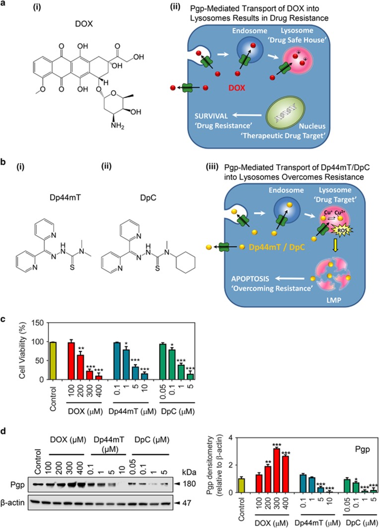
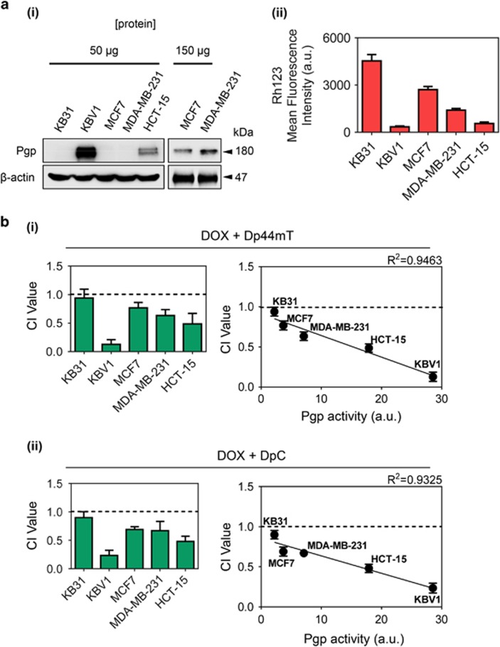
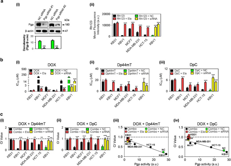
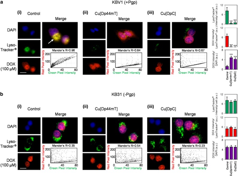
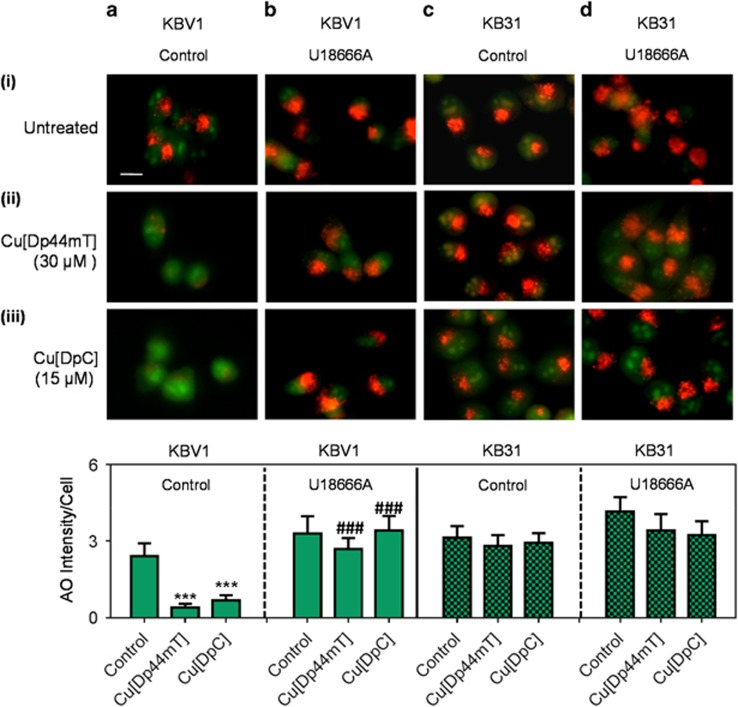
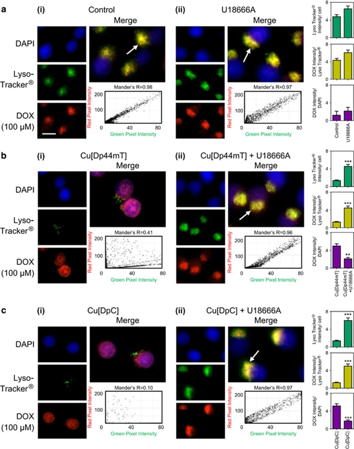
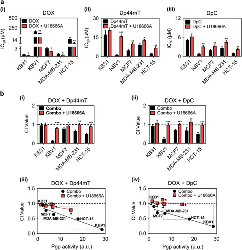
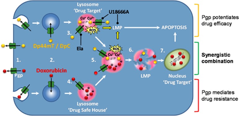
The intracellular distribution of a drug can cause significant variability in both activity and selectivity. Herein, we investigate the mechanism by which the anti-cancer agents, di-2-pyridylketone 4,4-dimethyl-3-thiosemicarbazone (Dp44mT) and the clinically trialed, di-2-pyridylketone 4-cyclohexyl-4-methyl-3-thiosemicarbazone (DpC), re-instate the efficacy of doxorubicin (DOX), in drug-resistant P-glycoprotein (Pgp)-expressing cells. Both Dp44mT and DpC potently target and kill Pgp-expressing tumors, while DOX effectively kills non-Pgp-expressing cancers. Thus, the combination of these agents should be considered as an effective rationalized therapy for potently treating advanced and resistant tumors that are often heterogeneous in terms of Pgp-expression. These studies demonstrate that both Dp44mT and DpC are transported into lysosomes via Pgp transport activity, where they induce lysosomal-membrane permeabilization to release DOX trapped within lysosomes. This novel strategy of loading lysosomes with DOX, followed by permeabilization with Dp44mT or DpC, results in the relocalization of stored DOX from its lysosomal 'safe house' to its nuclear targets, markedly enhancing cellular toxicity against resistant tumor cells. Notably, the combination of Dp44mT or DpC with DOX showed a very high level of synergism in multiple Pgp-expressing cell types, for example, cervical, breast and colorectal cancer cells. These studies revealed that the level of drug synergy was proportional to Pgp activity. Interestingly, synergism was ablated by inhibiting Pgp using the pharmacological inhibitor, Elacridar, or by inhibiting Pgp-expression using Pgp-silencing, demonstrating the importance of Pgp in the synergistic interaction. Furthermore, lysosomal-membrane stabilization inhibited the relocalization of DOX from lysosomes to the nucleus upon combination with Dp44mT or DpC, preventing synergism. This latter observation demonstrated the importance of lysosomal-membrane permeabilization to the synergistic interaction between these agents. The synergistic and potent anti-tumor efficacy observed between DOX and thiosemicarbazones represents a promising treatment combination for advanced cancers, which are heterogeneous and composed of non-Pgp- and Pgp-expressing tumor cells.


Conflict of interest statement

DRR is a stakeholder in Oncochel Therapeutics, and the remaining authors declare no conflict of interest.

Figures

Figure 1
Figure 1
Pgp expression is altered following treatment of cancer cells with increasing concentrations of DOX or the thiosemicarbazones, Dp44mT and DpC. (a) (i) Line drawing of the structure of doxorubicin (DOX). (a) (ii) Schematic showing that DOX is effluxed out of cells as it is a substrate of the drug efflux pump, P-glycoprotein (Pgp), but can also be transported by Pgp into endosomes and lysosomes upon endocytosis. Storage of DOX in the lysosome leads to multidrug resistance (MDR) as DOX is sequestered away from its major target, the nucleus (so-called lysosomal 'safe-house' effect). (b) (i,ii) Line drawings of the structures of Dp44mT and DpC. (b) (iii) Schematic demonstrating that Pgp facilitates Dp44mT and DpC transport out of the cells and also into endosomes/lysosomes., However, these agents overcome Pgp-mediated drug resistance by forming copper complexes that potently generate reactive oxygen species (ROS)., , The generation of ROS causes rapid lysosomal-membrane permeabilization (LMP), and apoptosis that leads to the death of the resistant cancer cell., , Hence, the lysosome is a novel drug target that can be implemented against Pgp-expressing cancers by utilizing their high levels of lysosomal Pgp. (c and d) KBV1 (+Pgp) cells were treated with DOX (72 h/37 °C; 100–400 μM), Dp44mT (24 h/37 °C; 0.1–10 μM) or DpC (24 h/37 °C; 0.05–5 μM) and assessed for (c) cell viability by trypan blue-staining and (d) Pgp protein expression by western blotting. *P<0.05, **P<0.01, ***P<0.001 relative to the control (no treatment). The western blot shown is a typical experiment of 3 performed. Densitometry is relative to β-actin and is mean±S.D. (three experiments)
Figure 2
Figure 2
Pgp activity enhances drug synergy between DOX and the thiosemicarbazones, Dp44mT and DpC. KB31, KBV1, MCF7, MDA-MB-231 and HCT-15 cell lines were assessed for: (a) (i) Pgp expression by western blotting and (ii) cellular Rh123 retention measured by flow cytometry. (b) Combination index (CI) values of the drug combinations between (i) DOX (72 h/37 °C) and Dp44mT (24 h/37 °C), (ii) DOX (72 h/37 °C) and DpC (24 h/37 °C), as measured by the Chou-Talalay method., CI>1 Antagonism, CI=1 Additive, CI<1 Synergistic. The correlation between the CI values (b) (i), (ii) and the Pgp activity (a) (ii) was plotted using linear regression. The western shown is a typical experiment of 3 performed. Results are shown as mean±S.D. (three experiments)
Figure 3
Figure 3
Inhibition of Pgp activity using the inhibitor, Elacridar (Ela), or Pgp expression using two Pgp siRNAs prevents synergy between DOX and Dp44mT or DpC. Western blotting was used to assess Pgp expression in: (a) (i) KBV1 (+Pgp) cells incubated for 72 h/37 °C with two different Pgp siRNAs or a negative control siRNA (NC siRNA); (ii) Pgp activity in all cell lines assessed by retention of the Pgp substrate, Rh123 (10 μM; 30 min/37 °C). (b) Cytotoxicity assays with the treatments: (i) DOX (72 h/37 °C), (ii) Dp44mT (24 h/37 °C) and (iii) DpC (24 h/37 °C), in the absence or presence of Ela (0.2 μM) or Pgp siRNA. (c) Combination index (CI) values of the drug combinations between: (i) DOX (72 h/37 °C) and Dp44mT (24 h/37 °C); or (ii) DOX (72 h/37 °C) and DpC (24 h/37 °C), as measured by the Chou-Talalay method., The correlation between the CI values (ciii,iv) and the cellular Pgp activity (aii) was plotted using linear regression. *P<0.05, **P<0.01, ***P<0.001 relative to respective non-Ela- or NC-siRNA-treated control (black=Combo (no Ela); red=Combo+Ela; green=Combo+negative control (NC) siRNA; yellow=Combo+siRNA (Pgp siRNA)). Western blots are typical of three experiments. Results are shown as mean±S.D. (three experiments)
Figure 4
Figure 4
Co-localization of DOX with LysoTracker Green-stained lysosomes decreases with treatment with Dp44mT or DpC. Immunofluorescence microscopy images of: (a) KBV1 (+Pgp) and (b) KB31 cells (expressing extremely low Pgp levels) incubated with: (i) no treatment (Control); (ii) Cu[Dp44mT] (30 μM); or (iii) Cu[DpC] (15 μM) for 30 min/37 °C following staining with DOX (2 h/37 °C; 100 μM) and LysoTracker Green (40 min/37 °C; 100 nM). The overlap between DOX and LysoTracker Green (yellow in the merge) is indicated by the Mander's overlap coefficient, (R). LysoTracker Green was quantified as fluorescence intensity/cell in arbitrary units (a.u.). DOX was quantified as fluorescence intensity co-localization with LysoTracker Green using ImageJ software. ***P<0.001, relative to Control (no treatment). Scale bar=10 μm. Photographs are typical of three experiments. Quantitation is mean±S.D. (three experiments)
Figure 5
Figure 5
The cholesterol transport inhibitor, U18666A, prevents lysosomal-membrane permeabilization (LMP) following treatment with Dp44mT or DpC. Live cell immunofluorescence microscopy images using the following conditions: (a) KBV1 cells (+Pgp); (b) KBV1 cells with U18666A (2.3 μg/ml); (c) KB31 cells (expressing extremely low Pgp levels) and (d) KB31 cells with U18666A (2.3 μg/ml). Cells were incubated for 30 min/37 °C with: (i) no treatment (untreated); (ii) Cu[Dp44mT] (30 μM); or (iii) Cu[DpC] (15 μM). Cells were then stained for 12 min/37 °C with acridine orange (AO; 5 μM). The AO staining was quantified as red fluorescence intensity/cell using ImageJ software. ***P<0.001 relative to the control (no treatment). ###P<0.001, relative to the respective non-U18666A-treated control. Scale bar=10 μm. Photographs are typical of three experiments. Quantitation is mean±S.D. (three experiments)
Figure 6
Figure 6
The cholesterol transport inhibitor, U18666A, prevents the release of DOX from LysoTracker Green-stained lysosomes following thiosemicarbazone treatment. Immunofluorescence microscopy images of KBV1 (+Pgp) cells treated with DOX (2 h/37 °C; 100 μM), LysoTracker Green (40 min/37 °C; 100 nM) and 30 min/37 °C after either: (a) no treatment (Control), (b) Cu[Dp44mT] (30 μM) or (c) Cu[DpC] (15 μM), in the (i) absence of U18666A or (ii) presence of U18666A (2.3 μg/ml). The overlap between DOX and LysoTracker Green (yellow merge; ac) is indicated by Mander's overlap coefficient (R). LysoTracker Green was quantified as fluorescence intensity/cell, while DOX was quantified as DOX fluorescence intensity co-distribution with LysoTracker Green or DAPI, using ImageJ software. **P<0.001, ***P<0.001 relative to respective non-U16666A-treated control. Scale bar=10 μm. White arrow=overlap between LysoTracker Green and DOX. Results in photographs are typical of three experiments. Quantitation is mean±S.D. (three experiments)
Figure 7
Figure 7
Inhibition of intracellular cholesterol transport by U18666A prevents drug synergy between DOX and the thiosemicarbazones, Dp44mT and DpC. (a) Cytotoxicity assays with the treatments: (i) DOX (72 h/37 °C), (ii) Dp44mT (24 h/37 °C) and (iii) DpC (24 h/37 °C), in the absence or presence of U18666A (2.3 μg/ml). (b) Combination index (CI) values of the drug combination between: (i) DOX (72 h/37 °C) and Dp44mT (24 h/37 °C); or (ii) DOX (72 h/37 °C) and DpC (24 h/37 °C), as measured by the Chou-Talalay method., The correlation between the CI values (biii and biv) and the cellular Pgp activity (as measured by Rh123 retention) was plotted using linear regression for: (biii) DOX (72 h/37 °C) and Dp44mT (24 h/37 °C); or (biv) DOX (72 h/37 °C) and DpC (24 h/37 °C). CI>1 Antagonism, CI=1 Additive, CI<1 Synergistic, *P<0.05, **P<0.01, ***P<0.001 relative to the respective non-U18666A-treated control. (Black=Combo; red=Combo+U18666A). Dotted arrows indicate the alteration in CI value upon U18666A treatment. Results in (a) and (b) are mean±S.D. (three experiments)
Figure 8
Figure 8
Schematic illustration of the synergistic interaction between Doxorubicin and Dp44mT/DpC. (1) Pgp on the plasma membrane actively pumps Pgp substrates out of cells. As part of endocytosis, the plasma membrane containing Pgp buds inwards to form early endosomes. (2) As a consequence of endocytosis, the topology of Pgp is inverted, and thus substrates are transported into the vesicle lumen. As the endosome matures into the lysosome, it becomes increasingly acidified. When a Pgp substrate, such as (3) Dp44mT/DpC, or (4) DOX, enters the cell, the drug is sequestered into the acidic lysosomes by Pgp-transport activity., , If the substrate is protonated at acidic pH (such as DOX and Dp44mT/DpC), it becomes trapped in lysosomes., , , The trapping of protonated drugs prevents substrates reaching their molecular targets (e.g., the nucleus for DOX). (3) However, once trapped in the lysosome, Dp44mT or DpC bind copper and redox cycle forming reactive oxygen species (ROS) that cause lysosomal-membrane permeabilization (LMP) and then apoptosis., , , (5) When added in combination with DOX, Dp44mT or DpC redox cycle in lysosomes containing trapped DOX. (6) Dp44mT- or DpC-induced LMP causes the release of trapped DOX. (7) Then DOX redistributes to its molecular target, the nucleus. Notably, the Pgp-transport inhibitor, Ela, prevents entrance of Dp44mT or DpC into lysosomes, blocking LMP and the release of DOX to the nucleus. On the other hand, U18666A partially inhibits Pgp activity and also stabilizes the lysosomal membrane by cholesterol loading and prevents LMP mediated by Dp44mT or DpC, inhibiting lysosomal DOX release

Similar articles

Cited by

References

    1. Germann UA. P-glycoprotein – a mediator of multidrug resistance in tumour cells. Eur J Cancer 1996; 32A: 927–944. - PubMed
    1. Cole SP, Bhardwaj G, Gerlach JH, Mackie JE, Grant CE, Almquist KC et al. Overexpression of a transporter gene in a multidrug-resistant human lung cancer cell line. Science 1992; 258: 1650–1654. - PubMed
    1. Gottesman MM. Multidrug resistance in cancer: role of ATP-dependent transporters. Nat Rev Cancer 2002; 2: 48–58. - PubMed
    1. Yamagishi T, Sahni S, Sharp DM, Arvind A, Jansson PJ, Richardson DR. P-glycoprotein mediates drug resistance via a novel mechanism involving lysosomal sequestration. J Biol Chem 2013; 288: 31761–31771. - PMC - PubMed
    1. Jansson PJ, Yamagishi T, Arvind A, Seebacher N, Gutierrez E, Stacy A et al. Di-2-pyridylketone 4,4-dimethyl-3-thiosemicarbazone (Dp44mT) overcomes multidrug-resistance by a novel mechanism involving the hijacking of lysosomal P-glycoprotein (Pgp). J Biol Chem 2015; 290: 9588–9603. - PMC - PubMed

MeSH terms

Substances