. 2017 Feb 1;158(2):264-276.
doi: 10.1210/en.2016-1867.

FIAT Deletion Increases Bone Mass But Does Not Prevent High-Fat-Diet-Induced Metabolic Complications

Affiliations

FIAT Deletion Increases Bone Mass But Does Not Prevent High-Fat-Diet-Induced Metabolic Complications

Bahareh Hekmatnejad et al. Endocrinology. .

Abstract

Factor inhibiting activating transcription factor 4 (ATF4)-mediated transcription (FIAT) interacts with ATF4 to repress its transcriptional activity. We performed a phenotypic analysis of Fiat-deficient male mice (Fiat-/Y) at 8 and 16 weeks of age. Microcomputed tomography analysis of the distal femur demonstrated 46% and 13% age-dependent increases in trabecular bone volume and thickness, respectively, in Fiat-/Y mice. Cortical bone measurements at the femoral midshaft revealed a substantial increase in cortical thickness in older Fiat-/Y mice. Bone gain was related to increased mineral apposition rate and increased osteoblast function. Femoral stiffness and strength were substantially increased in Fiat-/Y compared with wild-type (WT) mice. We also investigated whether FIAT contributes to metabolic function. When fed standard mouse chow, Fiat-/Y animals were glucose-tolerant. However, when fed a high-fat diet (HFD) for 8 weeks, Fiat-/Y mice gained more weight than control mice, with a specific increase in white adipose tissue fat mass. The increase in fat mass was due to reduced energy expenditure, which correlated with reduced fatty acid oxidation and lipolysis in the adipose tissue of mutant mice. The expression of the Scd1 gene, involved in lipogenesis, was upregulated in the subcutaneous adipose tissue of Fiat-/Y mice. Moreover, HFD-fed Fiat-/Y mice exhibited increased circulating leptin and insulin levels relative to WT mice, demonstrating that endocrine abnormalities are associated with the disturbance in energy balance. We conclude that Fiat-/Y mice exhibited an anabolic bone phenotype but displayed increased susceptibility to developing metabolic-related disorders when consuming an HFD.


Figures

Figure 1.
Figure 1.
Fiat expression and weight gain in FIAT-deficient mice. (A) Fiat mRNA and (B) FIAT protein were not detectable in knock-out male (Fiat−/Y) mice. β-Actin was used as a protein loading control; the arrowhead points to the FIAT protein in the Western blot. Bodyweight (BW) was comparable between genotypes at (C) 8 weeks and (D) 16 weeks of age.
Figure 2.
Figure 2.
Long bone trabecular phenotype of Fiat-null mice. (A, B) Three-dimensional reconstruction of 25-μm trabecular bone obtained by μCT from distal femur of WT and Fiat−/Y 16-week-old male littermates. μCT scans were used to measure (C) BV over TV (BV/TV), (D) trabecular thickness, (E) trabecular number, (F) trabecular separation, (G) tissue volume, and (H) SMI as a function of age by analysis of 250-μm of trabecular bone under the primary spongiosa of the distal femur. Analyses were performed on 9 to 14 mice of each genotype at 8 and 16 weeks of age. **P < 0.01.
Figure 3.
Figure 3.
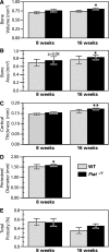
Cortical phenotype of Fiat-deficient mice. (A) Cortical bone volume, (B) area, (C) thickness, (D) periosteal diameter, and (E) cortical porosity were measured by μCT analysis in 8-week-old (n = 11 to 12) and 16-week-old (n = 9 to 14) WT or Fiat−/Y male mice. *P < 0.05, **P < 0.01.
Figure 4.
Figure 4.
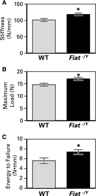
Bone biomechanical properties of 16-week-old Fiat-null mice. Three-point bending tests revealed substantial changes in (A) stiffness, (B) maximum load, and (C) energy to failure between genotypes. *P < 0.05.
Figure 5.
Figure 5.
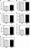
Fiat-deficient mice have enhanced osteoblast activity. qPCR of (A) osteocalcin (Bglap) and (B) Rankl (Tnfsf11) transcript levels in 16-week-old mice calvaria, normalized to β2-microglobulin. Measurement of the level of circulating (C) TRACP 5b and (D) CTX in the serum of 16-week-old Fiat−/Y and WT males (n = 9 to 13). (E) The mineral apposition rate (MAR), (F) bone formation rate per unit of tissue volume of trabecular bone (BFR/TV), and (G) bone formation rate per unit of bone surface (BFR/BS) were measured by double calcein staining (WT, n = 9; Fiat−/Y, n = 12). *P < 0.05; **P < 0.01.
Figure 6.
Figure 6.
Mesenchymal lineages differentiation of Fiat-deficient cells. (A) Formation of mineralized matrix was examined by alizarin red S staining of cultured primary osteoblasts isolated from long bone of WT (top) and Fiat−/Y (bottom) mice. (B) Cells were fixed after 24 days of culture in osteogenic medium, and alizarin red S staining was quantified by absorbance at 405 nm. Expression of osteoblast markers (C) Alpl, (D) Bglap, and (E) Runx2 in primary osteoblasts cultured in osteogenic medium for 14 and 24 days was measured by reverse transcription qPCR analysis, and the values were normalized to β2-microglobulin mRNA expression. (F) BMSCs from WT and Fiat−/Y mice (n = 3 per genotype) were induced to undergo adipogenic differentiation with staining for Oil Red O after 6 days. (G) mRNA levels for adipogenic markers Paprγ, adiponectin (Adipoq), CebpA, CebpB, and Fabp4 were measured by reverse transcription qPCR. *P < 0.05; **P < 0.01; ***P < 0.001.
Figure 7.
Figure 7.
HFD-fed Fiat−/Y mice had substantial increases in body weight and fat mass. (A) Body weight growth curves for Fiat−/Y mice (n = 8) and WT mice (n = 6) fed an HFD for 8 weeks, starting at 8 weeks of age. (B) Epididymal white adipose tissue (eWAT) and (C) sWAT were weighed in Fiat−/Y mice and control littermates. *P < 0.05; **P < 0.01; ***P < 0.001.
Figure 8.
Figure 8.
Fiat-deficiency decreased energy expenditure in mice fed a high-fat diet. (A, B) Metabolic rates and (C) heat production after 8 weeks of an HFD during dark and light 12-hour phases. (A) Volume of oxygen consumed (VO2). (B) Volume of carbon dioxide produced (VCO2). *P < 0.05; **P < 0.01.
Figure 9.
Figure 9.
Expression of genes involved in lipogenesis, lipolysis, and mitochondrial biogenesis. Reverse transcription qPCR of mRNA levels in WATs obtained from WT and Fiat−/Y mice after 8 weeks of HFD feeding. mRNA levels of (A) Prkaa2 and (B) Scd1 measured in sWAT and (C) Prkaa2 and (D) Pgc1α measured in epididymal WAT (eWAT). *P < 0.05.
Figure 10.
Figure 10.
HFD feeding deteriorated energy metabolism in Fiat-deficient mice. (A) Glucose tolerance tests were performed in WT and Fiat−/Y mice under standard diet (SD) and (B) HFD. Glucose was administered to overnight fasted mice, and the blood glucose levels were sampled from venous tail blood at the indicated time points. (C) Expression of Glut4 in adipose tissue was assessed by reverse transcription qPCR. Levels of (D) circulating insulin and (E) leptin were measured in SD- and HFD-fed mice. *P < 0.05; **P < 0.01; ***P < 0.001; ****P < 0.0001.

References

    1. Karsenty G, Kronenberg HM, Settembre C. Genetic control of bone formation. Annu Rev Cell Dev Biol. 2009;25:629–648. - PubMed
    1. Jochum W, David JP, Elliott C, Wutz A, Plenk H Jr, Matsuo K, Wagner EF. Increased bone formation and osteosclerosis in mice overexpressing the transcription factor Fra-1. Nat Med. 2000;6(9):980–984. - PubMed
    1. Sabatakos G, Sims NA, Chen J, Aoki K, Kelz MB, Amling M, Bouali Y, Mukhopadhyay K, Ford K, Nestler EJ, Baron R. Overexpression of DeltaFosB transcription factor(s) increases bone formation and inhibits adipogenesis. Nat Med. 2000;6(9):985–990. - PubMed
    1. Yang X, Matsuda K, Bialek P, Jacquot S, Masuoka HC, Schinke T, Li L, Brancorsini S, Sassone-Corsi P, Townes TM, Hanauer A, Karsenty G. ATF4 is a substrate of RSK2 and an essential regulator of osteoblast biology: implication for Coffin-Lowry syndrome. Cell. 2004;117(3):387–398. - PubMed
    1. Vinson C, Myakishev M, Acharya A, Mir AA, Moll JR, Bonovich M. Classification of human B-ZIP proteins based on dimerization properties. Mol Cell Biol. 2002;22(18):6321–6335. - PMC - PubMed