Review
. 2017 Apr;101(4):704-712.
doi: 10.1097/TP.0000000000001588.

Crosstalk Between T and B Cells in the Germinal Center After Transplantation

Affiliations
Review

Crosstalk Between T and B Cells in the Germinal Center After Transplantation

Jean Kwun et al. Transplantation. 2017 Apr.

Abstract

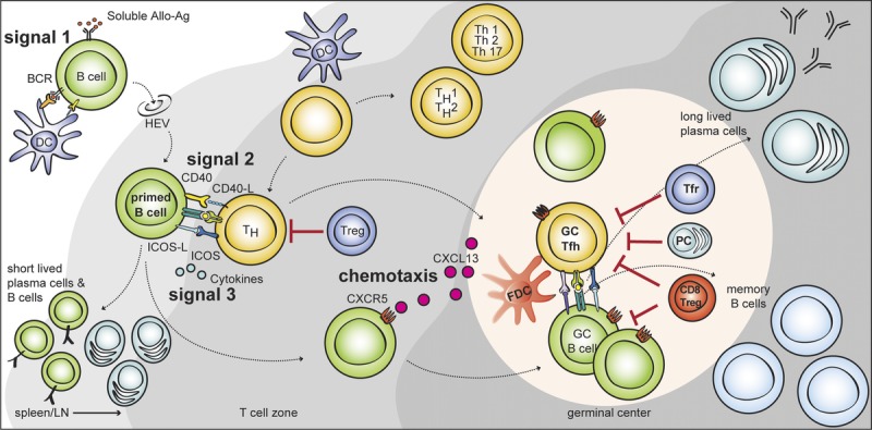
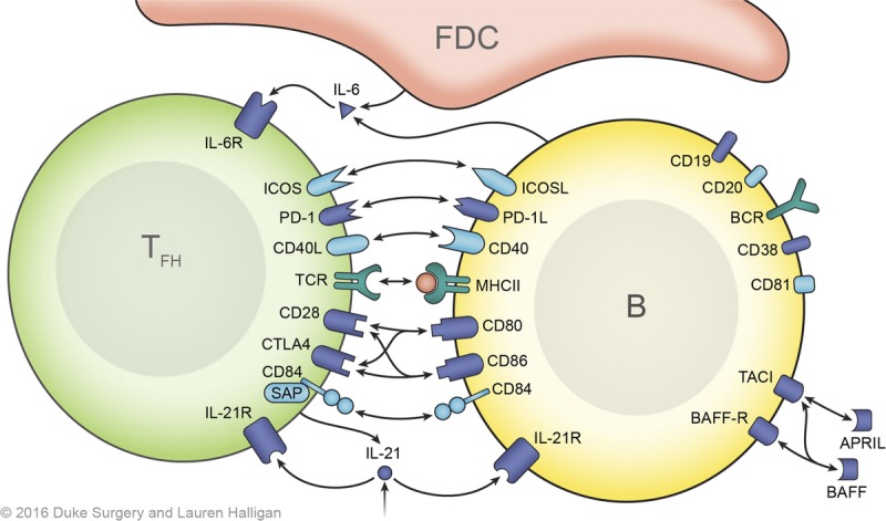
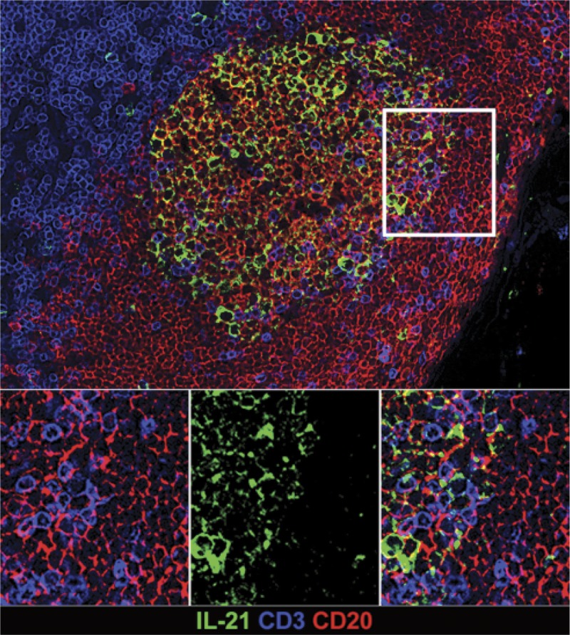
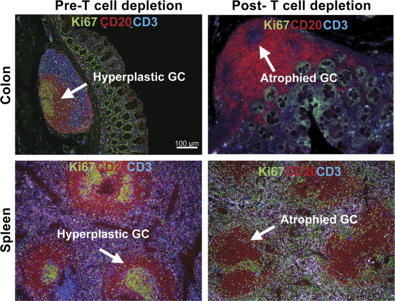
Crosstalk between B and T cells in transplantation is increasingly recognized as being important in the alloimmune response. T cell activation of B cells occurs by a 3-stage pathway, culminating with costimulation signals. We review the distinct T cell subtypes required for B-cell activation and discuss the formation of the germinal center (GC) after transplantation, with particular reference to the repopulation of the GC after depletional induction, and the subsequent effect of immunosuppressive manipulation of T cell-B cell interactions. In addition, ectopic GCs are seen in transplantation, but their role is not fully understood. Therapeutic options to target T cell-B cell interactions are of considerable interest, both as immunosuppressive tools, and to aid in the further understanding of these important alloimmune mechanisms.


Conflict of interest statement

The authors declare no conflicts of interest.

Figures

FIGURE 1.
FIGURE 1.
T cell–dependent B cell activation via multiple T-B interactions after allostimulation. Naive mature B cells are activated through BCR recognition (signal 1) and migrate to the T-B border via HEVs. Primed B cells receive further signals from costimulation (signal 2), and cytokines (signal 3) in the T-B border. Some activated CD4 T cells can acquire characteristics of Tfh cell lineage and migrate into the B cell follicle via CXCR5. These Tfh cells provide IL-21 and costimulation and induce the proliferation of cognate B cells, isotype switching, and somatic mutation. This massive B cell expansion and differentiation leads to the formation of hyperplastic GC in the B cell follicle. Tfr cells and CD8 Treg cells are thought to suppress this GC response either directly, by depleting B cells or indirectly by modulating Tfh cells. The GC response induces the differentiation of isotype-switched affinity mature B cells into memory B cells or into long-lived plasma cells. HEVs, high endothelial venules.
FIGURE 2.
FIGURE 2.
T-B cell interactions and signals. The initial B cell activation signal is provided by cognate BCR-alloantigen interaction and requires the CD19, CD21, CD81 complex for an optimal signaling. Cognate Tfh cells provide costimulation after presentation of antigenic peptides in MHC class II by the B cells in the GC. T-B interactions, especially ICOS-ICOSL, CD40-CD40L, and CD28-B7 interactions, are central to this process. Members of the SLAM (Signaling lymphocytic activation molecule) family of receptors, particularly CD84, are also important for stable T-B cell interactions, which also contribute to Tfh cell differentiation through SAP (SLAM associated protein)-mediated signaling. Cytokines, IL-6 and IL-21 contribute to Tfh cell differentiation via upregulation of BCL-6. The crosstalk between T and B cells in the GC is bidirectional mediated by ligand-receptor interactions, soluble mediators (cytokines). Biologics targeting these signals can modulate posttransplant humoral response.
FIGURE 3.
FIGURE 3.
In situ IL-21 staining in the GC. IL-21 expression is increased in hyperplastic follicles during AMR. Representative immunofluorescence image of GC staining with CD3 (blue), CD20 (red), IL-21 (green) in lymph node of rhesus macaque.
FIGURE 4.
FIGURE 4.
Eradication of GC in the lymph node after T cell depletion. Representative haematoxylin & eosin (left) and immunofluorescence image (right) of GC staining with CD3 (blue), CD20 (Red), Ki67 (green) in lymph node of rhesus macaque. Upper panels show sections of LN from healthy control with GCs, and lower panels show atrophied GC (white arrow) after T cell depletion.
FIGURE 5.
FIGURE 5.
Disrupted GC after T cell depletion in spleen and colon. Representative IHC panels show sections of colon (upper panel) and spleen (lower panel) from before and after T cell depletion with large lymphoid aggregation (Peyer patch) stained with Ki67, CD20, and CD3 antibodies. A, Similar to the lymph node, Peyer patch and spleen comprises T cell area and B-cell follicle with GC containing large proliferating B cells. B, GC in Peyer patch in the descending colon and spleen of rhesus macaques were disrupted after T cell depletion.
FIGURE 6.
FIGURE 6.
Ectopic GC in well-functioning graft. Representative GC response in the well-functioning kidney graft. Immunological profiles of Hoechst (left), CD20/CD3/Ki67 positive cells (right) within kidney graft section from renal transplant rhesus recipients. The sections were stained with Hoechst dye (white), CD3 (blue), CD20 (red), and Ki67 (green) antibodies.

References

    1. Lund FE, Randall TD. Effector and regulatory B cells: modulators of CD4+ T cell immunity Nat Rev Immunol 2010. 10236–247 - PMC - PubMed
    1. Kwun J, Knechtle SJ. Overcoming chronic rejection—can it B? Transplantation 2009. 88955–961 - PubMed
    1. van Zelm MC, Reisli I, van der Burg M. An antibody-deficiency syndrome due to mutations in the CD19 gene N Engl J Med 2006. 3541901–1912 - PubMed
    1. van Zelm MC, Smet J, Adams B. CD81 gene defect in humans disrupts CD19 complex formation and leads to antibody deficiency J Clin Invest 2010. 1201265–1274 - PMC - PubMed
    1. Thiel J, Kimmig L, Salzer U. Genetic CD21 deficiency is associated with hypogammaglobulinemia J Allergy Clin Immunol 2012. 129801–810.e6 - PubMed

MeSH terms