. 2016 Dec 1;12(12):e1006325.
doi: 10.1371/journal.pgen.1006325. eCollection 2016 Dec.

Structural and Functional Recovery of Sensory Cilia in C. elegans IFT Mutants upon Aging

Affiliations

Structural and Functional Recovery of Sensory Cilia in C. elegans IFT Mutants upon Aging

Astrid Cornils et al. PLoS Genet. .

Abstract

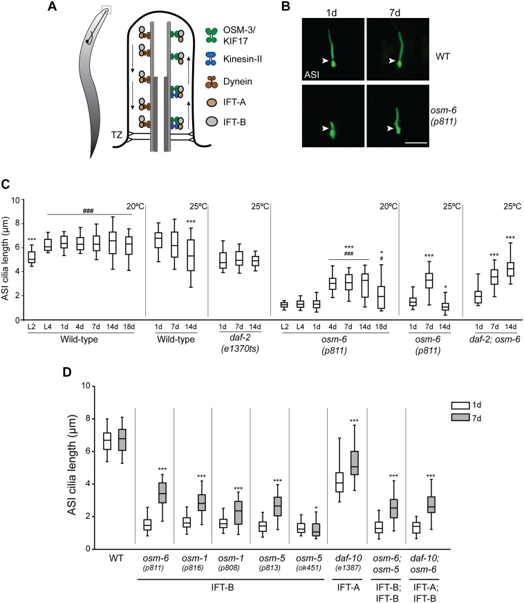
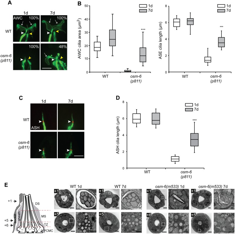
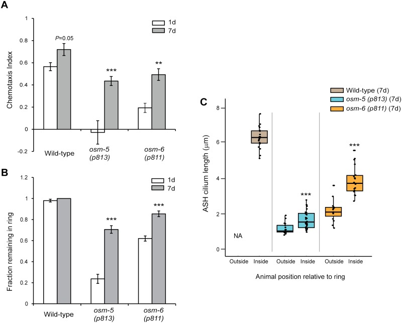
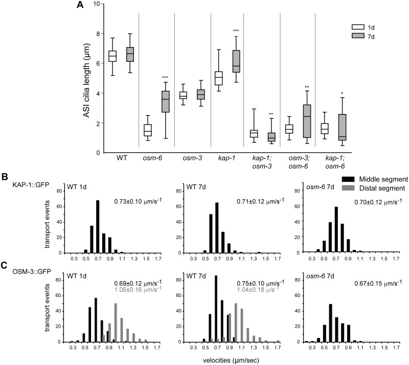
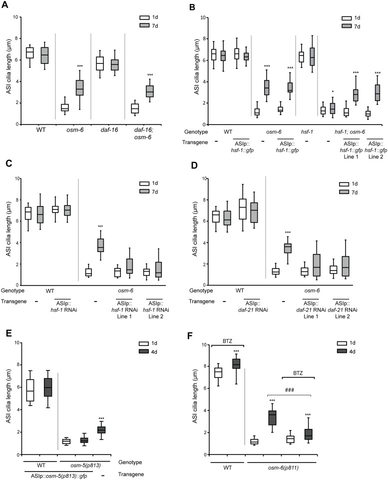
The majority of cilia are formed and maintained by the highly conserved process of intraflagellar transport (IFT). Mutations in IFT genes lead to ciliary structural defects and systemic disorders termed ciliopathies. Here we show that the severely truncated sensory cilia of hypomorphic IFT mutants in C. elegans transiently elongate during a discrete period of adult aging leading to markedly improved sensory behaviors. Age-dependent restoration of cilia morphology occurs in structurally diverse cilia types and requires IFT. We demonstrate that while DAF-16/FOXO is dispensable, the age-dependent suppression of cilia phenotypes in IFT mutants requires cell-autonomous functions of the HSF1 heat shock factor and the Hsp90 chaperone. Our results describe an unexpected role of early aging and protein quality control mechanisms in suppressing ciliary phenotypes of IFT mutants, and suggest possible strategies for targeting subsets of ciliopathies.


Conflict of interest statement

The authors have declared that no competing interests exist.

Figures

Fig 1
Fig 1. Cilia of the ASI sensory neurons elongate in aged IFT mutants.
(A) (Left) Cartoon of a worm showing a representative sensory neuron in the worm head. Cilia are present at the dendritic ends at the nose (box). (Right) Diagrammatic representation of the structure of a typical cilium and IFT in C. elegans. Arrows indicated direction of IFT. TZ—transition zone (showing Y-link microtubule-to-membrane connectors). (B) Representative images of ASI cilia in 1d and 7d old adult wild-type (WT) and osm-6(p811) mutants. Arrowheads indicate the cilia base. Anterior is at top. ASI cilia were visualized via expression of a GFP-tagged SRG-36 GPCR protein expressed under the ASI-specific str-3 promoter. Scale bar: 5 μm. (C) Quantification of ASI cilia length in animals of the indicated genetic backgrounds at different larval stages (L2, L4) or days of adulthood. Horizontal lines indicate 25th, 50th and 75th percentiles; bars indicate 5th and 95th percentiles. * and *** indicate different from 1d within a genotype at P<0.05 and 0.001, respectively; # and ### indicate different from L2 within a genotype at P<0.05 and 0.001, respectively (Kruskal-Wallis test with post hoc paired comparisons). n>30 for each; ≥3 independent experiments. Animals were grown at either 20°C or 25°C for each set of experiments (indicated at top right). (D) Quantification of ASI cilia length in 1d and 7d old animals of the indicated genotypes grown at 20°C. ASI cilia were visualized via expression of str-3p::srg-36::gfp. Alleles used in the double mutant strains were osm-6(p811), osm-5(p813), and daf-10(e1387). Horizontal lines indicate 25th, 50th and 75th percentiles; bars indicate 5th and 95th percentiles. * and *** indicate different from 1d within a genotype at P<0.05 and 0.001, respectively (Wilcoxon Mann-Whitney U test). n>30 for each; ≥3 independent experiments.
Fig 2
Fig 2. Cilia of multiple sensory neurons exhibit structural recovery in aged IFT mutants.
(A) Representative images of the fan-shaped AWC, and rod-like ASE, cilia in 1d and 7d old wild-type and osm-6(p811) mutants. Cilia were visualized via expression of ceh-36p::gfp which drives expression in AWC and ASE [44]. White and yellow arrowheads mark the bases of the AWC and ASE cilia, respectively. Arrows mark the AWC cilia membraneous expansions (‘fans’). Numbers indicate the percentage of animals exhibiting phenotypes similar to those shown; n≥12 for each. Anterior is at top. Scale bar: 5 μm. (B) Quantification of AWC cilia fan area (left) and ASE cilia length (right) in 1d and 7d old wild-type and osm-6(p811) mutants. *** indicates different from 1d within a genotype at P<0.001 (Wilcoxon Mann-Whitney U test). n>30 each; 3 independent experiments. (C) Representative images of ASH cilia in 1d and 7d old adult wild-type (WT) and osm-6(p811) mutants. Arrowheads indicate the cilia base. ASH neuronal processes including cilia are marked via expression of GFP under the sra-6 promoter. The cilium base is marked via localization of MKSR-2::TagRFP. Anterior is at top. Scale bar: 5 μm. (D) Quantification of ASH cilia length in wild-type and osm-6(p811) mutants at the indicated days of adulthood. *** indicates different from 1d within a genotype at P<0.001 (Wilcoxon Mann-Whitney U test). n>30 for each; ≥3 independent experiments. (E) Transmission electron microscopy of amphid channel sensory pores in adult 1d and 7d animals. Images acquired from serial cross sections of wild-type and osm-6(m533) mutant worms; each image pair consists of a low magnification image of the entire nose tip (left) and a high magnification image of an amphid pore (right; boxed regions in images at left). Numbers (microns) denote proximal positioning of section relative to the distal-most first section in the series; section positions also indicated in schematic. The schematic is a longitudinal representation of a wild-type amphid neuronal pore, enveloped by supporting sheath and socket glial cells. Pores consist of 10 ciliary axonemes (only 3 are shown), each with distal segment (DS; singlet microtubules), middle segment (MS; doublet microtubules), transition zone (TZ) and periciliary membrane (PCMC) subcompartments. Bars; 2 μm (low magnification images), 200 nm (high magnification images).
Fig 3
Fig 3. Cilia-dependent sensory behaviors are improved in aged IFT-B mutants.
(A) Chemotaxis responses of 1d and 7d old animals of the indicated genotypes to a point source of bacteria (see Materials and Methods). Positive chemotaxis indices indicate attraction. ** and *** indicate different from 1d within a genotype at P<0.01 and 0.001, respectively (Kruskal-Wallis non parametric test). Error bars are SEM. n>200 animals each from 8 independent assays. (B) Fraction of animals of the indicated genotypes and ages that remain within a ring of 8M glycerol after 2 minutes. Error bars are SEM. *** indicate different from 1d at P< 0.001 within a genotype (Kruskal-Wallis non-parametric test). n>100 animals each from 10 independent assays. (C) Length of ASH cilia in 7d old wild-type, osm-5(p813) and osm-6(p811) animals that remained within (inside), or escaped (outside), a ring of 8M glycerol after 2 minutes. Horizontal lines indicate 25th, 50th and 75th percentiles. *** indicates different from 1d within a genotype at P< 0.001 (Wilcoxon Mann-Whitney U test). n≥15 animals for each condition.
Fig 4
Fig 4. IFT motor proteins are necessary for age-dependent cilia recovery in IFT mutants.
(A) Quantification of ASI cilia length in 1d and 7d old animals of the indicated genotypes. ASI cilia were visualized via expression of str-3p::srg-36::gfp. Alleles used in the double mutant strains were osm-6(p811), kap-1(ok676) and osm-3(p802). Horizontal lines indicate 25th, 50th and 75th percentiles; bars indicate 5th and 95th percentiles. *, ** and *** indicate different from 1d within a genotype at P<0.05, 0.01 and 0.001, respectively (Wilcoxon Mann-Whitney U test). n>30 for each; ≥3 independent experiments. (B) Histograms of KAP-1::GFP and (C) OSM-3::GFP anterograde velocities in the ASH/ASI cilia of 1d and 7d old wild-type or osm-6(p811) mutants. kap-1:gfp and osm-3::gfp were expressed under the sra-6 promoter. IFT could not be reliably quantified in short cilia in 1d old osm-6 mutants. Anterograde velocities in the middle and distal segments are indicated by black and gray bars, respectively; average velocities are indicated at top right in each panel in corresponding colors. See S2 Table for statistical analyses.
Fig 5
Fig 5. Improved protein quality control mechanisms may underlie age-dependent cilia recovery in IFT mutants.
(A-D) Quantification of ASI cilia length in animals of the indicated genotypes and adult ages. Alleles used were osm-6(p811), daf-16(mu86), and hsf-1(sy441ts). ASI cilia were visualized via expression of str-3p::srg-36::gfp (A,C,D) or srg-47p::TagRFP (B). An hsf-1 cDNA tagged with gfp, and hsf-1 and daf-21 sense and antisense sequences were expressed in ASI under the srg-47 promoter. Lines 1 and 2 represent independent transgenic lines. Animals were grown at 20°C (A,C,D) or 25°C (B). Horizontal lines indicate 25th, 50th and 75th percentiles; bars indicate 5th and 95th percentiles. * and *** indicate different from 1d within a genotype at P<0.05 and 0.001, respectively (Wilcoxon Mann-Whitney U test). n>30 for each; ≥3 independent experiments. (E) Quantification of ASI cilia length in 1d and 4d old animals of the indicated genotypes, expressing srg-47p::osm-5(p813)::gfp. ASI cilia were visualized via expression of srg-47p::TagRFP. Horizontal lines indicate 25th, 50th and 75th percentiles; bars indicate 5th and 95th percentiles. *** indicates different from 1d of the same genotype at P<0.001 (Wilcoxon Mann-Whitney U test). n>50 each; 3 independent experiments. (F) Quantification of ASI cilia length in 1d and 4d old animals of the indicated genotypes. ASI cilia were visualized via expression of str-3p::srg-36::gfp. Animals were grown on 10 μM Bortezomib (BTZ). Horizontal lines indicate 25th, 50th and 75th percentiles; bars indicate 5th and 95th percentiles. *** indicates different from 1d within a genotype at P<0.001; ### indicates different between the indicated conditions at P<0.001 (Wilcoxon Mann-Whitney U test). n>50 each; 3 independent experiments.

Comment in

  • Middle Age Has Its Advantages.
    Barr MM. Barr MM. PLoS Genet. 2016 Dec 1;12(12):e1006426. doi: 10.1371/journal.pgen.1006426. eCollection 2016 Dec. PLoS Genet. 2016. PMID: 27906958 Free PMC article. No abstract available.

References

    1. Kozminski KG, Johnson KA, Forscher P, Rosenbaum JL. A motility in the eukaryotic flagellum unrelated to flagellar beating. Proc Natl Acad Sci USA. 1993;90: 5519–5523. - PMC - PubMed
    1. Pedersen LB, Rosenbaum JL. Intraflagellar transport (IFT) role in ciliary assembly, resorption and signalling. Curr Top Dev Biol. 2008;85: 23–61. 10.1016/S0070-2153(08)00802-8 - DOI - PubMed
    1. Cole DG, Diener DR, Himelblau AL, Beech PL, Fuster JC, Rosenbaum JL. Chlamydomonas kinesin-II-dependent intraflagellar transport (IFT): IFT particles contain proteins required for ciliary assembly in Caenorhabditis elegans sensory neurons. J Cell Biol. 1998;141: 993–1008. - PMC - PubMed
    1. Huangfu D, Liu A, Rakeman AS, Murcia NS, Niswander L, Anderson KV. Hedgehog signalling in the mouse requires intraflagellar transport proteins. Nature. 2003;426: 83–87. 10.1038/nature02061 - DOI - PubMed
    1. Murcia NS, Richards WG, Yoder BK, Mucenski ML, Dunlap JR, Woychik RP. The Oak Ridge Polycystic Kidney (orpk) disease gene is required for left-right axis determination. Development. 2000;127: 2347–2355. - PubMed

MeSH terms

Substances