. 2016 Dec 1;11(12):e0167366.
doi: 10.1371/journal.pone.0167366. eCollection 2016.

Silver Nanoparticle-Directed Mast Cell Degranulation Is Mediated through Calcium and PI3K Signaling Independent of the High Affinity IgE Receptor

Affiliations

Silver Nanoparticle-Directed Mast Cell Degranulation Is Mediated through Calcium and PI3K Signaling Independent of the High Affinity IgE Receptor

Nasser B Alsaleh et al. PLoS One. .

Abstract

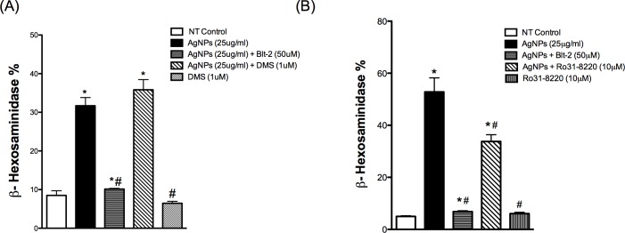
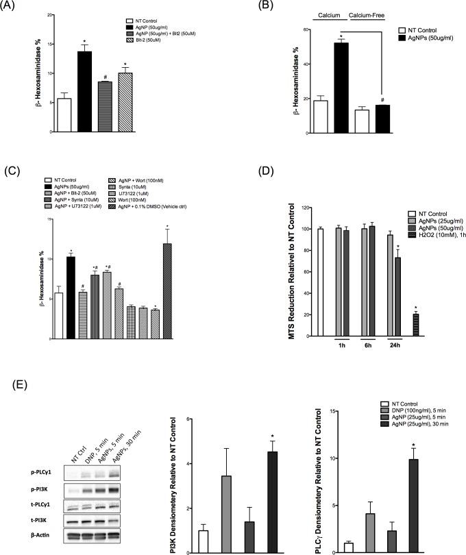
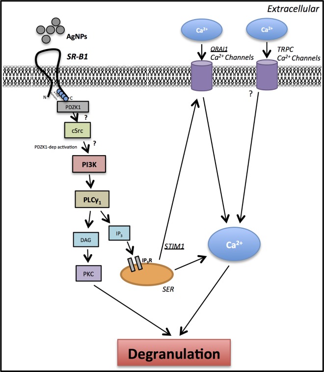
Engineered nanomaterial (ENM)-mediated toxicity often involves triggering immune responses. Mast cells can regulate both innate and adaptive immune responses and are key effectors in allergic diseases and inflammation. Silver nanoparticles (AgNPs) are one of the most prevalent nanomaterials used in consumer products due to their antimicrobial properties. We have previously shown that AgNPs induce mast cell degranulation that was dependent on nanoparticle physicochemical properties. Furthermore, we identified a role for scavenger receptor B1 (SR-B1) in AgNP-mediated mast cell degranulation. However, it is completely unknown how SR-B1 mediates mast cell degranulation and the intracellular signaling pathways involved. In the current study, we hypothesized that SR-B1 interaction with AgNPs directs mast cell degranulation through activation of signal transduction pathways that culminate in an increase in intracellular calcium signal leading to mast cell degranulation. For these studies, we utilized bone marrow-derived mast cells (BMMC) isolated from C57Bl/6 mice and RBL-2H3 cells (rat basophilic leukemia cell line). Our data support our hypothesis and show that AgNP-directed mast cell degranulation involves activation of PI3K, PLCγ and an increase in intracellular calcium levels. Moreover, we found that influx of extracellular calcium is required for the cells to degranulate in response to AgNP exposure and is mediated at least partially via the CRAC channels. Taken together, our results provide new insights into AgNP-induced mast cell activation that are key for designing novel ENMs that are devoid of immune system activation.


Conflict of interest statement

The authors have declared that no competing interests exist.

Figures

Fig 1
Fig 1. Time Course of Mast Cell Association of AgNPs
(A) Representative Transmission Electron Microcopy (TEM) image demonstrating AgNP shape and size. (B) Representative TEM images of mast cells following exposure to 20 nm AgNPs over time. Mast cells were treated with AgNPs (25 μg/ml) for 10, 20, 30 and 60 min and AgNPs uptake by mast cells was assessed. Arrow indicates AgNPs that were being taken up by a mast cell (inset). A representative image was obtained from at least 5 different images.
Fig 2
Fig 2. Mast cell degranulation following exposure to AgNPs
Mast cell degranulation was assessed by measuring the release of β-hexosaminidase into supernatants. (A) Cells were pre-treated with the SR-B1 inhibitor Blt-2 (50 μM), SR-B1 specific antibodies (1:100 dilution) 30 min prior to AgNP (25 μg/ml) exposure for 1h and release of β-hexosaminidase was assessed. (B) Cells were sensitized with anti-DNP IgE overnight and then exposed to AgNPs (25 μg/ml) for 1 h and release of β-hexosaminidase was assessed. (C) Cells were pre-treated with Blt-2 (50 μM) for 30 min then activated with either DNP (30 min) or AgNP (1 h) and release of β-hexosaminidase was assessed. (D) Representative immunoblots for global p-Tyr and p-Ser/Thr following DNP (100 ng/ml) or AgNPs (25 μg/ml) exposure. Values are expressed as mean ± SEM of at least 3 independent experiments. *Indicates significant difference from controlled group (p≤0.05). #Indicates significant difference from AgNP-treated group (p≤0.05)
Fig 3
Fig 3. Calcium signal in mast cell following exposure to AgNPs
Mast cell degranulation was assessed by measuring the release of β-hexosaminidase into supernatants. (A) Mast cells degranulation was measured following exposure to AgNPs in the presence and absence of calcium. (B–left panel) Cells were stained with Fluo-4 AM (5 μM) and mean fluorescence intensity was assessed before (baseline NT control, solid line) and after exposure to ionomycin (1 μM) or AgNPs (50 μg/ml) (dotted line) for 2 min. (B–right panel) A representative graph of 3 independent experiments showing fold change of mean fluorescence intensity relative to NT control. (C) Cells were pre-treated with the CRAC calcium channels inhibitor Synta (10 μM) 30 min prior to AgNP (25 μg/ml) exposure for 1 h and release of β-hexosaminidase was assessed. Values are expressed as mean ± SEM of at least 3 independent experiments. *Indicates significant difference from controlled group (p≤0.05). #Indicates significant difference from indicated groups (p≤0.05)
Fig 4
Fig 4. PLC and PI3K signaling in response to AgNPs
Mast cell degranulation was assessed by measuring the release of β-hexosaminidase into supernatants. Cells were pre-treated with (A) the PLCγ inhibitor U73122 (1 μM) or (B) the PI3K inhibitor wortmannin (100 nM) 30 min prior to AgNP (25 μg/ml) exposure for 1 h and release of β-hexosaminidase was assessed. (C) Representative immunoblots for p-PI3K in samples pretreated with or without Blt-2 (50 μM) and followed by AgNP exposure for 1 h. Values are expressed as mean ± SEM of at least 3 independent experiments. *Indicates significant difference from controlled group (p≤0.05). #Indicates significant difference from AgNP-treated group (p≤0.05)
Fig 5
Fig 5. Involvement of other signaling pathways in response to AgNPs
Mast cell degranulation was assessed by measuring the release of β-hexosaminidase into supernatants. Cells were pre-treated with (A) the sphingosine kinase inhibitor DMS (1 μM) or (B) the PKC inhibitor Ro31-8220 (10 μM) for 30 min prior to AgNP (25 μg/ml) exposure for 1 h and release of β-hexosaminidase was assessed. Values are expressed as mean ± SEM of at least 3 independent experiments. *Indicates significant difference from controlled group (p≤0.05). #Indicates significant difference from AgNP-treated group (p≤0.05)
Fig 6
Fig 6. Confirmation of BMMCs results in RBL-2H3
(A) Cells were pre-treated with Blt-2 (50 μM) 30 min prior to AgNP (50 μg/ml) exposure for 1 h and release of β-hexosaminidase was assessed into supernatants. (B) Mast cells degranulation was measured following exposure to AgNPs (50 μg/ml) in the presence and absence of calcium. (C) Cells were pre-treated with the indicated inhibitors 30 min prior to AgNP (50 μg/ml) exposure for 1 h and release of β-hexosaminidase was assessed into supernatants. (D) Cells were treated with AgNPs (25 or 50 μg/ml) for 1, 6, and 24 h and cell viability was assessed by measuring the conversion of MTS into formazan. (E) Representative immunoblots for p-PLCγ and p-PI3K of mast cell in the presence or absence of DNP (100 ng/ml) for 5 min or AgNPs (25 μg/ml) for 5 and 30 min. Values are expressed as mean ± SEM of at least 3 independent experiments. *Indicates significant difference from controlled group (p≤0.05). #Indicates significant difference from AgNP-treated group (p≤0.05)
Fig 7
Fig 7. A schematic representation of proposed signaling pathway involved in activation of mast cells by AgNPs
We propose that AgNPs interact with SR-B1 leading to recruitment of PDZK1 (SR-B1 adaptor protein), which activates downstream signaling cascade involving PI3K and PLCγ. Inositol 1,4,5-triphosphate (IP3), which is released following activation of PLCγ, interacts with its receptor IP3R on smooth endoplasmic reticulum (SER) leading to the release of Ca2+ from ER stores. As a result of a drop in Ca2+ levels in SER, the CRAC Ca2+ channels (cell membrane) are activated leading to influx of extracellular Ca2+. Increasing intracellular Ca2+ levels ([Ca2+]i) is ultimately culminated in mast cell degranulation. PKC: Protein kinase C; IP3 (InsP3): inositol triphosphate; PtdIns(4,5)P2: phosphatidylinositol-4,5-bisphosphate (PIP2); PtdIns(3,4,5)P3: phosphatidylinositol-3,4,5-Triphosphate (PIP3); PIP2 and PIP3 are membrane phospholipids; DAG: diacylglycerol. cSrc: cellular sarcoma (protein tyrosine kinase); TRPC: transient receptor potential cation channels.

References

    1. Vance ME, Kuiken T, Vejerano EP, McGinnis SP, Hochella MF Jr., Rejeski D, et al. Nanotechnology in the real world: Redeveloping the nanomaterial consumer products inventory. Beilstein journal of nanotechnology. 2015;6:1769–80. PubMed Central PMCID: PMC4578396. doi: 10.3762/bjnano.6.181 - DOI - PMC - PubMed
    1. Oberdorster G, Oberdorster E, Oberdorster J. Nanotoxicology: an emerging discipline evolving from studies of ultrafine particles. Environmental health perspectives. 2005;113(7):823–39. PubMed Central PMCID: PMC1257642. doi: 10.1289/ehp.7339 - DOI - PMC - PubMed
    1. Dobrovolskaia MA, Shurin M, Shvedova AA. Current understanding of interactions between nanoparticles and the immune system. Toxicology and applied pharmacology. 2016;299:78–89. PubMed Central PMCID: PMC4811709. doi: 10.1016/j.taap.2015.12.022 - DOI - PMC - PubMed
    1. Chen X, Schluesener HJ. Nanosilver: a nanoproduct in medical application. Toxicology letters. 2008;176(1):1–12. doi: 10.1016/j.toxlet.2007.10.004 - DOI - PubMed
    1. Christensen FM, Johnston HJ, Stone V, Aitken RJ, Hankin S, Peters S, et al. Nano-silver—feasibility and challenges for human health risk assessment based on open literature. Nanotoxicology. 2010;4(3):284–95. doi: 10.3109/17435391003690549 - DOI - PubMed