. 2016 Dec 20;7(51):85502-85514.
doi: 10.18632/oncotarget.13453.

Expression analysis and clinical significance of eIF4E, VEGF-C, E-cadherin and MMP-2 in colorectal adenocarcinoma

Affiliations

Expression analysis and clinical significance of eIF4E, VEGF-C, E-cadherin and MMP-2 in colorectal adenocarcinoma

Minna Gao et al. Oncotarget. .

Abstract

The underlying mechanisms of colorectal carcinoma (CRC) metastasis remain to be elucidated. The aim of this study is to investigate clinical significance and the expression of eIF4E, VEGF-C, MMP-2, and E-cadherin in the CRC metastasis. We investigated their expressions in 108 patients, analyzed the relationships between their expressions in CRC and evaluated the relationships between their expressions and clinical pathogenic parameters. Furthermore, their roles in patient survival and in CRC metastasis were also investigated. We found that eIF4E, VEGF-C and MMP-2 were up-regulated in CRC, and their expression frequencies (EFs) were higher in cancerous tissues than in adjacent normal tissues. The EF of E-cadherin is lower in cancerous tissues than in adjacent normal tissues. Totally, their EFs were not associated with sex and age of patient, however, their EFs were associated with tumor differentiation, the depth of invasion, lymph node metastasis and tumor stages. Furthermore, eIF4E, VEGF-C, and MMP-2 shortened and E-cadherin prolonged survival in patient-derived CRC xenografts. Similarly, eIF4E, VEGF-C, and MMP-2 promoted and E-cadherin suppressed the lung metastasis of CRC cells. In addition, knockdown of eIF4E inhibited migration of CRC cells, downregulated VEGF-C, MMP-2 and upregulated E-cadherin. In conclusion, eIF4E promoted CRC metastasis via up-regulating the expression of VEGF-C, MMP-2 and suppressing E-cadherin.

Keywords: E-cadherin; MMP-2; VEGF-C; colorectal cancer; eIF4E.


Conflict of interest statement

CONFLICTS OF INTEREST

The authors declare no conflicts interest.

Figures

Figure 1
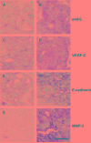
Figure 1. The expression of eIF4E, VEGF-C, E-cadherin and MMP-2 in colon cancerous tissues
A. The expression of eIF4E in normal colon tissues around cancer. B. The negative expression of eIF4E in colon cancerous tissues. C. The positive expression of eIF4E in colon cancerous tissues. D. The expression of VEGF-C in normal colon tissues around cancer. E. The negative expression of VEGF-C in colon cancerous tissues. F. The positive expression of VEGF-C in colon cancerous tissues. G. The expression of E-cadherin (E-cad) in normal colon tissues around cancer. H. The negative expression of E-cad in colon cancerous tissues. I. The positive expression of E-cad in colon cancerous tissues. J. The expression of MMP-2 in normal colon tissues around cancer. K. The negative expression of MMP-2 in colon cancerous tissues. L. The positive expression of MMP-2 in colon cancerous tissues. Bar = 50 μM.
Figure 2
Figure 2. Kaplan-Meier survival analysis for xenograft mice
Log rank test χ2= 75.0054. p< 0.0001. Fragments of human colon tumors were implanted subcutaneously (s.c.) into nude mice, and mice with tumors that reached 4–5 mm in diameter (n=5 per group) were applied to survival statistics. A. Kaplan–Meier survival curves of mice bearing eIF4E negative and positive tumors. B. Kaplan–Meier survival curves of mice bearing VEGF-C negative and positive tumors. C. Kaplan–Meier survival curves of mice bearing MMP-2 negative and positive tumors. D. Kaplan–Meier survival curves of mice bearing E-cadherin negative and positive tumors.
Figure 3
Figure 3. The expression of eIF4E, VEGF-C, MMP-2 and E-cadherin in primary site of patient-derived colon cancer xenografts
A. The negative expression of eIF4E in colon cancerous tissues. B. The positive expression of eIF4E in colon cancerous tissues. C. The negative expression of VEGF-C in colon cancerous tissues. D. The positive expression of VEGF-C in colon cancerous tissues. E. The negative expression of E-cadherin in colon cancerous tissues. F. The positive expression of E-cadherin in colon cancerous tissues. G. The negative expression of MMP-2 in colon cancerous tissues. H. The positive expression of MMP-2 in colon cancerous tissues. Bar = 25 μM.
Figure 4
Figure 4. The growth rates of colon cancer cell lines HCT-15/Rluc with overexpression of E-cadherin, MMP-2, VEGF-c and eIF4E and parental HCT-15/Rluc
A. Western blotting showed that the stable cell lines including HCT-15/Rluc/E-cadherin, HCT-15/Rluc/VEGF-C, HCT-15/Rluc/MMP-2, and HCT-15/Rluc/eIF4E were constructed successfully. B. The stable colon cancer cell lines with overexpression of E-cadherin, MMP-2, VEGF-c and eIF4E as well as parental HCT-15/Rluc cell line showed different growth rates.
Figure 5
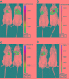
Figure 5. RL bioluminescence from HCT-15/Rluc cells present in Lung tissue in living mice
A. The HCT-15/Rluc/eIF4E cells (1.0 × 10⁶) and corresponding HCT-15-Rluc were injected via tail-vein 3 days later. The bioluminescence seen represents the thorax region of the mouse where HCT-15/Rluc and HCT-15/Rluc/eIF4E cells are trapped in the lungs. B. The HCT-15/Rluc/VEGF-C cells (1.0 × 10⁶) and corresponding HCT-15/Rluc were injected via tail-vein 3 days later. The bioluminescence seen represents the thorax region of the mouse where HCT-15/Rluc and HCT-15/Rluc/VEGF-C cells are trapped in the lungs. C. The HCT-15/Rluc/MMP-2 cells (1.0 × 10⁶) and corresponding HCT-15/Rluc were injected via tail-vein 3 days later. The bioluminescence seen represents the thorax region of the mouse where HCT-15/Rluc and HCT-15/Rluc/MMP-2 cells are trapped in the lungs. D. The HCT/15-Rluc/E-cadherin cells (1.0 × 10⁶) and corresponding HCT-15-Rluc were injected via tail-vein 3 days later. The bioluminescence seen represents the thorax region of the mouse where HCT-15/Rluc and HCT-15/Rluc/E-cadherin cells are trapped in the lungs.
Figure 6
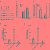
Figure 6. The expression of VEGF-C, MMP-2 and E-cadherin in the stable SW480 cell lines with the overexpression and Knockdown of eIF4E
A. Western blotting showed the expression of eIF4E, VEGF-C, MMP-2, and E-cadherin in the corresponding stable SW 480 cell lines with the overexpression and knockdown of eIF4E. Control, Control stable SW480 cell lines by use of lentiviral infection packaged with control empty vectors. Overexpression, the stable SW480 cell line with the overexpression of eIF4E. sheIF4E, the stable SW480 cell lines with knockdown of eIF4E. The cells were lysed for loading on SDS-PAGE. The blotting were performed by use of indicated antibodies. B. Q-PCR showed the expression of eIF4E, VEGF-C, MMP-2, and E-cadherin in the stable SW 480 cell lines with the overexpression of eIF4E. C. Q-PCR showed the expression of eIF4E, VEGF-C, MMP-2, and E-cadherin in the stable SW 480 cell lines with the knockdown of eIF4E. Control, Control stable SW480 cell lines by use of lentiviral infection packaged with control empty vectors. Overexpression of eIF4E, the stable SW480 cell line with the overexpression of eIF4E. sheIF4E, the stable SW480 cell lines with knockdown of eIF4E. *, p<0.05. D. Relative number of migrating cells (y-axis) in a transwell migration assay is shown for Control, eIF4E-overexpression, and eIF4E-knockdown SW 480 cells. *, p<0.05. E. Number of mammospheres (y-axis) formed by Control, eIF4E-overexpression and eIF4E-knockdown SW480 cells. *, p<0.05.

Similar articles

Cited by

References

    1. American Cancer Association. Cancer Facts & Figures 2016. American Cancer Association.
    1. Sunavala-Dossabhoy G, Palaniyandi S, Clark C, Nathan CO, Abreo FW, Caldito G. Analysis of eIF4E and 4EBP1 mRNAs in head and neck cancer. The Laryngoscope. 2011;121:2136–2141. - PubMed
    1. Joukov V, Pajusola K, Kaipainen A, Chilov D, Lahtinen I, Kukk E, Saksela O, Kalkkinen N, Alitalo K. A novel vascular endothelial growth factor, VEGF-C, is a ligand for the Flt4 (VEGFR-3) and KDR (VEGFR-2) receptor tyrosine kinases. The EMBO journal. 1996;15:1751. - PMC - PubMed
    1. de Oliveira AT, Reis RM, Afonso J, Martinho O, Matos D, Carvalho AL, Vazquez VL, Silva TB, Scapulatempo C, Saad SS, Longatto-Filho A. Lymphangiogenic VEGF-C and VEGFR-3 expression in genetically characterised gastrointestinal stromal tumours. Histology and histopathology. 2011;26:1499–1507. - PubMed
    1. Beavon IR. The E-cadherin-catenin complex in tumour metastasis: structure, function and regulation. European journal of cancer. 2000;36:1607–1620. - PubMed

MeSH terms