. 2016 Dec 6;68(22):2395-2407.
doi: 10.1016/j.jacc.2016.08.073.

Aβ Amyloid Pathology Affects the Hearts of Patients With Alzheimer's Disease: Mind the Heart

Affiliations

Aβ Amyloid Pathology Affects the Hearts of Patients With Alzheimer's Disease: Mind the Heart

Luca Troncone et al. J Am Coll Cardiol. .

Abstract

Background: Individually, heart failure (HF) and Alzheimer's disease (AD) are severe threats to population health, and their potential coexistence is an alarming prospect. In addition to sharing analogous epidemiological and genetic profiles, biochemical characteristics, and common triggers, the authors recently recognized common molecular and pathological features between the 2 conditions. Whereas cognitive impairment has been linked to HF through perfusion defects, angiopathy, and inflammation, whether patients with AD present with myocardial dysfunction, and if the 2 conditions bear a common pathogenesis as neglected siblings are unknown.

Objectives: Here, the authors investigated whether amyloid beta (Aβ) protein aggregates are present in the hearts of patients with a primary diagnosis of AD, affecting myocardial function.

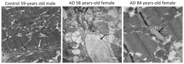
Methods: The authors examined myocardial function in a retrospective cross-sectional study from a cohort of AD patients and age-matched controls. Imaging and proteomics approaches were used to identify and quantify Aβ deposits in AD heart and brain specimens compared with controls. Cell shortening and calcium transients were measured on isolated adult cardiomyocytes.

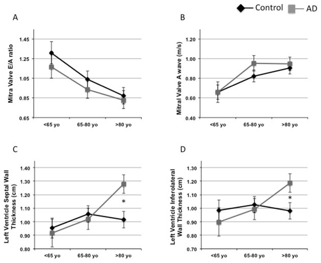
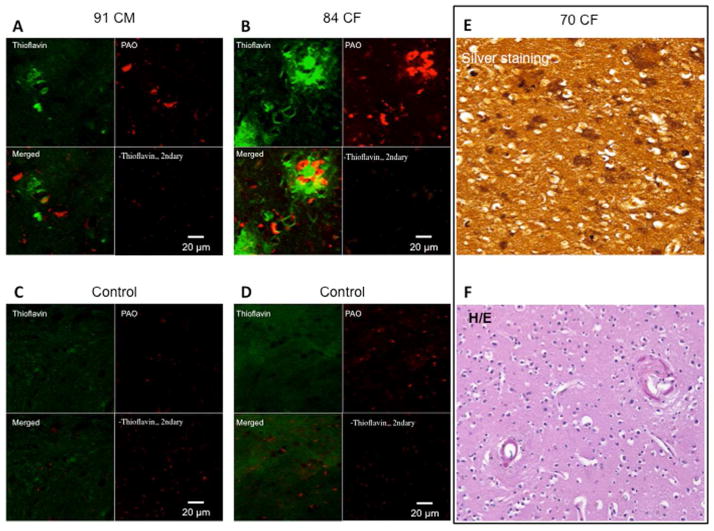
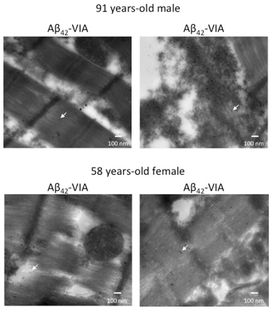
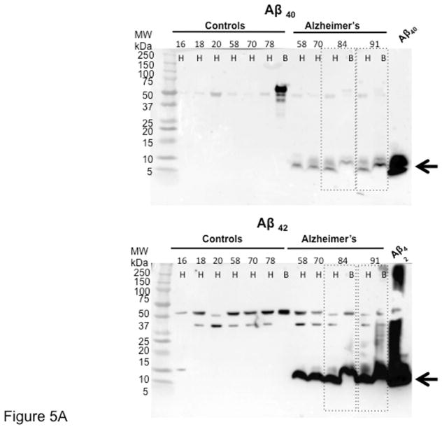
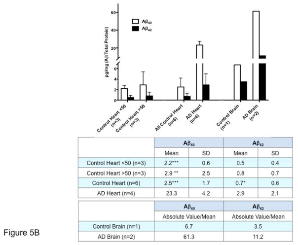
Results: Echocardiographic measurements of myocardial function suggest that patients with AD present with an anticipated diastolic dysfunction. As in the brain, Aβ40 and Aβ42 are present in the heart, and their expression is increased in AD.

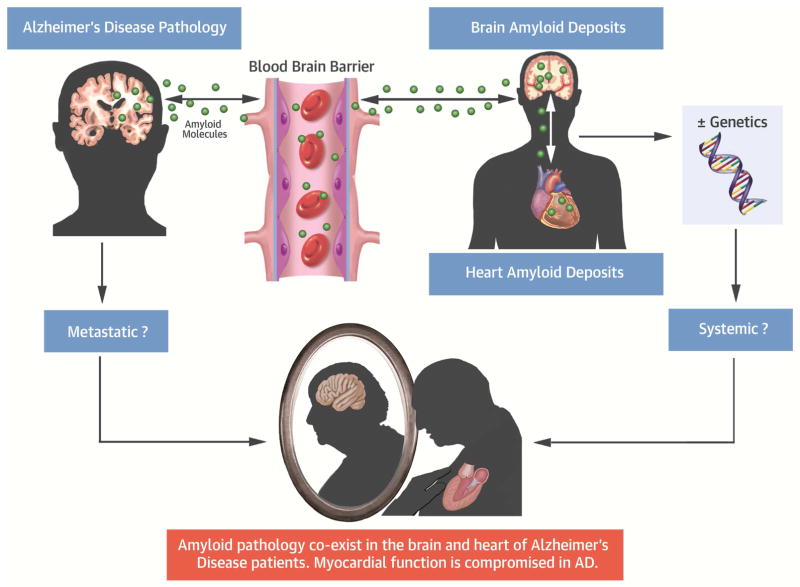
Conclusions: Here, the authors provide the first report of the presence of compromised myocardial function and intramyocardial deposits of Aβ in AD patients. The findings depict a novel biological framework in which AD may be viewed either as a systemic disease or as a metastatic disorder leading to heart, and possibly multiorgan failure. AD and HF are both debilitating and life-threatening conditions, affecting enormous patient populations. Our findings underline a previously dismissed problem of a magnitude that will require new diagnostic approaches and treatments for brain and heart disease, and their combination.

Keywords: amyloidosis; cardiomyopathy; dementia; heart failure; protein aggregates.


Figures

Figure 1
Figure 1. Crude Means of Cardiac Ultrasound Parameters for Groups and Age Ranges Show Worse Myocardial Function in the AD Clinical Cohort Compared With Controls
Crude means of cardiac ultrasound parameters for age tertiles and groups corresponding to the linear regression. The top left panel shows how the value of the MVE/A ratio is lower in younger AD subjects and progressively overlaps with the controls at advanced ages. The mean value of the MVE/A ratio of AD patients in the first tertile appears intermediate between the value of the first and the second tertile of controls. An analogous pattern is shown for AD patients in the second tertile (A). (B) Notably, the atrial (A) component alone showed no differences in all groups, suggesting that the LV compliance is the main contributor to the diastolic dysfunction in AD patients. (C and D) show the increased LV septal and inferior wall thickness, respectively, in the older AD subjects. *p < 0.05. Black diamonds: controls; gray squares: AD cases. Aβ = amyloid beta; AD = Alzheimer’s disease; LV = left ventricle/ventricular; MVE/A = mitral valve E/A ratio.
Figure 2
Figure 2. Aβ is present in AD Brain Specimens at Histopathology
Immunohistochemistry images of brain plaques stained with Thioflavin S and structural anti-oligo A11 antibodies in the brain of the (A) 91-year-old man and (B) the 84-year-old woman. The images shows minimal overlap of the A11 anti-oligo antibodies with the fibrillar mature plaques, as described by Kayed et al. (33) (C–D) Control brains. (E) Silver staining of the occipital cortex of the 70-year-old woman, showing amyloid plaques and amyloid angiopathy by Hirano silver staining. (F) Hematoxylin-eosin staining for the brain structure of the 70-year-old woman. Magnification 200×. (G) Gross pathology of the brain of the 70-year-old woman. The middle image shows the entire brain, with the hemorrhagic frontal lobe of the right hemisphere. The left and right images show the brain sections of the posterior and anterior halves of the brain, respectively. The left hemisphere appears hypotrophic, a common feature of AD. Magnification 100×. CF = Caucasian female; CM = Caucasian male; H/E = hematoxylin and eosin; PAO = pre-amyloid oligomer. Other abbreviations as in Figure 1.
Figure 3
Figure 3. Transmission Electron Microscopy Identified Myocardial Aggregates
Representative transmission electron micrographs of myocardium from the 58- and 84-year-old female donors, showing interstitial aggregates (black arrows). The white arrow indicates the interstitial tissue in the normal heart to distinguish it from the aggregates indicated by the black arrows in the diseased hearts. Images of the full set of samples are in Online Figure 2. AD = Alzheimer’s disease.
Figure 4
Figure 4. Aβ-PAO Can Be Detected in the Myocytes and Interstitial tissue
Gold-immunolabeled electron micrographs of the myocardium from the oldest and youngest AD donors. Anti-Aβ42 antibodies were conjugated with 10-nm gold beads and structural antibodies against Aβ42-PAO (VIA) were conjugated with 15-nm gold beads. The arrows indicate representative gold-labeled beads. The images show the presence of positive staining for the Aβ42-PAO detected by the VIA antibodies, but minimal to no staining for the nonoligomerized Aβ42 inside and outside the myocytes. Abbreviations as in Figures 1 and 2.
Figure 5
Figure 5. Aβ is Increased in AD Hearts
(A) Immunoblotting of AD and young and age/sex/ethnicity-matched control hearts and brains. Samples are ordered by age, from younger to older. The blot demonstrated the presence of both Aβ40 (upper panel) and Aβ42 (lower panel) in the hearts and brains of AD patients. Aβ fragments of multiple molecular weights (≅30, 40, 50 kDa) could be detected both in the heart and the brain. Of note, both Aβ40 and Aβ42 high molecular weight bands were also present in old cases without diagnosis of AD, and also in young patients, although at lower levels, as previously described in the brain. (B) Quantification of the levels of Aβ40 and Aβ42 in AD and donor samples by ELISA, showing increased levels of both fragments in the hearts and brains of patients affected by AD. Controls were divided by age ≥ or ≤50 years. Student t-test: *p < 0.05, **p < 0.01 ***p < 0.005 compared with AD. A 1-way analysis of variance (ANOVA) was also performed in all groups, followed by Bonferroni post hoc analysis. ANOVA was significant for Aβ40 (p < 0.0001) and Aβ42 (p < 0.05). Post-test for Aβ40 was significant (p < 0.001) by age group comparisons, whereas p = 0.074 for all groups for Aβ42. The Levene test was performed for equal variances. A larger sample size may be necessary to establish the significance of the differences. The axis of ordinates is separated to better visualize the control values. Values indicate pg/mg Aβ/total protein. Abbreviations as in Figure 1.
Central Illustration
Central Illustration. Myocardial Aβ Amyloid Deposits in Alzheimer Disease
Aβ aggregates extend outside the central nervous system to the myocardium in AD. Patients with a diagnosis of AD present with clinical signs of a functional defect. Therefore, in the context of increasing life expectancy, these findings raise an alarming threat for public health, as they suggest that the 2 maladies may coexist, either sharing a common etiology or as phenotypes of a multiorgan syndrome. Aβ = amyloid beta; AD = Alzheimer’s disease; BBB = blood-brain barrier.

Comment in

References

    1. National Institute on Aging. [Accessed September 20, 2016];Alzheimer’s Disease Progress Report 2014–2015: Advancing Research Toward a Cure. 2015 Available at: https://www.nia.nih.gov/alzheimers/publication/2014-2015-alzheimers-dise....
    1. Ames BN, Shigenaga MK, Hagen TM. Oxidants, antioxidants, and the degenerative diseases of aging. Proc Natl Acad Sci U S A. 1993;90:7915–22. - PMC - PubMed
    1. Moreira PI, Nunomura A, Honda K, et al. The Key Role of Oxidative Stress in Alzheimer’s Disease. In: Ali Qureshi G, Hassan Parvez S, editors. Oxidative Stress and Neurodegenerative Disorders. Amsterdam, The Netherlands: Elsevier; 2007. pp. 267–82.
    1. Moreira PI, Smith MA, Zhu X, et al. Oxidative stress and neurodegeneration. Ann N Y Acad Sci. 2005;1043:545–52. - PubMed
    1. Tan MS, Yu JT, Tan L. Bridging integrator 1 (BIN1): form, function, and Alzheimer’s disease. Trends Mol Med. 2013;19:594–603. - PubMed

MeSH terms

Substances