. 2016 Dec 6;68(22):2454-2464.
doi: 10.1016/j.jacc.2016.09.925.

Pim1 Kinase Overexpression Enhances ckit+ Cardiac Stem Cell Cardiac Repair Following Myocardial Infarction in Swine

Affiliations

Pim1 Kinase Overexpression Enhances ckit+ Cardiac Stem Cell Cardiac Repair Following Myocardial Infarction in Swine

Shathiyah Kulandavelu et al. J Am Coll Cardiol. .

Abstract

Background: Pim1 kinase plays an important role in cell division, survival, and commitment of precursor cells towards a myocardial lineage, and overexpression of Pim1 in ckit+ cardiac stem cells (CSCs) enhances their cardioreparative properties.

Objectives: The authors sought to validate the effect of Pim1-modified CSCs in a translationally relevant large animal preclinical model of myocardial infarction (MI).

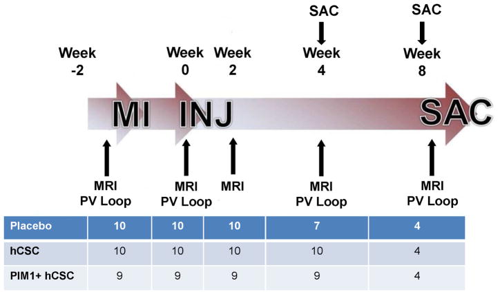
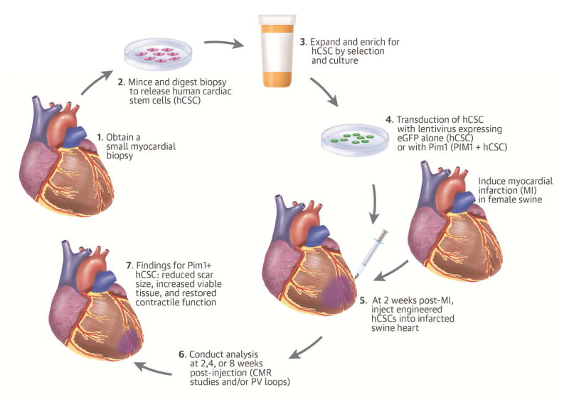
Methods: Human cardiac stem cells (hCSCs, n = 10), hckit+ CSCs overexpressing Pim1 (Pim1+; n = 9), or placebo (n = 10) were delivered by intramyocardial injection to immunosuppressed Yorkshire swine (n = 29) 2 weeks after MI. Cardiac magnetic resonance and pressure volume loops were obtained before and after cell administration.

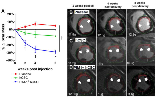
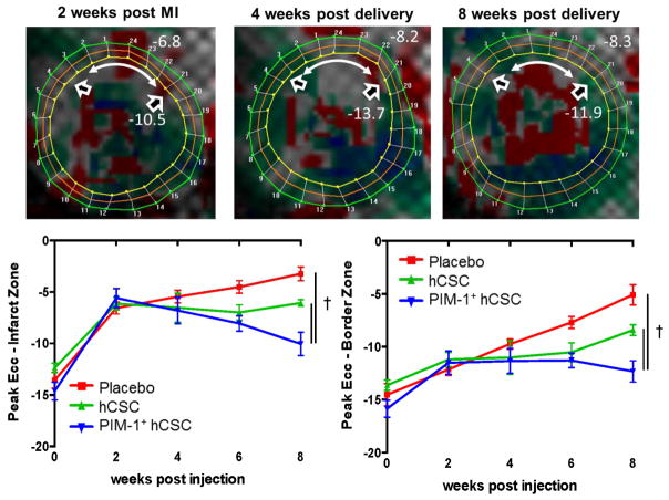
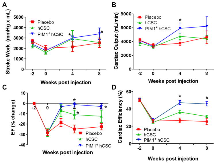
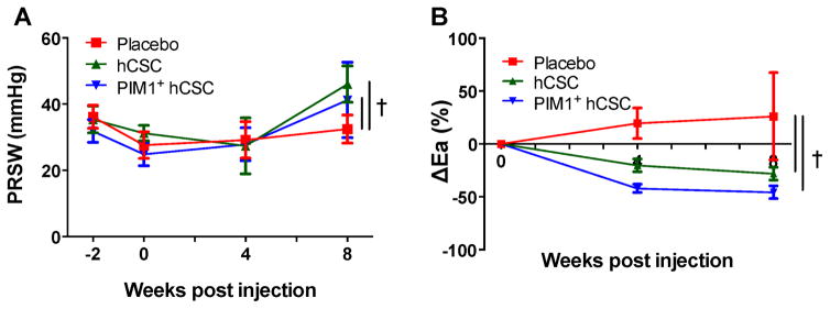
Results: Whereas both hCSCs reduced MI size compared to placebo, Pim1+ cells produced a ∼3-fold greater decrease in scar mass at 8 weeks post-injection compared to hCSCs (-29.2 ± 2.7% vs. -8.4 ± 0.7%; p < 0.003). Pim1+ hCSCs also produced a 2-fold increase of viable mass compared to hCSCs at 8 weeks (113.7 ± 7.2% vs. 65.6 ± 6.8%; p <0.003), and a greater increase in regional contractility in both infarct and border zones (both p < 0.05). Both CSC types significantly increased ejection fraction at 4 weeks but this was only sustained in the Pim1+ group at 8 weeks compared to placebo. Both hCSC and Pim1+ hCSC treatment reduced afterload (p = 0.02 and p = 0.004, respectively). Mechanoenergetic recoupling was significantly greater in the Pim1+ hCSC group (p = 0.005).

Conclusions: Pim1 overexpression enhanced the effect of intramyocardial delivery of CSCs to infarcted porcine hearts. These findings provide a rationale for genetic modification of stem cells and consequent translation to clinical trials.

Keywords: heart failure; human cardiac progenitor cells; injection; pressure volume.


Figures

Figure 1
Figure 1. Antifibrotic Effects
(A) At 4 and 8 weeks post-injection, both cell-treated groups exhibited decreased scar formation. In the PIM1+ human cardiac stem cell (hCSC) group, the antifibrotic effect was enhanced 3-fold compared to hCSC group. Representative examples of delayed enhancement of the myocardium using gadolinium cardiac magnetic resonance (CMR) images from placebo (B), hCSC (C), or Pim1+ hCSC (D) treated animals depict the progress of the delayed enhancement at the same location throughout different time points during the study. *p < 0.05 vs. pre-injection time; p < 0.05 PIM1+ hCSC vs. hCSC and placebo; p < 0.05 PIM1+ hCSC vs. hCSC and placebo and hCSC vs. placebo.
Figure 2
Figure 2. Study Timeline
The study was composed of 2 branches, with either a 4- or 8-week follow-up after injection (INJ). A series of CMR studies and pressure volume (PV) loop studies were conducted throughout the study as indicated. The table represents the number of animals per group at each time point. MI = myocardial infarction; SAC = sacrifice; other abbreviations as in Figure 1.
Figure 3
Figure 3. Improved Contractility Measured by Systolic Strain
(A) Eulerian circumferential strain (Ecc) is a measure of contractility; the more negative the value, the better the contractility. At 2 weeks post-MI (pre-injection) the contractility at the anteroseptal wall (in red) was greatly impaired (white double-headed arrow = infarct zone; open arrowheads = bordering zone). At 4 weeks post-delivery of Pim1+ hCSCs, contractility started to improve (in gray) and at 8 weeks, areas at the infarct zone approached near normal contractility levels (in green). In both the infarct (B) and border (C) zones, the Pim1+ hCSC group demonstrated greater improvement in contractility at 8 weeks than the other groups. p < 0.05 PIM1+ hCSC vs. hCSC and placebo. Abbreviations as in Figures 1 and 2.
Figure 4
Figure 4. Cardiac Function
At 8 weeks post-injection, (A) stroke work and (B) cardiac output both improved in the Pim1+ hCSCs treated animals compared to pre-injection levels while the hCSC- and placebo-treated animals remained unchanged. (C) Left ventricular ejection fraction (EF) improved in both cell groups at 4 weeks compared to placebo, but this improvement was sustained only in the Pim1+ hCSCs group at 8 weeks. (D) Cardiac efficiency, the ratio of used mechanical energy to chemical energy consumed by the myocardium per beat, was restituted to near normal levels at 4 and at 8 weeks post-injection in the Pim1+ hCSC-treated animals while remaining unchanged in the placebo group. *p < 0.05 vs. pre-injection time. Abbreviations as in Figure 1.
Figure 5
Figure 5. PRSW and Arterial Elastance
Injection of hCSCs improved (A) preload recruitable stroke work (PRSW) and (B) change in arterial elastance (Ea) versus placebo. p < 0.05 PIM1+ hCSC vs. hCSC and placebo. Abbreviations as in Figure 1.
Figure 6
Figure 6. Pim1+ hCSCs Engraft and Differentiate
Localization of human mitochondrial marker is shown: (A) Confocal image depicts human mitochondria (red) costained with myosin light chain (green) and DAPI in Pim1+ hCSC animals 8 weeks after injection. Colocalization of human mitochondria (green), PIM1 (red), and nuclei (blue) is seen in hCSC animals (B) and PIM1+ hCSC-treated animals (C) at 8 weeks post-injection (arrows point to cells costained for both human mitochondria and PIM1). Scale bar = 10 μm. DAPI = 4’,6-diamidino-2- phenylindole; hMito = human mitochondria; other abbreviations as in Figure 1.
Figure 7
Figure 7. Mitotic Activity in Cardiomyocytes
(A) Confocal image depicts proliferating cells phospho-histone H3 (pH3) costained with myosin light chain (MLC) and DAPI. (B) Mitotic activity of endogenous cardiomyocytes (pH3) increased in the border zone in Pim1+ hCSC-injected animals as compared to placebo 8 weeks post-injection (p < 0.05). (C) There were no significant differences in mitotic activity within noncardiomyocyte cells. Scale bar = 20 μm. *p < 0.05 Pim1+ hCSCs vs. placebo. Abbreviations as in Figures 1 and 6.
Central Illustration
Central Illustration. Pim1 Overexpression Enhances Cardiac Repair
Following ischemia/reperfusion injury in swine, genetically engineered human cardiac stem cell (hCSC) with Pim1+ reduced scar size, increased viable tissue, and restored contractile function towards normal 8 weeks post-injection. Cardiac magnetic resonance imaging and pressure volume (PV) loop analyses showed that PIM1+ overexpression enhanced the cardioreparative capabilities of hCSCs. eGFP = enhanced green fluorescent protein.

Comment in

References

    1. Golpanian S, Wolf A, Hatzistergos KE, Hare JM. Rebuilding the Damaged Heart: Mesenchymal Stem Cells, Cell-Based Therapy, and Engineered Heart Tissue. Physiol Rev. 2016;96:1127–68. - PMC - PubMed
    1. Fischer KM, Cottage CT, Wu W, et al. Enhancement of myocardial regeneration through genetic engineering of cardiac progenitor cells expressing Pim-1 kinase. Circulation. 2009;120:2077–87. - PMC - PubMed
    1. Muraski JA, Rota M, Misao Y, et al. Pim-1 regulates cardiomyocyte survival downstream of Akt. Nat Med. 2007;13:1467–75. - PubMed
    1. Mohsin S, Khan M, Toko H, et al. Human cardiac progenitor cells engineered with Pim-I kinase enhance myocardial repair. J Am Coll Cardiol. 2012;60:1278–87. - PMC - PubMed
    1. Fischer KM, Cottage CT, Konstandin MH, et al. Pim-1 kinase inhibits pathological injury by promoting cardioprotective signaling. J Mol Cell Cardiol. 2011;51:554–8. - PMC - PubMed

Substances