. 2017 Feb 2;129(5):609-618.
doi: 10.1182/blood-2016-08-735365. Epub 2016 Dec 1.

An anti-CD3/anti-CLL-1 bispecific antibody for the treatment of acute myeloid leukemia

Affiliations

An anti-CD3/anti-CLL-1 bispecific antibody for the treatment of acute myeloid leukemia

Steven R Leong et al. Blood. .

Abstract

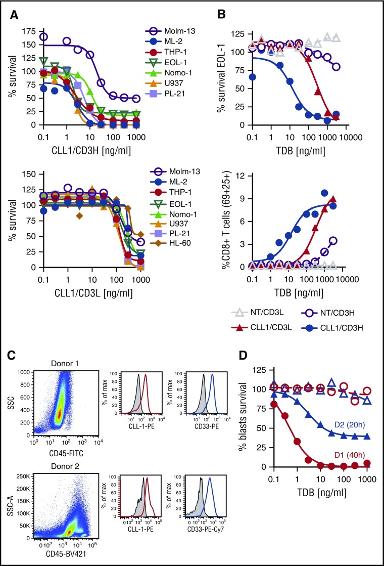
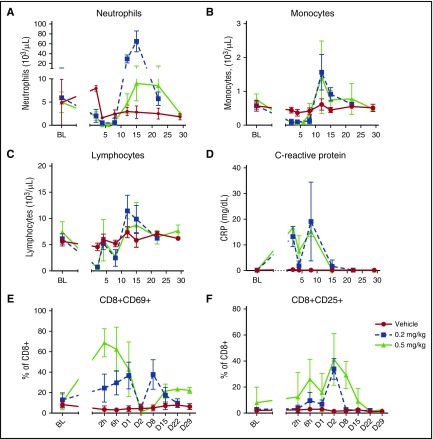
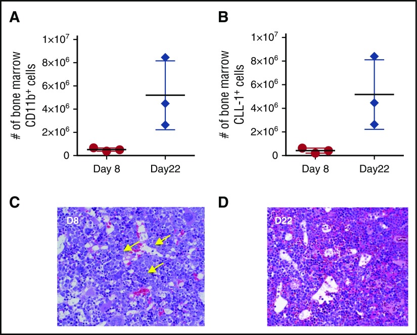
Acute myeloid leukemia (AML) is a major unmet medical need. Most patients have poor long-term survival, and treatment has not significantly changed in 40 years. Recently, bispecific antibodies that redirect the cytotoxic activity of effector T cells by binding to CD3, the signaling component of the T-cell receptor, and a tumor target have shown clinical activity. Notably, blinatumomab is approved to treat relapsed/refractory acute lymphoid leukemia. Here we describe the design, discovery, pharmacologic activity, pharmacokinetics, and safety of a CD3 T cell-dependent bispecific (TDB) full-length human IgG1 therapeutic antibody targeting CLL-1 that could potentially be used in humans to treat AML. CLL-1 is prevalent in AML and, unlike other targets such as CD33 and CD123, is not expressed on hematopoietic stem cells providing potential hematopoietic recovery. We selected a high-affinity monkey cross-reactive anti-CLL-1 arm and tested several anti-CD3 arms that varied in affinity, and determined that the high-affinity CD3 arms were up to 100-fold more potent in vitro. However, in mouse models, the efficacy differences were less pronounced, probably because of prolonged exposure to TDB found with lower-affinity CD3 TDBs. In monkeys, assessment of safety and target cell depletion by the high- and low-affinity TDBs revealed that only the low-affinity CD3/CLL1 TDB was well tolerated and able to deplete target cells. Our data suggest that an appropriately engineered CLL-1 TDB could be effective in the treatment of AML.


Figures

Figure 1.
Figure 1.
AML tumor cell growth suppression is dependent on affinity to CD3ε and effects of CLL1/CD3H TDB on AML BMMNCs. (A) Dose-dependent survival curves determined by Prism-6 using a nonlinear regression with sigmoidal dose response where X is the log [ ]. Percent survival is calculated by dividing the treatment live events by its untreated control replicate live events, and multiplying by 100. HL60 was performed in a separate experiment from the other cell lines (data not shown for CLL1/CD3H). (B) EOL-1 dose-dependent survival and percent of CD8+CD69+CD25+ effector cells. One of 3 experiments, each using a different blood donor. Similar results were observed for other AML cell lines. (C-D) CD8+ T cells were purified from peripheral blood of a healthy human donor (ALLCells, Alameda, CA) for donor 1 and a Genentech donor for donor 2 by Ficoll density gradient centrifugation and a human CD8+ T cell isolation kit from Miltenyi (130-096-495). Patient AML bone marrow was obtained from the Stanford Cancer Center and purified by Ficoll centrifugation. The AML blasts were preincubated for ∼6 hours with hIgG (EU = 0.07/mg) to reduce nonspecific binding to FcγRs. The E:T ratio was 3:1 (150 000 CD8 T cells to 50 000 blasts). Medium used for the assay was RPMI 1640 supplemented with 10% heat-inactivated fetal bovine serum, 2 mM glutamine, Pen-Strep, 1X cytokine bullet (IL-3, IL-6, SCF, Flt3; StemCell Technologies, Vancouver, Canada), 0.1 μg/mL GM-CSF and 0.1 μg/mL G-CSF. The test articles were NT/CD3H and CLL1/CD3H diluted serially in threefold steps. The assay was set up in triplicate with either a 20-hour (donor 2; blue lines) or 40-hour (donor 1; red lines) exposure to test article. (C) Dot plot shows SSC/CD45 profile of AML patient donor 1 and donor 2. Histogram shows positive staining of CD11b/CD34+ AML blasts for human CD33 (red) and CLL-1 (blue) compared with isotype control antibody. (D) Allogeneic CD8+ effector T cells in the presence of threefold serially diluted CLL1/CD3H shows concentration-dependent killing of AML blasts with an EC50 ∼0.45 ng/mL for donor 1 compared with the NT/CD3H TDB. Dotted line (NT/CD3H) and solid line (CLL1/CD3H). Donor 1 phenotype was consistent with AML LSCs—CD45+/CD34+/CD33+/CLL-1+/CD38 (99%) with <1% CD38+. Donor 2 phenotype was consistent with AML progenitors—CD45+/CD34+/CD33+/CLL-1+/CD38+ (∼97%).
Figure 2.
Figure 2.
Depletion of human CLL-1+ 2xBAC-Tg mouse cells from blood and bone marrow after single IV dose administration of 0.5 mg/kg CLL-1 TDB. (A) CLL1/CD3VH, (B) CLL1/CD3H, and (C) CLL1/CD3L. Percent of hCLL-1+ cells were derived from the average naïve untreated mice hCLL-1+ cells; blood (n = 39), bone marrow and spleen (n = 4). Upregulation of CD69 on CD8+ and CD4+ T cells by (D) CLL1/CD3VH, (E) CLL1/CD3H, and (F) CLL1/CD3L. Time points are terminal bleeds. The mean and standard deviation are represented by horizontal and vertical lines, respectively.
Figure 3.
Figure 3.
PK profiles for CLL-1/CD3 TDBs in target-deficient and target-expressing mice. Animals were administered a single IV dose of 0.5 mg/kg of either nontargeting (NT) TDBs or CLL-1–specific TDBs over a period of 14 to 21 days. (A) Target-deficient SCID.Beige mice PK values, (B) target-expressing 2xBACT-Tg (human CLL-1 and human CD3ε) PK values.
Figure 4.
Figure 4.
Cytokine levels in cynomolgus monkeys receiving CLL1/CD3H (HA) and CLL1/CD3L (LA). (A) G-CSF, (B) tumor necrosis factor-α (TNFα), (C) IL-2, and (D) IL-6. Cytokine release typically occurred within 2 to 6 hours of administration of either molecule and was markedly increased in those animals that received 0.5 mg/kg CLL1/CD3H compared with those that received CLL1/CD3L. All animals that required unscheduled euthanasia had comparatively elevated cytokine levels compared with those that survived to scheduled necropsy. The one animal that received 0.5 mg/kg CLL1/CD3L and required unscheduled necropsy had perimortem increases in G-CSF, IL-6, and other innate cytokines such as MCP-1 (data not shown).
Figure 5.
Figure 5.
Clinical pathology and peripheral blood flow cytometry from cynomolgus monkeys receiving CLL1/CD3L TDB. Target cell depletion of neutrophils (A) and monocytes (B) is similar between both dose groups, with nadirs between day 4 and 8 and 2 and 8 post dose, respectively. Rebound neutrophilia between days 10 and 15 is marked and greater in the 0.2 mg/kg dose group. Note, the mild decrease in neutrophil count on day 2 in the vehicle group is likely secondary to repeat blood sampling. Reduction of lymphocytes (C) occurred in 2 waves (days 2-8) concurrent to target cell depletion, elevation of acute phase proteins such as C-reactive protein (D), and increased activation of CD8+ cytotoxic T cells as measured by CD69 (E) and CD25 (F) expression. The immediate increase in percentage of CD69+ CD8+ T cells was greater (peak 71.3%-80.7% vs 25.2%-57.0%), and the percentage of CD25+ T cells was elevated for longer (return to baseline D15-D22 vs D8-D15) in the 0.5 mg/kg group.
Figure 6.
Figure 6.
Myeloid depletion in the bone marrow of cynomolgus monkeys receiving 0.2 mg/kg CLL1-CD3L. Myeloid cell number, assessed by either CD11b reactivity (A) or CLL-1 reactivity (B), were markedly attenuated on day 8 (nadir of peripheral neutrophils) compared with day 22 (peripheral neutrophil recovery). Horizontal and vertical lines represent mean and standard deviation, respectively. (C) Bone marrow histology at day 8 revealed marked depletion of late-stage myeloid cells with a relative increase in early myeloid progenitors (arrows), suggesting that hematopoietic stem cells were not affected at this dose. (D) By day 22, late-stage myeloid cells were recovered and minimally increased in relative abundance (asterisks). Images of FFPE 5-µm sections stained with hematoxylin and eosin were acquired on an Olympus BX53 (Olympus, Burlingame, CA) camera, objective UPlanSApo 40x/0.95 at 20-23°C, and photographed with an Infinity 2 camera and analyzed with Infinity Analyze software (Lumenera, Ottawa, Ontario, Canada).

References

    1. Freireich EJ, Wiernik PH, Steensma DP. The leukemias: a half-century of discovery. J Clin Oncol. 2014;32(31):3463-3469. - PubMed
    1. Wander SA, Levis MJ, Fathi AT. The evolving role of FLT3 inhibitors in acute myeloid leukemia: quizartinib and beyond. Ther Adv Hematol. 2014;5(3):65-77. - PMC - PubMed
    1. Topp MS, Gökbuget N, Zugmaier G, et al. . Long-term follow-up of hematologic relapse-free survival in a phase 2 study of blinatumomab in patients with MRD in B-lineage ALL. Blood. 2012;120(26):5185-5187. - PubMed
    1. Topp MS, Kufer P, Gökbuget N, et al. . Targeted therapy with the T-cell-engaging antibody blinatumomab of chemotherapy-refractory minimal residual disease in B-lineage acute lymphoblastic leukemia patients results in high response rate and prolonged leukemia-free survival. J Clin Oncol. 2011;29(18):2493-2498. - PubMed
    1. Kung Sutherland MS, Walter RB, Jeffrey SC, et al. . SGN-CD33A: a novel CD33-targeting antibody-drug conjugate using a pyrrolobenzodiazepine dimer is active in models of drug-resistant AML. Blood. 2013;122(8):1455-1463. - PubMed

MeSH terms