Review
. 2017 Feb;37(2):191-204.
doi: 10.1161/ATVBAHA.116.306256. Epub 2016 Dec 1.

Pathology of Human Coronary and Carotid Artery Atherosclerosis and Vascular Calcification in Diabetes Mellitus

Affiliations
Review

Pathology of Human Coronary and Carotid Artery Atherosclerosis and Vascular Calcification in Diabetes Mellitus

Kazuyuki Yahagi et al. Arterioscler Thromb Vasc Biol. 2017 Feb.

Abstract

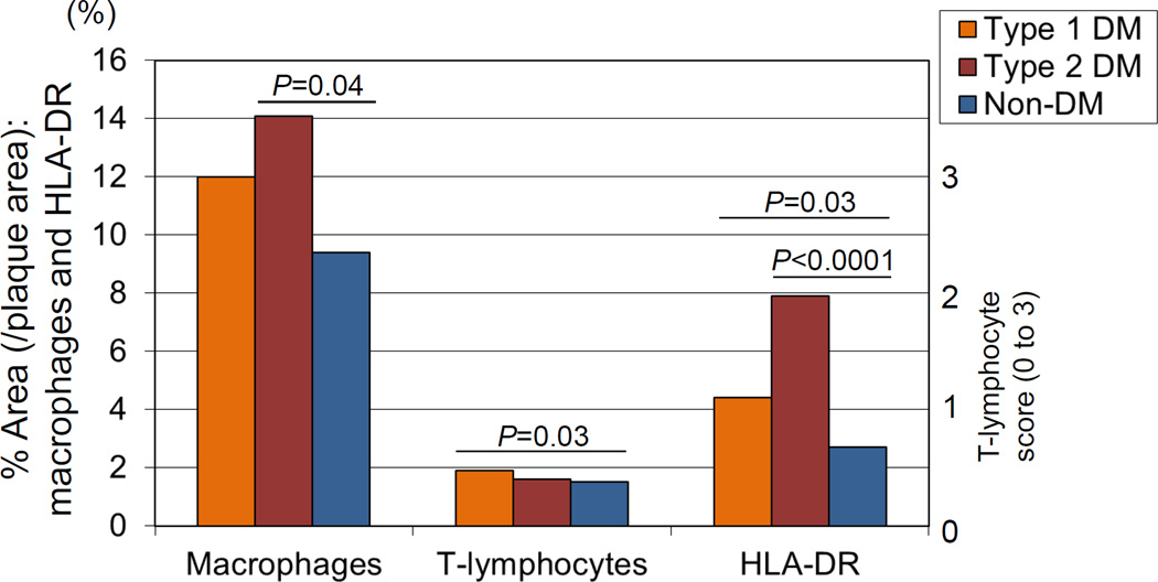
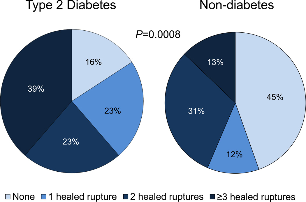
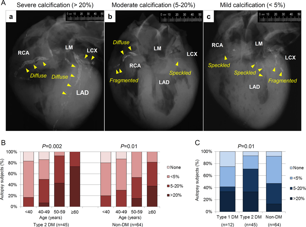
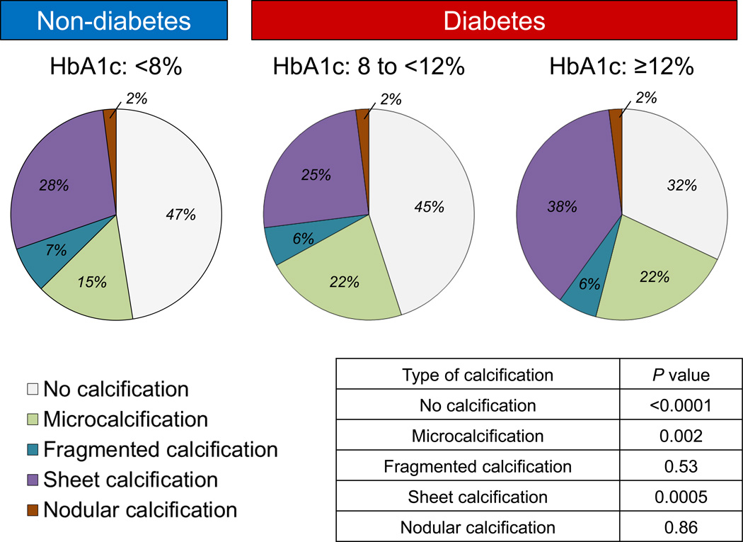
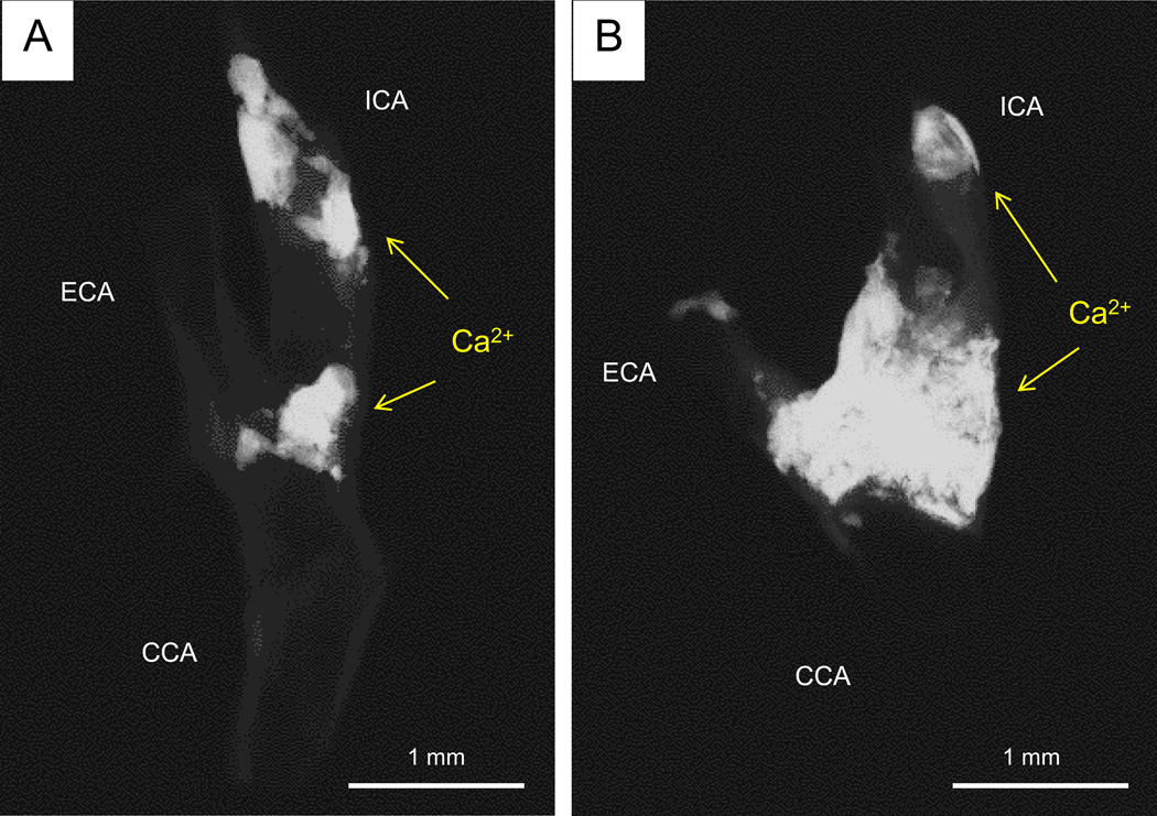
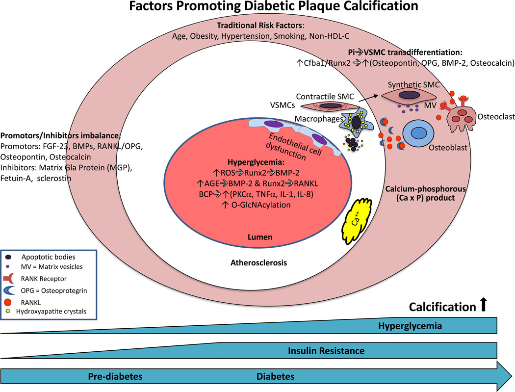
The continuing increase in the prevalence of diabetes mellitus in the general population is predicted to result in a higher incidence of cardiovascular disease. Although the mechanisms of diabetes mellitus-associated progression of atherosclerosis are not fully understood, at clinical and pathological levels, there is an appreciation of increased disease burden and higher levels of arterial calcification in these subjects. Plaques within the coronary arteries of patients with diabetes mellitus generally exhibit larger necrotic cores and significantly greater inflammation consisting mainly of macrophages and T lymphocytes relative to patients without diabetes mellitus. Moreover, there is a higher incidence of healed plaque ruptures and positive remodeling in hearts from subjects with type 1 diabetes mellitus and type 2 diabetes mellitus, suggesting a more active atherogenic process. Lesion calcification in the coronary, carotid, and other arterial beds is also more extensive. Although the role of coronary artery calcification in identifying cardiovascular disease and predicting its outcome is undeniable, our understanding of how key hormonal and physiological alterations associated with diabetes mellitus such as insulin resistance and hyperglycemia influence the process of vascular calcification continues to grow. Important drivers of atherosclerotic calcification in diabetes mellitus include oxidative stress, endothelial dysfunction, alterations in mineral metabolism, increased inflammatory cytokine production, and release of osteoprogenitor cells from the marrow into the circulation. Our review will focus on the pathophysiology of type 1 diabetes mellitus- and type 2 diabetes mellitus-associated vascular disease with particular focus on coronary and carotid atherosclerotic calcification.

Keywords: atherosclerosis; carotid arteries; coronary artery disease; death, sudden; diabetes mellitus.


Figures

Figure 1
Figure 1
Inflammation in diabetic coronary arteries. Coronary fibroatheromas illustrating the extent of macrophages (CD68), T cells (CD45RO), and HLA-DR expression in patients with type 1 and 2 diabetes mellitus (DM) with control, nondiabetic subjects. Reproduced with permission from Burke AP, et al. Arterioscler Thromb Vasc Biol. 2004;24:1266-1271.
Figure 2
Figure 2
The pie charts reflect the percentage of healed ruptures (HPR) per heart relative to diabetic status at autopsy. Type 2 diabetes had higher numbers of HPRs compared to non-diabetics (p=0.0008). The data constitutes a re-analysis of 142 sudden coronary death cases, published in Burke AP, et al. Circulation. 2001;103:934-940, and stratified by with or without diabetes mellitus.
Figure 3
Figure 3
Coronary artery calcification in sudden coronary death evaluated by post-mortem radiography. (A) Representative post-mortem radiographs showing various patterns of calcification. The severity of calcification was assessed based on the percentage of calcification area: (a) severe artery calcification (>20%), (b) moderate calcification (5–20%), and (c) mild calcification (<5%). Arrows indicate type of calcification (speckled [<2mm in length], fragmented [2–5mm] or diffuse [≥5mm] calcification). (B) Percentage of total calcified area, divided into mild, moderate and severe in sudden coronary death patients with type 2 DM and non-DM stratified by decade. (C) Percentage of total calcified area in sudden coronary death comparing type 1 DM, type 2 DM, and non-DM.
Figure 4
Figure 4
Percentage of sudden deaths based on coronary artery calcification type (none, micro-, fragmented, sheet, and nodular, stratified by HbA1c level (<8%) non-diabetics and diabetic (8% to <12% and ≥12%). 1630 histologic sections (<8%, n=776 sections; 8% to <12%, n=548; and ≥12%, n=306) from 57 patients with stable coronary artery disease were examined. Note the declining shift in the number of lesions without calcification and significant reciprocal increase sheet calcification with escalating HbA1C levels.
Figure 5
Figure 5
Representative digital radiography of calcified carotid endarterectomy specimens from diabetic and non-diabetic patients. (A) Focal calcification at the bifurcation and distal internal carotid artery (ICA) in a non-diabetic patient (calcified area 57.6 mm2) (B) The extent of severe calcification in a type 2 diabetic patient involving the ICA and external carotid artery (ECA) (calcified area 196.1 mm2). Total calcification area and percent calcification area in patients with or without diabetes are shown in the table. Abbreviations: CCA= common carotid artery.
Figure 6
Figure 6
Mechanisms of plaque calcification in diabetes. The earliest form of calcification, microcalcification occurs in apoptotic vascular smooth muscle cells (vSMCs) and macrophages in conjunction with an increase in serum calcium-phosphorous (CaxP) product. Increases in phosphate concentration in vSMCs induce a switch towards an osteoblast-like phenotype, particularly in states of Pi excess. Osteogenic-primed vSMCs express alkaline phosphatase (ALP) and under the control of Cfba-1, secrete bone-associated proteins such as osteopontin, collagen type 1, osteoprotegerin, bone morphogenic protein-2 and osteocalcin, accompanied by the release of mineralization-competent matrix vesicles (MVs). Changes in promoters or inhibitors of mineralization also affect calcification. Hyperglycemia may also affect calcification through multiple mechanisms such as oxidative stress, AGEs, BCP, O-GlcNAcylation, and endothelial dysfunction as discussed in the text. Abbreviations: BMP=bone morphogenic protein; EPCs=endothelial progenitor cells; FGF=fibroblast growth factor; HDL-C=high density lipoprotein-Cholesterol; IL=interleukin; MCCs=myeloid calcifying cells; OPG=osteoprotegrin; ROC=reactive oxygen series; Runx2=Runt-related transcription factor-2;TNF=tumor necrosis factor; TGF=transforming growth factor; BCP=basic calcium phosphate crystals.

References

    1. Malmberg K, Yusuf S, Gerstein HC, Brown J, Zhao F, Hunt D, Piegas L, Calvin J, Keltai M, Budaj A. Impact of diabetes on long-term prognosis in patients with unstable angina and non-Q-wave myocardial infarction: results of the OASIS (Organization to Assess Strategies for Ischemic Syndromes) Registry. Circulation. 2000;102:1014–1019. - PubMed
    1. Mukamal KJ, Nesto RW, Cohen MC, Muller JE, Maclure M, Sherwood JB, Mittleman MA. Impact of diabetes on long-term survival after acute myocardial infarction: comparability of risk with prior myocardial infarction. Diabetes Care. 2001;24:1422–1427. - PubMed
    1. Sarwar N, Gao P, Seshasai SR, et al. Diabetes mellitus, fasting blood glucose concentration, and risk of vascular disease: a collaborative meta-analysis of 102 prospective studies. Lancet. 2010;375:2215–2222. - PMC - PubMed
    1. Lam DW, LeRoith D. The worldwide diabetes epidemic. Curr Opin Endocrinol Diabetes Obes. 2012;19:93–96. - PubMed
    1. Group IDFDA. Update of mortality attributable to diabetes for the IDF Diabetes Atlas: Estimates for the year 2013. Diabetes Res Clin Pract. 2015;109:461–465. - PubMed

Publication types

MeSH terms