. 2017 Jan 20;292(3):814-825.
doi: 10.1074/jbc.M116.761734. Epub 2016 Dec 1.

Identification of a Substrate-selective Exosite within the Metalloproteinase Anthrax Lethal Factor

Affiliations

Identification of a Substrate-selective Exosite within the Metalloproteinase Anthrax Lethal Factor

Allison B Goldberg et al. J Biol Chem. .

Abstract

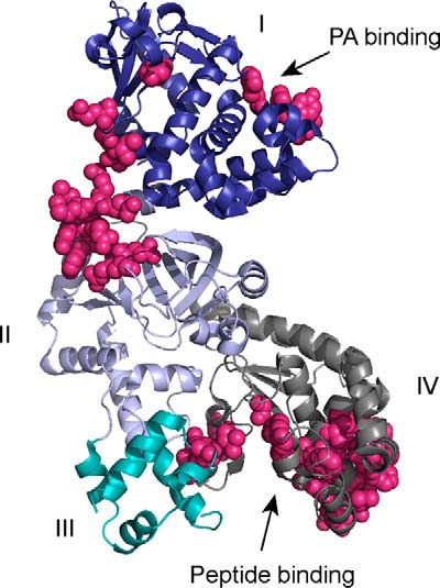
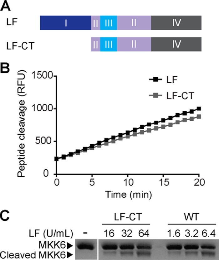
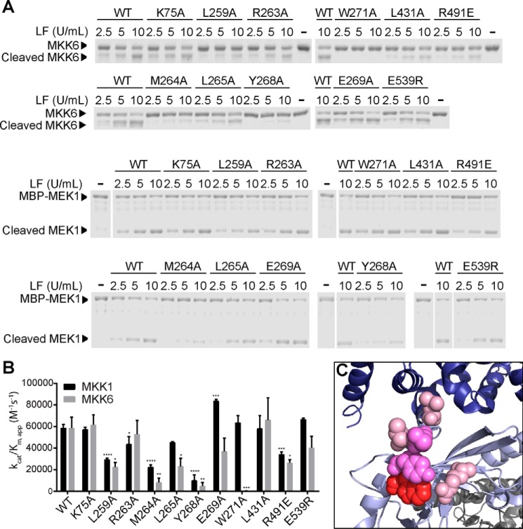
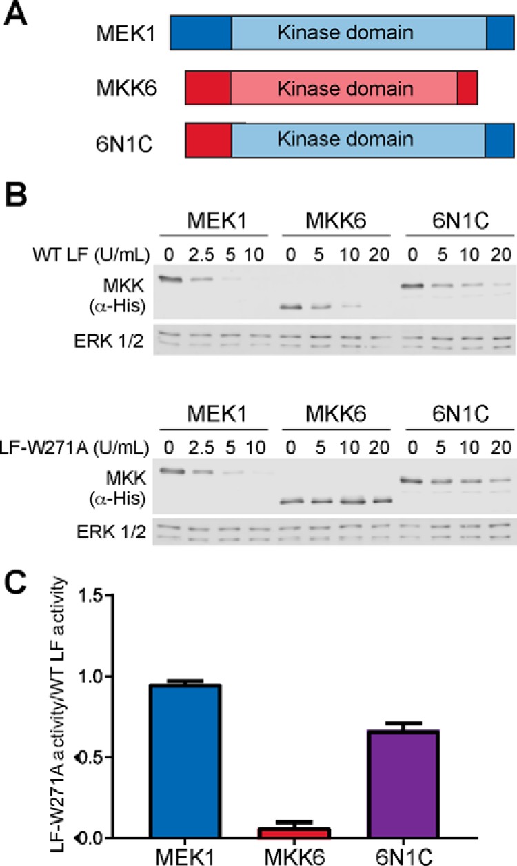
The metalloproteinase anthrax lethal factor (LF) is secreted by Bacillus anthracis to promote disease virulence through disruption of host signaling pathways. LF is a highly specific protease, exclusively cleaving mitogen-activated protein kinase kinases (MKKs) and rodent NLRP1B (NACHT leucine-rich repeat and pyrin domain-containing protein 1B). How LF achieves such restricted substrate specificity is not understood. Previous studies have suggested the existence of an exosite interaction between LF and MKKs that promotes cleavage efficiency and specificity. Through a combination of in silico prediction and site-directed mutagenesis, we have mapped an exosite to a non-catalytic region of LF. Mutations within this site selectively impair proteolysis of full-length MKKs yet have no impact on cleavage of short peptide substrates. Although this region appears important for cleaving all LF protein substrates, we found that mutation of specific residues within the exosite differentially affects MKK and NLRP1B cleavage in vitro and in cultured cells. One residue in particular, Trp-271, is essential for cleavage of MKK3, MKK4, and MKK6 but dispensable for targeting of MEK1, MEK2, and NLRP1B. Analysis of chimeric substrates suggests that this residue interacts with the MKK catalytic domain. We found that LF-W271A blocked ERK phosphorylation and growth in a melanoma cell line, suggesting that it may provide a highly selective inhibitor of MEK1/2 for use as a cancer therapeutic. These findings provide insight into how a bacterial toxin functions to specifically impair host signaling pathways and suggest a general strategy for mapping protease exosite interactions.

Keywords: NACHT leucine-rich repeat and pyrin domain containing protein (NLRP); anthrax toxin; bacterial pathogenesis; host-pathogen interaction; macrophage; melanoma; mitogen-activated protein kinase kinase; protein kinase; proteinase.


Figures

FIGURE 1.
FIGURE 1.
LF structure showing protein interface residues predicted by CPORT. The four domains of LF as described in the text are labeled and color-coded blue (domain I), lavender (domain II), cyan (domain III), and gray (domain IV). Residues predicted by CPORT to be involved in protein interaction interfaces are shown in magenta. The figure was made from Protein Data Bank entry 1J7N using PyMOL (54).
FIGURE 2.
FIGURE 2.
Removal of domain I specifically reduces cleavage of a full-length MKK substrate by LF. A, scheme showing LF-CT protein, an LF construct lacking the N-terminal domain I. B, peptide cleavage assay comparing full-length LF with LF-CT. C, MKK6 cleavage assay comparing full-length LF with LF-CT. Recombinant MKK6 protein substrate was subjected to proteolysis by the indicated units of LF and LF-CT as defined by activity against the peptide substrate. MKK6 cleavage was visualized by SDS-PAGE followed by Coomassie staining.
FIGURE 3.
FIGURE 3.
MKK cleavage assays with LF mutants. A, recombinant MKK6 or MEK1 substrate was incubated with increasing concentrations of WT LF or the indicated LF point mutants for 30 min at room temperature. Reactions were fractionated by SDS-PAGE followed by Coomassie staining. A representative gel from at least three separate experiments is shown. B, apparent catalytic efficiencies were calculated from quantified data corresponding to the experiment shown in A and its replicates. The error bars show S.E. (n ≥ 3). Statistical significance was determined by testing for differences with a one-way analysis of variance, followed by Fisher's least significant difference post hoc test to compare with WT. *, p < 0.05; **, p < 0.01; ***, p < 0.001; ****, p < 0.0001. C, residues mutated that had a statistically significant impact on LF cleavage of MKK6 are shown in space fill representation: Trp-271 (red), Met-264 and Tyr-268 (bright pink), and Leu-259 and Arg-491 (light pink).
FIGURE 4.
FIGURE 4.
Cleavage of a chimeric MEK1/MKK6 substrate by LF. A, schematic showing MEK1 (blue), MKK6 (red), and the chimeric construct 6N1C. B, WT LF and LF-W271A cleavage of MEK1, MKK6, and 6N1C in mammalian cell lysates. A representative immunoblot from one of three replicates is shown. C, the relative activity of LF-W271A as a fraction of WT LF cleavage of the same substrate was calculated from the experiment shown in B and replicates. The error bars show S.E. (n = 3).
FIGURE 5.
FIGURE 5.
LF shows no evidence of dimerization or dimerization-dependent proteolytic activity. A, WT LF was subjected to SEC, and the molecular weight of protein in the eluate was calculated by MALS analysis. B, titration of LF using MEK1 and MKK6 substrates indicates that proteolytic activity does not increase exponentially with LF concentration.
FIGURE 6.
FIGURE 6.
LF exosite mutants differentially impact cleavage of MKKs in in cultured macrophage cells. J774A.1 mouse macrophage cells were treated with 0.5 μg/ml PA and increasing concentrations of WT or mutant LF for 90 min. The cells were harvested, and cleavage of MKK3 (A), MKK4 (B), and MEK2 (C) was assessed by immunoblotting of cell lysates. Representative immunoblots are shown at left, and quantified results are reported as percentages of uncleaved MKK remaining. The error bars show S.E. (n = 3). Statistical significance was calculated as in Fig. 3 comparing the value corresponding to the highest concentration of a given mutant to that of WT LF. ns, not significant; *, p < 0.05; ***, p < 0.001; ****, p < 0.0001.
FIGURE 7.
FIGURE 7.
Activity of LF exosite mutants in cleaving NLRP1B and causing macrophage death. A, HEK293T cell lysates containing exogenously expressed GFP-NLRP1B were incubated with WT or mutant LF for 30 min at room temperature. A representative immunoblot (top panel) shows the levels of uncleaved GFP-NLRP1B and GFP-tagged cleavage product. The asterisk indicates a cross-reacting background band. B, activity of each mutant is shown as a percentage of WT LF activity. The error bars show S.E. (n = 3). B, J774A.1 cells were subjected to the indicated concentrations WT and mutant LF in combination with 0.5 μg/ml PA for 4 h, and cell viability was measured by resazurin assay. Results show cell viability as a percentage of untreated cells. The error bars show S.E. (n = 3). Statistical significance was calculated as in Fig. 3. *, p < 0.05; ***, p < 0.001.
FIGURE 8.
FIGURE 8.
Effect of exosite mutations on LF activity in melanoma cells. A, A375 cells were treated with 0.5 μg/ml PA and the indicated concentrations of WT or mutant LF for 72 h, and cell viability was measured by resazurin assay. Measurements from three independent experiments are plotted as percentages of cell number in the untreated control group. The error bars indicate S.E. B, IC50 values calculated from A375 cell growth assays. The means and S.E. are shown (n = 3). C–E. A375 cells were treated with 0.5 μg/ml PA and 0.2 ng/ml LF or LF mutant for 16 h, and cells were harvested. Cell lysates were prepared and immunoblotted to detect phospho-ERK1/2 (C), phospho-p38 (D), and MKK3 (E). Representative immunoblots are shown at left, and mean quantified signals relative to the untreated control are shown at right. The error bars indicate standard errors (n = 3). Statistical significance was calculated as for Fig. 3. *, p < 0.05; **, p < 0.01; ***, p < 0.001; ****, p < 0.0001.

References

    1. Alouf J. E. (2000) Bacterial protein toxins: an overview. Methods Mol. Biol. 145, 1–26 - PubMed
    1. Blasi J., Chapman E. R., Link E., Binz T., Yamasaki S., De Camilli P., Südhof T. C., Niemann H., and Jahn R. (1993) Botulinum neurotoxin A selectively cleaves the synaptic protein SNAP-25. Nature 365, 160–163 - PubMed
    1. Schiavo G., Benfenati F., Poulain B., Rossetto O., Polverino deLaureto P., DasGupta B. R., and Montecucco C. (1992) Tetanus and botulinum-B neurotoxins block neurotransmitter release by proteolytic cleavage of synaptobrevin. Nature 359, 832–835 - PubMed
    1. Turk B. E. (2007) Manipulation of host signalling pathways by anthrax toxins. Biochem. J. 402, 405–417 - PubMed
    1. Brossier F., Lévy M., Landier A., Lafaye P., and Mock M. (2004) Functional analysis of Bacillus anthracis protective antigen by using neutralizing monoclonal antibodies. Infect. Immun. 72, 6313–6317 - PMC - PubMed

Publication types

MeSH terms

LinkOut - more resources