Castration alters protein balance after high-frequency muscle contraction
- PMID: 27909227
- PMCID: PMC5338601
- DOI: 10.1152/japplphysiol.00740.2016
Castration alters protein balance after high-frequency muscle contraction
Abstract
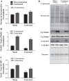
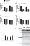
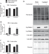
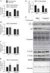
Resistance exercise increases muscle mass by shifting protein balance in favor of protein accretion. Androgens independently alter protein balance, but it is unknown whether androgens alter this measure after resistance exercise. To answer this, male mice were subjected to sham or castration surgery 7-8 wk before undergoing a bout of unilateral, high-frequency, electrically induced muscle contractions in the fasted or refed state. Puromycin was injected 30 min before euthanasia to measure protein synthesis. The tibialis anterior was analyzed 4 h postcontraction. In fasted mice, neither basal nor stimulated rates of protein synthesis were affected by castration despite lower phosphorylation of mechanistic target of rapamycin in complex 1 (mTORC1) substrates [p70S6K1 (Thr389) and 4E-BP1 (Ser65)]. Markers of autophagy (LC3 II/I ratio and p62 protein content) were elevated by castration, and these measures remained elevated above sham values after contractions. Furthermore, in fasted mice, the protein content of Regulated in Development and DNA Damage 1 (REDD1) was correlated with LC3 II/I in noncontracted muscle, whereas phosphorylation of uncoordinated like kinase 1 (ULK1) (Ser757) was correlated with LC3 II/I in the contracted muscle. When mice were refed before contractions, protein synthesis and mTORC1 signaling were not affected by castration in either the noncontracted or contracted muscle. Conversely, markers of autophagy remained elevated in the muscles of refed, castrated mice even after contractions. These data suggest the castration-mediated elevation in baseline autophagy reduces the absolute positive shift in protein balance after muscle contractions in the refed or fasted states.
New & noteworthy: In the absence of androgens, markers of autophagy were elevated, and these could not be normalized by muscle contractions. In the fasted state, REDD1 was identified as a potential contributor to autophagy in noncontracted muscle, whereas phosphorylation of ULK1 may contribute to this process in the contracted muscle. In the refed state, markers of autophagy remain elevated in both noncontracted and contracted muscles, but the relationship with REDD1 and ULK1 (Ser757) no longer existed.
Keywords: autophagy; protein degradation; protein synthesis; resistance exercise.
Copyright © 2017 the American Physiological Society.
Figures


Similar articles
-
Nutrient-induced stimulation of protein synthesis in mouse skeletal muscle is limited by the mTORC1 repressor REDD1.J Nutr. 2015 Apr;145(4):708-13. doi: 10.3945/jn.114.207621. Epub 2015 Feb 25. J Nutr. 2015. PMID: 25716553 Free PMC article.
-
Loss of REDD1 augments the rate of the overload-induced increase in muscle mass.Am J Physiol Regul Integr Comp Physiol. 2016 Sep 1;311(3):R545-57. doi: 10.1152/ajpregu.00159.2016. Epub 2016 Jul 27. Am J Physiol Regul Integr Comp Physiol. 2016. PMID: 27465734 Free PMC article.
-
Reduced REDD1 expression contributes to activation of mTORC1 following electrically induced muscle contraction.Am J Physiol Endocrinol Metab. 2014 Oct 15;307(8):E703-11. doi: 10.1152/ajpendo.00250.2014. Epub 2014 Aug 26. Am J Physiol Endocrinol Metab. 2014. PMID: 25159324 Free PMC article.
-
Exercise-mediated modulation of autophagy in skeletal muscle.Scand J Med Sci Sports. 2018 Mar;28(3):772-781. doi: 10.1111/sms.12945. Epub 2017 Aug 4. Scand J Med Sci Sports. 2018. PMID: 28685860 Review.
-
Androgen-mediated regulation of skeletal muscle protein balance.Mol Cell Endocrinol. 2017 May 15;447:35-44. doi: 10.1016/j.mce.2017.02.031. Epub 2017 Feb 22. Mol Cell Endocrinol. 2017. PMID: 28237723 Free PMC article. Review.
Cited by
-
Exercise as a therapy for cancer-induced muscle wasting.Sports Med Health Sci. 2020 Dec 3;2(4):186-194. doi: 10.1016/j.smhs.2020.11.004. eCollection 2020 Dec. Sports Med Health Sci. 2020. PMID: 35782998 Free PMC article. Review.
-
Understanding the Role of Exercise in Cancer Cachexia Therapy.Am J Lifestyle Med. 2017 Aug 17;13(1):46-60. doi: 10.1177/1559827617725283. eCollection 2019 Jan-Feb. Am J Lifestyle Med. 2017. PMID: 30627079 Free PMC article.
-
Dynamic resistance exercise increases skeletal muscle-derived FSTL1 inducing cardiac angiogenesis via DIP2A-Smad2/3 in rats following myocardial infarction.J Sport Health Sci. 2021 Sep;10(5):594-603. doi: 10.1016/j.jshs.2020.11.010. Epub 2020 Nov 24. J Sport Health Sci. 2021. PMID: 33246164 Free PMC article.
-
A clinically relevant decrease in contractile force differentially regulates control of glucocorticoid receptor translocation in mouse skeletal muscle.J Appl Physiol (1985). 2021 Apr 1;130(4):1052-1063. doi: 10.1152/japplphysiol.01064.2020. Epub 2021 Feb 18. J Appl Physiol (1985). 2021. PMID: 33600283 Free PMC article.
-
Inflammatory signalling regulates eccentric contraction-induced protein synthesis in cachectic skeletal muscle.J Cachexia Sarcopenia Muscle. 2018 Apr;9(2):369-383. doi: 10.1002/jcsm.12271. Epub 2017 Dec 7. J Cachexia Sarcopenia Muscle. 2018. PMID: 29215198 Free PMC article.
References
-
- Axell AM, MacLean HE, Plant DR, Harcourt LJ, Davis JA, Jimenez M, Handelsman DJ, Lynch GS, Zajac JD. Continuous testosterone administration prevents skeletal muscle atrophy and enhances resistance to fatigue in orchidectomized male mice. Am J Physiol Endocrinol Metab 291: E506–E516, 2006. doi:10.1152/ajpendo.00058.2006. - DOI - PubMed
MeSH terms
Substances
LinkOut - more resources
Full Text Sources
Other Literature Sources

