Review
. 2017 Jan 1;130(1):63-70.
doi: 10.1242/jcs.198630. Epub 2016 Dec 1.

3D culture models of tissues under tension

Affiliations
Review

3D culture models of tissues under tension

Jeroen Eyckmans et al. J Cell Sci. .

Abstract

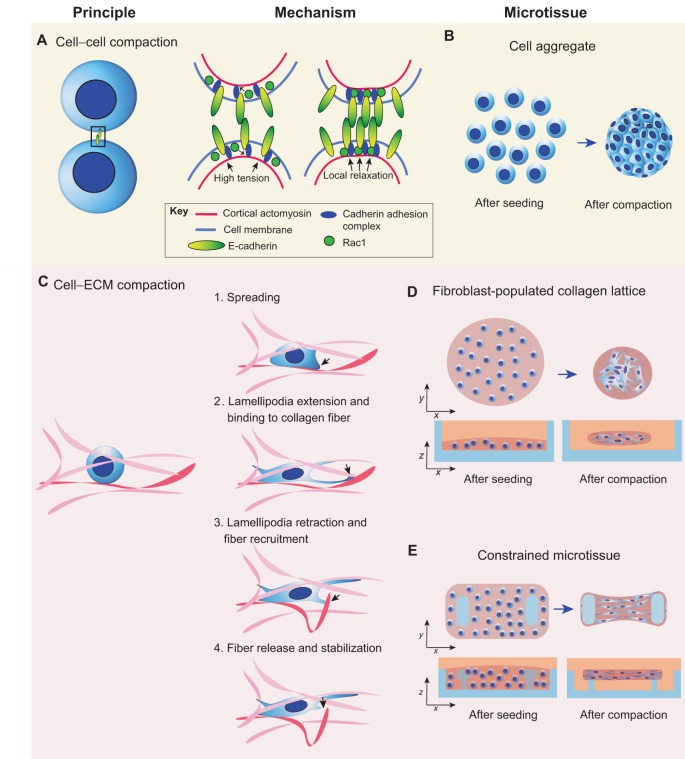
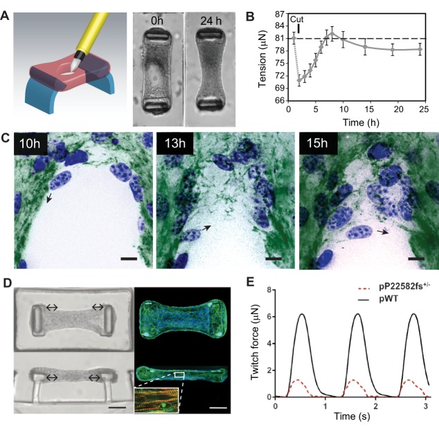
Cells dynamically assemble and organize into complex tissues during development, and the resulting three-dimensional (3D) arrangement of cells and their surrounding extracellular matrix in turn feeds back to regulate cell and tissue function. Recent advances in engineered cultures of cells to model 3D tissues or organoids have begun to capture this dynamic reciprocity between form and function. Here, we describe the underlying principles that have advanced the field, focusing in particular on recent progress in using mechanical constraints to recapitulate the structure and function of musculoskeletal tissues.

Keywords: 3D model; Contractility; Extracellular matrix; Microtissue; TFM.


Conflict of interest statement

Competing interests

The authors declare no competing or financial interests.

Figures

Fig. 1.
Fig. 1.
Mechanical basis of microtissue formation coupled with high-level microtissue architecture. (A) Schematic overview of cell aggregate formation. Epithelial cell–cell contact is initiated by engagement of E-cadherin adhesion proteins. Upon binding of the extracellular domain of E-cadherin to other cadherins, the intracellular cadherin adhesion complex connects the intracellular domain of E-cadherin to cortical actin. At the same time, activated Rac1 suppresses cortical actomyosin activity, which locally decreases the cortical tension at the junction. This decrease in cortical tension flattens the cell membrane, thus increasing the adhesive surface between two cells, and more juxtapositional cadherins are activated, reinforcing cell–cell contact. When multiple cells are involved, this mechanism drives the formation of aggregates (B). (C) Schematic overview of cell–ECM interactions during microtissue formation. Fibroblasts seeded in a fibrous collagen matrix (fibers are pink, red fiber is fiber of interest) spread in the matrix. After extension of lamellipodia (black arrow) and binding to a collagen fiber (red), the lamellipodia retracts and the fiber is recruited and released once it reaches the cell body, where it is crosslinked with the surrounding matrix. This mechanism drives the compaction of (D) unconstrained fibroblast-populated collagen lattices (blue, culture dish) and (E) constrained microtissues (blue, culture device with pillars).
Fig. 2.
Fig. 2.
Application of constrained microtissues in wound healing and cardiac morphogenesis studies. Microtissues under tension exhibit entire tissue contractions and matrix remodeling during wound closure. (A) Using a microsurgical knife, full-thickness wounds are made in microtissues that are anchored to two flexible pillars (blue). Within 24 h, NIH/3T3 fibroblasts close the open gaps and restore tissue integrity. (B) Quantification of tissue contractility by measuring the deflection of the pillars shows that wound closure in microtissues is a staged process comprising relaxation after damage that is followed by tissue contraction and a steady-state sub-baseline tension stage. Error bars, s.e.m.; dashed line represents baseline tension before microtissue damage. (C) During the latter stage, fibroblasts (blue, nuclei) tow fibronectin (green) from the microtissue and assemble a provisional fibronectin template to bridge the gap, which serves as a substrate on which cells migrate further into the gap area (black arrows indicate migration direction of the cell). Scale bars: 25 µm. Panels A–C are adapted from Sakar et al., 2016 under CC BY 4.0 licence. In addition to wound healing, constrained microtissues have been employed to model cardiac function of human iPSC-differentiated cardiomyocytes. (D) Microscopy images [brightfield (left) and fluorescence (right; blue, nuclei; green, phalloidin)] of iPSC-cardiac microtissue (iPSC-CMT) suspended between two flexible pillars from top-down (upper) and side (lower) views. Upon maturation, iPSC-CMT forms aligned sarcomere structures (inset D; red, F-actin; green, α-actinin A) that beat synchronously and dynamically pull the caps of the pillars to the center of the tissue (double-headed arrows). Scale bars: 50 µm. (E) By measuring the deflection of the pillars, twitch force, a metric for cardiac function, can be assessed in iPSC-CMTs constituted from wild-type human iPSC-cardiomyocytes (pWT) or from individuals with mutations in titin, a structural sarcomere protein (pP22582fs+/−). Panels D and E are adapted from Hinson et al., 2015 with permission from AAAS.

References

    1. Abhilash A. S., Baker B. M., Trappmann B., Chen C. S. and Shenoy V. B. (2014). Remodeling of fibrous extracellular matrices by contractile cells: predictions from discrete fiber network simulations. Biophys. J. 107, 1829-1840. 10.1016/j.bpj.2014.08.029 - DOI - PMC - PubMed
    1. Baker B. M., Trappmann B., Wang W. Y., Sakar M. S., Kim I. L., Shenoy V. B., Burdick J. A. and Chen C. S. (2015). Cell-mediated fibre recruitment drives extracellular matrix mechanosensing in engineered fibrillar microenvironments. Nat. Mater. 14, 1262-1268. 10.1038/nmat4444 - DOI - PMC - PubMed
    1. Barcellos-Hoff M. H., Aggeler J., Ram T. G. and Bissell M. J. (1989). Functional differentiation and alveolar morphogenesis of primary mammary cultures on reconstituted basement membrane. Development 105, 223-235. - PMC - PubMed
    1. Beauchamp P., Moritz W., Kelm J. M., Ullrich N. D., Agarkova I., Anson B. D., Suter T. M. and Zuppinger C. (2015). Development and characterization of a scaffold-free 3D spheroid model of induced pluripotent stem cell-derived human cardiomyocytes. Tissue Eng. Part C Methods 21, 852-861. 10.1089/ten.tec.2014.0376 - DOI - PubMed
    1. Bell E., Ivarsson B. and Merrill C. (1979). Production of a tissue-like structure by contraction of collagen lattices by human fibroblasts of different proliferative potential in vitro. Proc. Natl. Acad. Sci. USA 76, 1274-1278. 10.1073/pnas.76.3.1274 - DOI - PMC - PubMed

Publication types