Review
. 2016 Nov 17:10:536.
doi: 10.3389/fnins.2016.00536. eCollection 2016.

The Eye As a Biomarker for Alzheimer's Disease

Affiliations
Review

The Eye As a Biomarker for Alzheimer's Disease

Jeremiah K H Lim et al. Front Neurosci. .

Abstract

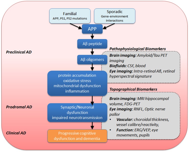
Alzheimer's disease (AD) is a progressive neurodegenerative disorder resulting in dementia and eventual death. It is the leading cause of dementia and the number of cases are projected to rise in the next few decades. Pathological hallmarks of AD include the presence of hyperphosphorylated tau and amyloid protein deposition. Currently, these pathological biomarkers are detected either through cerebrospinal fluid analysis, brain imaging or post-mortem. Though effective, these methods are not widely available due to issues such as the difficulty in acquiring samples, lack of infrastructure or high cost. Given that the eye possesses clear optics and shares many neural and vascular similarities to the brain, it offers a direct window to cerebral pathology. These unique characteristics lend itself to being a relatively inexpensive biomarker for AD which carries the potential for wide implementation. The development of ocular biomarkers can have far implications in the discovery of treatments which can improve the quality of lives of patients. In this review, we consider the current evidence for ocular biomarkers in AD and explore potential future avenues of research in this area.

Keywords: Alzheimer's disease; biomarker; eye; neurodegeneration; ocular; retina.


Figures

Figure 1
Figure 1
Dementia world prevalence by age. Data based on meta-analyzed estimates of dementia prevalence (%) generated from Poisson random effects models from WHO (2012).
Figure 2
Figure 2
Risk factors associated with Alzheimer's disease. This figure shows known associations with Alzheimer's disease.
Figure 3
Figure 3
Intra- and extra-cellular Alzheimer's disease hallmark formation. Amyloid precursor protein (APP) transmembrane protein contains between 365 and 770 amino acids beginning with the N- and ending with the C-terminus. β-secretase cleavage leads to the formation of a 99-chain amino acid at the C terminus (C99). It undergoes further cleavage via γ-secretase to form either Aβ-40 or Aβ-42 monomers. These monomers clump together, taking on complex formations eventually leading to Aβ plaque formation. Similarly, tau monomers clump to form complex oligomers and eventual neurofibrillary tangles, though this process is less well understood. Non-pathological APP processing via α-secretase is not shown in the diagram.
Figure 4
Figure 4
Summary of eye and brain biomarkers of AD. APP, amyloid precursor protein; PS1/PS2, presenilin-1 −2, ERG, electroretinography; CSF, cerebrospinal fluid; MRI, magnetic resonance imaging; PET, positron emission tomography; FDG-PET, fluorodeoxyglucose-PET; RNFL, retinal nerve fiber layer; VEP, visual evoked potential.
Figure 5
Figure 5
Anatomical representation of Ocular biomarkers. Figure illustrates ocular biomarkers which have shown changes in AD patients as well as potential future ocular biomarkers as marked by location in the eye. Images are broadly representative of the techniques employed in studies. Red arrows denote where measurements are typically taken.

References

    1. Abbott C. J., Choe T. E., Lusardi T. A., Burgoyne C. F., Wang L., Fortune B. (2013). Imaging axonal transport in the rat visual pathway. Biomed. Optics Express 4, 364–386. 10.1364/BOE.4.000364 - DOI - PMC - PubMed
    1. Alberdi A., Aztiria A., Basarab A. (2016). On the early diagnosis of Alzheimer's Disease from multimodal signals: a survey. Artif. Intell. Med. 71, 1–29. 10.1016/j.artmed.2016.06.003 - DOI - PubMed
    1. Aloe L., Rocco M. L., Bianchi P., Manni L. (2012). Nerve growth factor: from the early discoveries to the potential clinical use. J. Transl. Med. 10:239. 10.1186/1479-5876-10-239 - DOI - PMC - PubMed
    1. Anderson D. H., Talaga K. C., Rivest A. J., Barron E., Hageman G. S., Johnson L. V. (2004). Characterization of β amyloid assemblies in drusen: the deposits associated with aging and age-related macular degeneration. Exp. Eye Res. 78, 243–256. 10.1016/j.exer.2003.10.011 - DOI - PubMed
    1. Ascaso F. J., Cruz N., Modrego P. J., Lopez-Anton R., Santabárbara J., Pascual L. F., et al. . (2014). Retinal alterations in mild cognitive impairment and Alzheimer's disease: an optical coherence tomography study. J. Neurol. 261, 1522–1530. 10.1007/s00415-014-7374-z - DOI - PubMed