Review
. 2016 Nov 17:7:547.
doi: 10.3389/fphys.2016.00547. eCollection 2016.

Mechanosensitive Molecular Networks Involved in Transducing Resistance Exercise-Signals into Muscle Protein Accretion

Affiliations
Review

Mechanosensitive Molecular Networks Involved in Transducing Resistance Exercise-Signals into Muscle Protein Accretion

Emil Rindom et al. Front Physiol. .

Abstract

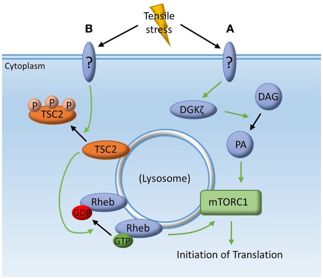
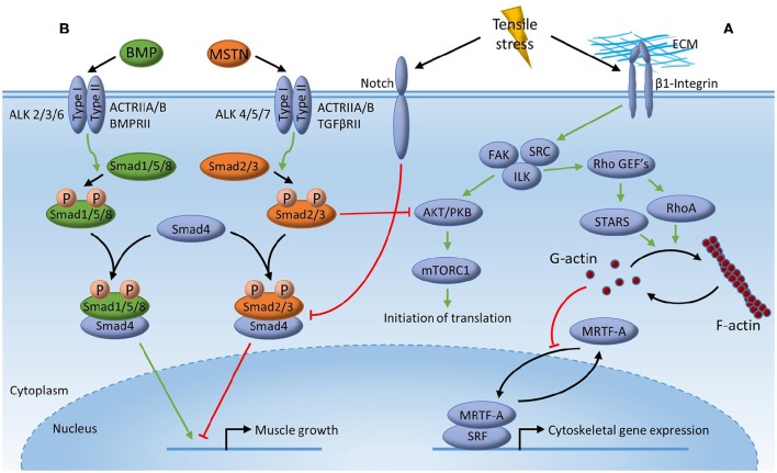
Loss of skeletal muscle myofibrillar protein with disease and/or inactivity can severely deteriorate muscle strength and function. Strategies to counteract wasting of muscle myofibrillar protein are therefore desirable and invite for considerations on the potential superiority of specific modes of resistance exercise and/or the adequacy of low load resistance exercise regimens as well as underlying mechanisms. In this regard, delineation of the potentially mechanosensitive molecular mechanisms underlying muscle protein synthesis (MPS), may contribute to an understanding on how differentiated resistance exercise can transduce a mechanical signal into stimulation of muscle accretion. Recent findings suggest specific upstream exercise-induced mechano-sensitive myocellular signaling pathways to converge on mammalian target of rapamycin complex 1 (mTORC1), to influence MPS. This may e.g. implicate mechanical activation of signaling through a diacylglycerol kinase (DGKζ)-phosphatidic acid (PA) axis or implicate integrin deformation to signal through a Focal adhesion kinase (FAK)-Tuberous Sclerosis Complex 2 (TSC2)-Ras homolog enriched in brain (Rheb) axis. Moreover, since initiation of translation is reliant on mRNA, it is also relevant to consider potentially mechanosensitive signaling pathways involved in muscle myofibrillar gene transcription and whether some of these pathways converge with those affecting mTORC1 activation for MPS. In this regard, recent findings suggest how mechanical stress may implicate integrin deformation and/or actin dynamics to signal through a Ras homolog gene family member A protein (RhoA)-striated muscle activator of Rho signaling (STARS) axis or implicate deformation of Notch to affect Bone Morphogenetic Protein (BMP) signaling through a small mother of decapentaplegic (Smad) axis.

Keywords: BMP-Smad; PLD-PA; Rheb; Rho-STARS; mechanotransduction.


Figures

Figure 1
Figure 1
Mechanotransduction for muscle protein synthesis. Tensile stress inherent of mechanical deformation may stimulate muscle protein synthesis through; (A) yet unidentified mechanosensing proteins acting on the zeta isoform of diacylglycerol kinase (DGKζ), resulting in the conversion of diacylglycerol (DAG) to phosphatidic acid (PA) which then directly activates the mechanistic target of rapamycin complex 1 (mTORC1); (B) an unidentified kinase phosphorylating the tuberous sclerosis complex-2 (TSC2) which is then translocated away from the lysosome allowing Ras homolog enriched in brain (Rheb) to be in its guanosine triphosphate (GTP) bound state which can then directly activate mTORC1.
Figure 2
Figure 2
Mechanotransduction for muscle mRNA transcription. Tensile stress inherent of mechanical deformation may stimulate muscle mRNA transcription through; (A) deformation of membrane-associated β1-Integrin activating focal adhesion kinase (FAK), integrin-linked kinase (ILK) and SRC, which then promotes activation of striated muscle activator of Rho signaling (STARS) and Ras homolog gene family member A (RhoA) through Rho guanine nucleotide exchange factors (GEFs) leading to polymerization of globular actin (G-actin) into filamentous actin (F-actin). Release of cytoplasmic G-actin from myocardin-related transcription factor (MRTF) then allows MRTF to translocate to the nucleus to act as a co-transcription factor with transcription factor serum response factor (SRF), leading to gene expression of multiple muscle myofibrillar and cytoskeletal genes; (B) competitive inhibition of Myostatin (MSTN) signaling by Bone Morphogenetic Protein (BMP) signaling through the common mediator small mother of decapentaplegic 4 (Smad4). Binding of MSTN to its receptor, leads to phosphorylation of Smad2/3 enabling formation of a transcriptional complex with Smad4, which then translocate to the nucleus to modulate transcriptional events resulting in impaired muscle growth. BMP leads to phosphorylation of Smad1/5/8 resulting in the possible formation of a Smad1/5/8-Smad4 transcriptional complex resulting in expression of genes important for muscle growth. Tensile stress inherent of mechanical deformation limits smad2/3 signaling through the membrane-associated protein Notch thereby allowing Smad1/5/8 signaling resulting in muscle accretion.

References

    1. Adams G. R., Haddad F. (1996). The relationships among IGF-1, DNA content, and protein accumulation during skeletal muscle hypertrophy. J. Appl. Physiol. (1985) 81, 2509–2516. - PubMed
    1. Arai A., Spencer J. A., Olson E. N. (2002). STARS, a striated muscle activator of Rho signaling and serum response factor-dependent transcription. J. Biol. Chem. 277, 24453–24459. 10.1074/jbc.M202216200 - DOI - PubMed
    1. Aspuria P. J., Tamanoi F. (2004). The Rheb family of GTP-binding proteins. Cell. Signal. 16, 1105–1112. 10.1016/j.cellsig.2004.03.019 - DOI - PubMed
    1. Atherton P. J., Smith K. (2012). Muscle protein synthesis in response to nutrition and exercise. J. Physiol. 590, 1049–1057. 10.1113/jphysiol.2011.225003 - DOI - PMC - PubMed
    1. Avila-Flores A., Santos T., Rincón E., Mérida I. (2005). Modulation of the mammalian target of rapamycin pathway by diacylglycerol kinase-produced phosphatidic acid. J. Biol. Chem. 280, 10091–10099. 10.1074/jbc.M412296200 - DOI - PubMed

LinkOut - more resources