

The novel laparoscopic training 3D model in urology with surgical anatomic remarks: Fresh-frozen cadaveric tissue
- PMID: 27909613
- PMCID: PMC5125734
- DOI: 10.5152/tud.2016.84770
The novel laparoscopic training 3D model in urology with surgical anatomic remarks: Fresh-frozen cadaveric tissue
Abstract
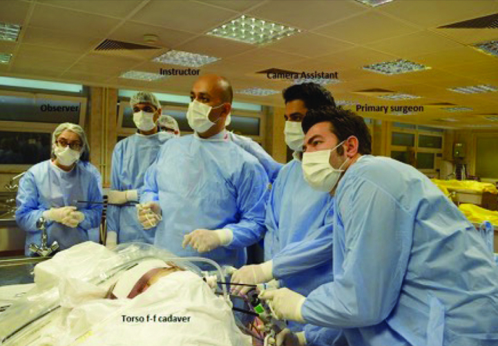
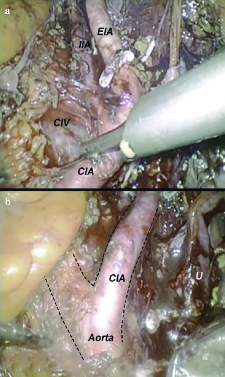
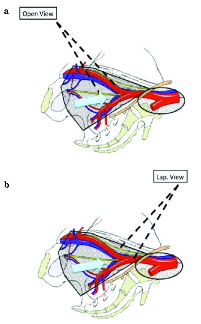
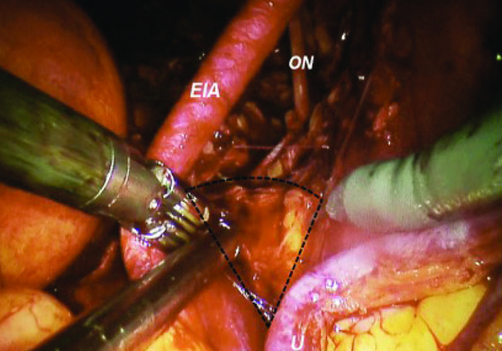
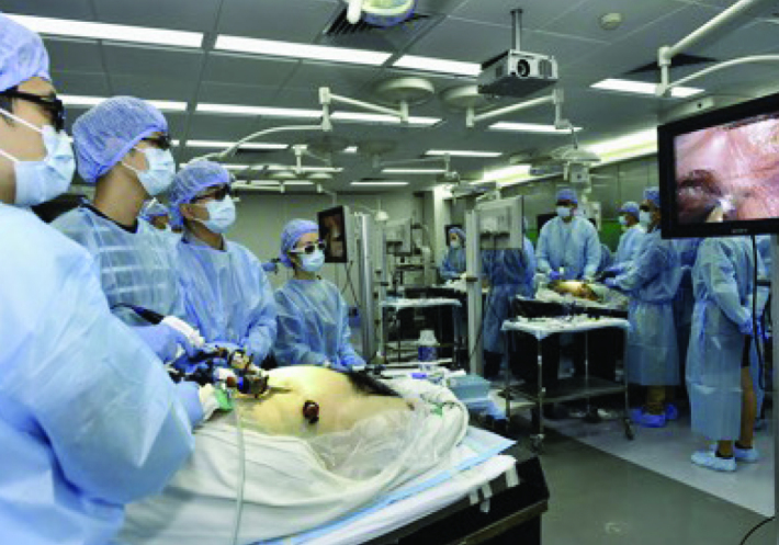
Laparoscopic surgery is routinely used to treat many urological conditions and it is the gold standard treatment option for many surgeries such as radical nephrectomy. Due to the difficulty of learning, laparoscopic training should start outside the operating room. Although it is a very different model of laparoscopic training; the aim of this review is to show the value of human cadaveric model for laparoscopic training and present our experience in this area. Fresh frozen cadaveric model in laparoscopic training, dry lab, cadaveric model, animal models and computer based simulators are the most commonly used models for laparoscopic training. Cadaveric models mimic the live setting better than animal models. Also, it is the best way in demonstrating important anatomic landmarks like prostate, bladder, and pelvic lymph nodes templates. However, cadaveric training is expensive, and should be used by multiple disciplines for higher efficiency. The laparosopic cadaveric training starts with didactic lectures with introduction of pelvic surgical anatomy. It is followed by hands-on dissection. A typical pelvic dissection part can be completed in 6 hours. Surgical robot and some laparoscopy platforms are equipped with 3-D vision. In recent years, we have use the stereoscopic laparoscopy system for training purposes to show exact anatomic landmarks. Cadavers are removed from their containers 3 to 5 days prior to training session to allow enough time for thawing. Intracorporeal suturing is an important part of laparoscopic training. We believe that suturing must be practiced in the dry lab, which is significantly cheaper than cadaveric models. Cadaveric training model should focus on the anatomic dissection instead. In conclusion, fresh-frozen cadaveric sample is one of the best 3D simulation models for laparoscopic training purposes. Major aim of cadaveric training is not only mimicking the surgical technique but also teaching true anatomy. Lack of availability and higher financial cost are the two setbacks for the use of cadavers. Surgeon should learn basic laparoscopic skills with box trainers prior to cadaveric skill training.
Keywords: Cadaver; laparoscopic surgery; training.
Conflict of interest statement
No conflict of interest was declared by the authors.
Figures
References
-
- Furriel FT, Laguna MP, Figueiredo AJ, Nunes PT, Rassweiler JJ. Training of European urology residents in laparoscopy: results of a pan-European survey. BJU Int. 2013;112:1223–8. https://doi.org/10.1111/bju.12410. - DOI - PubMed
-
- Albala DM, Kavoussi LR, Clayman RV. Laparoscopic nephrectomy. Semin Urol. 1992;10:146–51. - PubMed
-
- Ljungberg B, Bensalah K, Canfield S, Dabestani S, Hofmann F, Hora M, et al. EAU guidelines on renal cell carcinoma: 2014 update. Eur Urol. 2015;67:913–24. https://doi.org/10.1016/j.eururo.2015.01.005. - DOI - PubMed
-
- ten Brinke B, Klitsie PJ, Timman R, Busschbach JJ, Lange JF, Kleinrensink GJ. Anatomy education and classroom versus laparoscopic dissection-based training: a randomized study at one medical school. Acad Med. 2014;89:806–10. https://doi.org/10.1097/ACM.0000000000000223. - DOI - PubMed
-
- Martin JA, Regehr G, Reznick R, MacRae H, Murnaghan J, Hutchison C, et al. Objective structured assessment of technical skill (OSATS) for surgical residents. Br J Surg. 1997;84:273–8. https://doi.org/10.1046/j.1365-2168.1997.02502.x. - DOI - PubMed
Publication types
LinkOut - more resources
Full Text Sources
Other Literature Sources
Miscellaneous