

Distribution of interstitial cells of Cajal in the bladders of fetal rats with retinoic acid induced myelomeningocele
- PMID: 27909623
- PMCID: PMC5125744
- DOI: 10.5152/tud.2016.98474
Distribution of interstitial cells of Cajal in the bladders of fetal rats with retinoic acid induced myelomeningocele
Abstract
Objective: Myelomeningocele (MMC) is one of the most common reason of neurogenic bladder dysfunction in children. Although neurogenic bladder dysfunction occurrence is related with bladder innervation, also there are some changes seen in the smooth muscle and neural cells of the bladder. Interstitial cells of Cajal (ICC) are the pacemaker cells found in organs with peristaltic activity. Although it has been shown that ICC are diminished in the rat urinary bladder with traumatic spinal cord injury, there is no data about ICC in fetal rat bladders with MMC. This study has been conducted to investigate the ICC in the bladders of fetal rats with retinoic acid induced MMC.
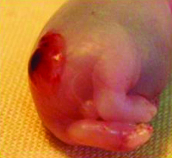
Materials and methods: Time dated pregnant Wistar albino rats were divided into 3 groups. In MMC group, dams were fed with gavage solution containing 60 mg/kg all-trans retinoic acid dissolved in olive oil on 10. embryologic day. Sham group animals were fed only olive oil. Control group dams were fed with standard rat chow. Fetuses were delivered by cesarean section and harvested on 22. embryologic day. MMC was identified by observing MMC sacs at the back of the fetuses. Distribution of ICCs were evaluated using immunohistochemical staining.
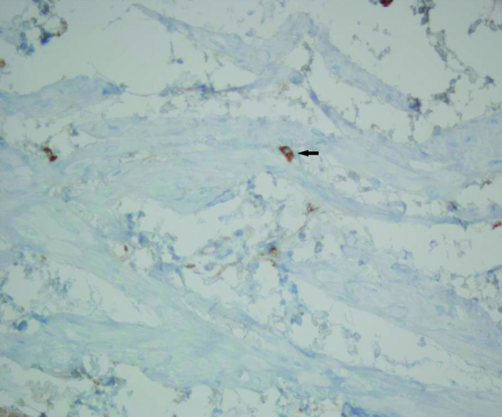
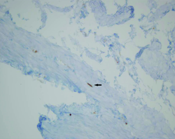
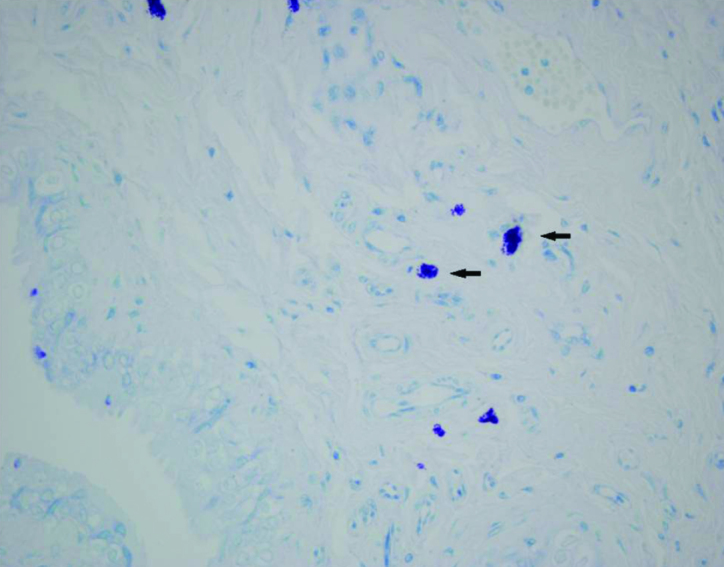
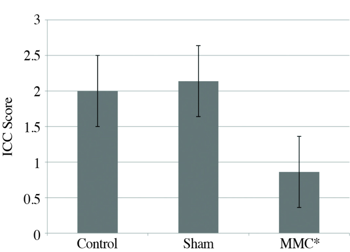
Results: ICCs were found in all groups, which have the same morphological features that had been described earlier in the gastrointestinal tract and the bladder. The density of the ICC in the MMC group was found to be significantly decreased when compared with the control and the sham groups (p<0.05).
Conclusion: The density of the ICC in the urinary bladder decreased in the neurogenic bladder developed in MMC.
Keywords: Bladder contractility; bladder dysfunction; interstitial cells of Cajal; myelomeningocele; neurogenic bladder.
Conflict of interest statement
No conflict of interest was declared by the authors.
Figures
References
-
- Yeung CK, Barker GM, Läckgren G. Pathophysiology of bladder dysfunction. In: Gearhart JP, Rink R, Mouriquand PDE, editors. Pediatric Urology. 2 Edition. Philadelphia: Saunders Elsevier; 2010. pp. 353–65. https://doi.org/10.1016/B978-1-4160-3204-5.00027-X. - DOI
-
- Rien JM, Nijman ST. Pathophysiology of neurogenic bladder dysfunction. In: Ciro Esposito JMG, Gough D, Savanelli A, editors. Pediatric Neurogenic Bladder Dysfunction: Diagnosis, Treatment, Long-Term Follow-up. Berlin: Springer; 2006. pp. 33–8.
-
- Brading AF. A myogenic basis for the overactive bladder. Urology. 1997;50:57–67. https://doi.org/10.1016/S0090-4295(97)00591-8. - DOI - PubMed
-
- Andersson KE, Arner A. Urinary bladder contraction and relaxation: physiology and pathophysiology. Physiol Rev. 2004;84:935–86. https://doi.org/10.1152/physrev.00038.2003. - DOI - PubMed
-
- Davidson RA, McCloskey KD. Morphology and localization of interstitial cells in the guinea pig bladder: structural relationships with smooth muscle and neurons. J Urol. 2005;173:1385–90. https://doi.org/10.1097/01.ju.0000146272.80848.37. - DOI - PubMed
LinkOut - more resources
Full Text Sources
Other Literature Sources