

doi: 10.1016/j.eats.2016.05.002.
eCollection 2016 Oct.
Arthroscopic Capsular Reconstruction of the Hip With Acellular Dermal Extracellular Matrix: Surgical Technique
Affiliations
- PMID: 27909667
- PMCID: PMC5124028
- DOI: 10.1016/j.eats.2016.05.002
Item in Clipboard
Arthroscopic Capsular Reconstruction of the Hip With Acellular Dermal Extracellular Matrix: Surgical Technique
Arthrosc Tech.
.
Abstract
Atraumatic instability of the hip has become an increasingly studied occurrence in recent years. There are several established surgical techniques that help restore stability of the native hip joint. In some cases, these procedures are not an option. As the phenomenon has become recognized more frequently, a greater number of revision surgeries are warranted in patients with ligamentous laxity. A durable solution for irreparable microinstability needs to be formulated to address this vulnerable patient demographic. We describe the surgical technique for capsular reconstruction with acellular dermal extracellular matrix.
Figures

The Suture Tak anchor (Arthrex) is implanted into the femoral neck using the distal lateral accessory portal while being visualized with the camera in the anterolateral portal. This anchor will be used for the attachment of the Arthroflex (Arthrex) graft to the capsular defect zone. (A, Knotless Suture Tak Anchor [Arthrex]; DC, distal capsule; FN, femoral neck.)
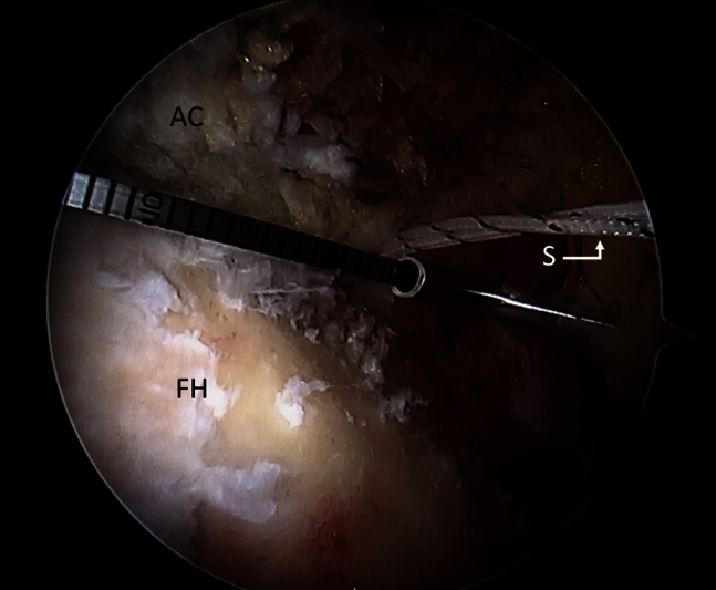
Distance between the anchors is measured by the use of an arthroscopic measuring tool (Arthex) to plan for appropriate allograft dimensions. (AC, acetabulum; FH, femoral head; S, suture.)

The Arthroflex graft (Arthrex) is prepared according to the measurements taken arthroscopically. The graft is cut to be 1 cm beyond the distances between the anchors.

The Arthroflex (Arthex) graft is shuttled through the DLAP into the left hip joint while pulling the posts of the proximal sliding knots. (AL, anterolateral portal into which the camera is inserted to visualize the procedure; DLAP, distal lateral accessory portal; LH, left hip.)

The graft is sutured to the anchors placed in each of the 4 corners of the capsular defect. (AG, Arthroflex graft [Arthrex]; CR, capsular remnant.)

The reconstructed capsule is then assessed on all facets. (AG, Arthroflex graft [Arthrex].)
Similar articles
-
Arthroscopic Technique of Capsular Plication for the Treatment of Hip Instability.Arthrosc Tech. 2015 Apr 13;4(2):e163-7. doi: 10.1016/j.eats.2015.01.004. eCollection 2015 Apr. Arthrosc Tech. 2015. PMID: 26052494 Free PMC article.
-
Posterior Glenohumeral Capsular Reconstruction Using an Acellular Dermal Allograft.Arthrosc Tech. 2018 Jun 18;7(7):e739-e745. doi: 10.1016/j.eats.2018.03.011. eCollection 2018 Jul. Arthrosc Tech. 2018. PMID: 30094145 Free PMC article.
-
Anterior Capsule Augmentation and Posterior Glenohumeral Capsular Reconstruction With Human Dermal Allograft for Multidirectional Shoulder Instability.Arthrosc Tech. 2020 Apr 25;9(5):e657-e662. doi: 10.1016/j.eats.2020.01.020. eCollection 2020 May. Arthrosc Tech. 2020. PMID: 32489841 Free PMC article.
-
Arthroscopic versus open Bankart repair for traumatic anterior shoulder instability.Clin Sports Med. 2000 Jan;19(1):19-48. doi: 10.1016/s0278-5919(05)70294-5. Clin Sports Med. 2000. PMID: 10652663 Review.
-
The hip joint: arthroscopic procedures and postoperative rehabilitation.J Orthop Sports Phys Ther. 2006 Jul;36(7):516-25. doi: 10.2519/jospt.2006.2138. J Orthop Sports Phys Ther. 2006. PMID: 16881468 Review.
Cited by
-
Clinical applications of acellular dermal matrices: A review.Scars Burn Heal. 2022 Jan 19;8:20595131211038313. doi: 10.1177/20595131211038313. eCollection 2022 Jan-Dec. Scars Burn Heal. 2022. PMID: 35083065 Free PMC article. Review.
-
Hip Capsular Reconstruction Made Easy: The Timing and the Technique.Arthrosc Tech. 2020 Dec 19;10(1):e73-e78. doi: 10.1016/j.eats.2020.09.012. eCollection 2021 Jan. Arthrosc Tech. 2020. PMID: 33532211 Free PMC article.
-
Is there enough evidence to support hip capsular reconstruction? A systematic review of biomechanical studies.J Hip Preserv Surg. 2021 Aug 26;8(2):156-163. doi: 10.1093/jhps/hnab059. eCollection 2021 Jul. J Hip Preserv Surg. 2021. PMID: 35145712 Free PMC article.
-
Anterior Capsule Reconstruction of the Native Hip: A Technique Guide.Arthrosc Tech. 2019 Sep 26;8(10):e1247-e1253. doi: 10.1016/j.eats.2019.06.014. eCollection 2019 Oct. Arthrosc Tech. 2019. PMID: 32042580 Free PMC article.
-
Arthroscopic Triple Reconstruction in the Hip Joint: Restoration of Soft-Tissue Stabilizers in Revision Surgery for Gross Instability.Arthrosc Tech. 2021 Apr 12;10(5):e1239-e1248. doi: 10.1016/j.eats.2021.01.018. eCollection 2021 May. Arthrosc Tech. 2021. PMID: 34141538 Free PMC article.
References
-
- Bharam S. Labral tears, extra-articular injuries, and hip arthroscopy in the athlete. Clin Sports Med. 2006;25:279–292. ix. - PubMed
-
- Telleria J.J.M., Lindsey D.P., Giori N.J., Safran M.R. A quantitative assessment of the insertional footprints of the hip joint capsular ligaments and their spanning fibers for reconstruction. Clin Anat. 2014;27:489–497. - PubMed
-
- Shindle M.K., Ranawat A.S., Kelly B.T. Diagnosis and management of traumatic and atraumatic hip instability in the athletic patient. Clin Sports Med. 2006;25:309–326. ix-x. - PubMed
-
- Bellabarba C., Sheinkop M.B., Kuo K.N. Idiopathic hip instability. An unrecognized cause of coxa saltans in the adult. Clin Orthop. 1998;355:261–271. - PubMed
LinkOut - more resources
Full Text Sources
Other Literature Sources
Research Materials
