

doi: 10.1016/j.eats.2016.05.009.
eCollection 2016 Oct.
Arthroscopic Primary Repair of Proximal Anterior Cruciate Ligament Tears
Affiliations
- PMID: 27909675
- PMCID: PMC5124221
- DOI: 10.1016/j.eats.2016.05.009
Item in Clipboard
Arthroscopic Primary Repair of Proximal Anterior Cruciate Ligament Tears
Arthrosc Tech.
.
Abstract
In a select group of patients with proximal anterior cruciate ligament (ACL) tears, primary repair can be a useful technique. Preservation of the native ACL may be advantageous for proprioceptive function and is thought to restore normal knee joint kinematics. The procedure is a less morbid and more conservative surgical approach to restore knee stability. Primary repair is preferably performed in the acute setting because of better healing capacity and tissue quality. We present the surgical technique of arthroscopic primary ACL repair with suture anchors in patients with proximal tears and excellent tissue quality.
Figures

(A) Arthroscopic view of a left knee, viewed from the anterolateral portal with the patient supine and the knee in 90° flexion. A proximal tear (asterisk) is visible, but the exact location of the tear needs to be assessed by a probe. Excellent tissue quality of the anteromedial (left arrowhead) and posterolateral bundle (right arrowhead) is seen. (B) Arthroscopic view of a left knee, viewed from the anterolateral portal with the patient supine and the knee in 90° flexion. A proximal type I tear (arrowhead) with good tissue quality is shown after a probe (asterisk) is used to remove the remnant from the femoral wall.
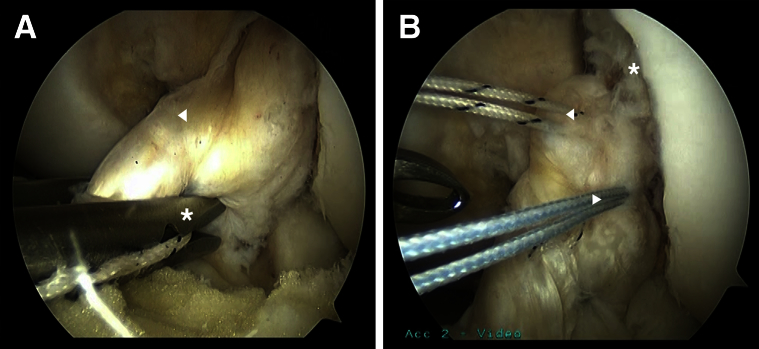
(A) Arthroscopic view of a left knee, viewed from the anterolateral portal with the patient supine and the knee in 90° flexion. A suture passer (asterisk) is used to pass a No. 2 TigerWire suture through the anteromedial bundle (arrowhead). The suture is passed in an alternating, interlocking Bunnell-type pattern. (B) Arthroscopic view of a left knee, viewed from the anterolateral portal with the patient supine and the knee in 90° flexion. Two different sutures have been passed through both the anteromedial (left arrowhead) and posterolateral bundle (right arrowhead). The proximal tear is visible (asterisk).
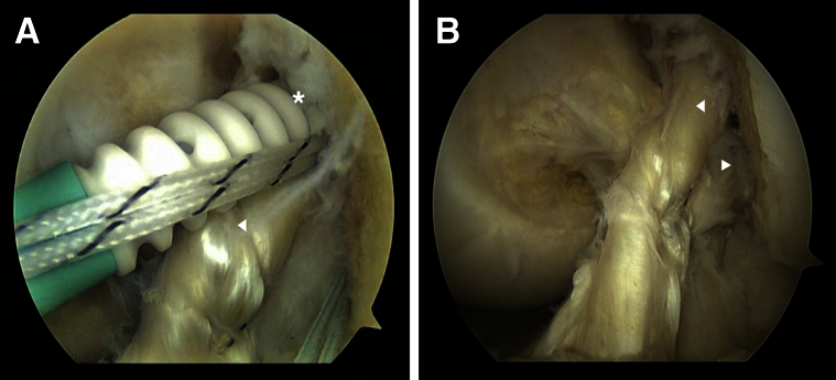
(A) Arthroscopic view of a left knee, viewed from the anterolateral portal with the patient supine and the knee in 90° flexion. After creating a 4.5 mm × 20 mm hole in the anteromedial bundle origin of the femoral wall, the suture anchor of the anteromedial bundle is deployed into the femur toward the hole (asterisk), whereas the anteromedial bundle of the anterior cruciate ligament (ACL) remnant (arrowhead) is tensioned to the wall. (B) Arthroscopic view of a left knee, viewed from the anterolateral portal with the patient supine and the knee in 90° flexion. A completed primary ACL repair is seen after both the anteromedial (left arrowhead) and posterolateral bundle (right arrowhead) are deployed into the femoral wall at their anatomic origin.
References
-
- Sherman M.F., Lieber L., Bonamo J.R., Podesta L., Reiter I. The long-term followup of primary anterior cruciate ligament repair. Defining a rationale for augmentation. Am J Sports Med. 1991;19:243–255. - PubMed
-
- Feagin J.A., Jr., Curl W.W. Isolated tear of the anterior cruciate ligament: 5-year follow-up study. Am J Sports Med. 1976;4:95–100. - PubMed
-
- Odensten M., Lysholm J., Gillquist J. Suture of fresh ruptures of the anterior cruciate ligament. A 5-year follow-up. Acta Orthop Scand. 1984;55:270–272. - PubMed
-
- Genelin F., Trost A., Primavesi C., Knoll P. Late results following proximal reinsertion of isolated ruptured ACL ligaments. Knee Surg Sports Traumatol Arthrosc. 1993;1:17–19. - PubMed
-
- DiFelice G.S., Villegas C., Taylor S.A. Anterior cruciate ligament preservation: Early results of a novel arthroscopic technique for suture anchor primary anterior cruciate ligament repair. Arthroscopy. 2015;31:2162–2171. - PubMed
LinkOut - more resources
Full Text Sources
Other Literature Sources
Medical
