

Inhibition of CDH17 gene expression via RNA interference reduces proliferation and apoptosis of human MKN28 gastric cancer cells
- PMID: 27909714
- PMCID: PMC5182006
- DOI: 10.3892/ijo.2016.3783
Inhibition of CDH17 gene expression via RNA interference reduces proliferation and apoptosis of human MKN28 gastric cancer cells
Abstract
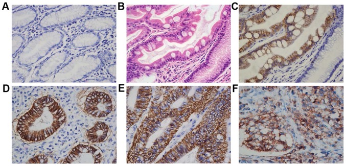
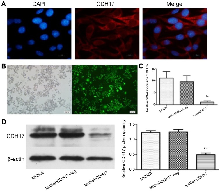
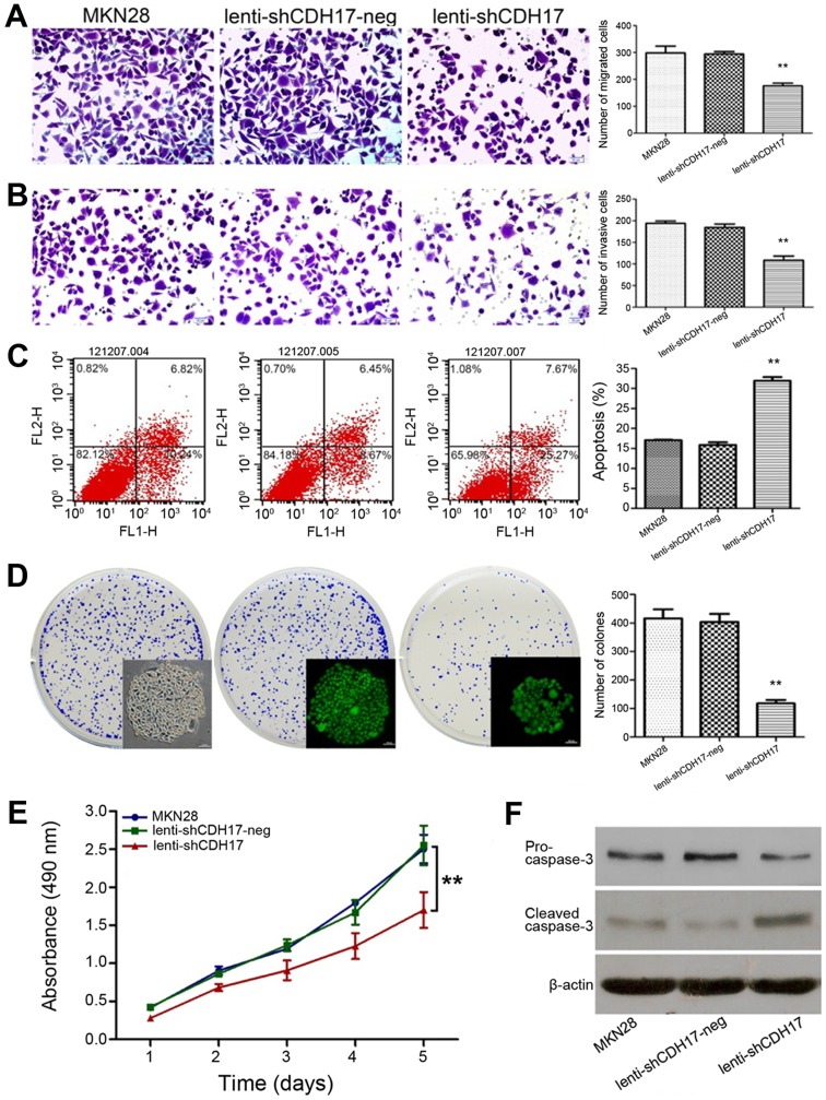
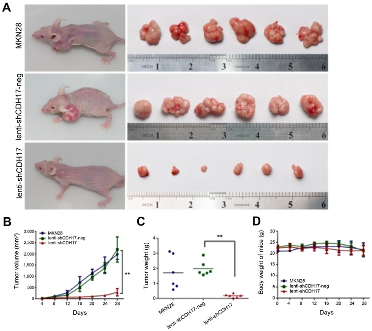
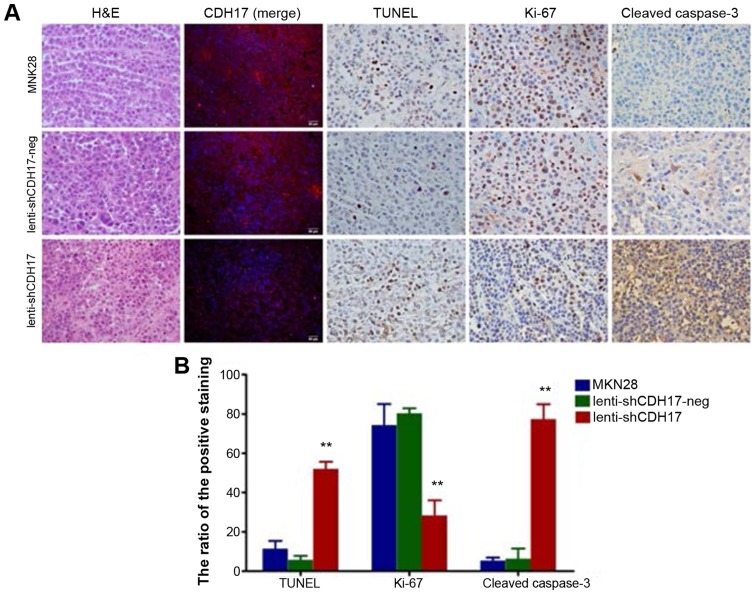
Gastric cancer is the fourth most common type of cancer and the second cause of cancer‑related mortalities worldwide despite the use of multimodal therapy. Cadherins are transmembrane glycoproteins that are involved in tumorigenesis. CDH17 has been found to be over‑expressed in gastric cancer and its overexpression was associated with lymph node metastasis and tumor‑node‑metastasis stage of the patients, yet the exact role and molecular mechanism of CDH17 in gastric cancer have not been determined. Using a lentiviral system as a delivery mediator of RNA interference, we found that inhibition of CDH17 can lead to reduce proliferation and increase apoptosis of gastric cancer cell line MKN28 in vitro and significantly diminish their tumorigenicity in vivo. Our results of the present study suggest that CDH17 may be a promising candidate for the therapeutic targeting of gastric cancer.
Figures
Similar articles
-
Targeting CDH17 suppresses tumor progression in gastric cancer by downregulating Wnt/β-catenin signaling.PLoS One. 2013;8(3):e56959. doi: 10.1371/journal.pone.0056959. Epub 2013 Mar 15. PLoS One. 2013. PMID: 23554857 Free PMC article. Clinical Trial.
-
Cadherin-17 induces tumorigenesis and lymphatic metastasis in gastric cancer through activation of NFκB signaling pathway.Cancer Biol Ther. 2013 Mar;14(3):262-70. doi: 10.4161/cbt.23299. Epub 2013 Jan 8. Cancer Biol Ther. 2013. PMID: 23298905 Free PMC article.
-
Lentiviral vector-mediated survivin shRNA delivery in gastric cancer cell lines significantly inhibits cell proliferation and tumor growth.Oncol Rep. 2015 Aug;34(2):859-67. doi: 10.3892/or.2015.4033. Epub 2015 Jun 5. Oncol Rep. 2015. PMID: 26043753
-
Molecular events in gastric carcinogenesis.J Med Life. 2014 Sep 15;7(3):375-8. Epub 2014 Sep 25. J Med Life. 2014. PMID: 25408758 Free PMC article. Review.
-
Search for new biomarkers of gastric cancer through serial analysis of gene expression and its clinical implications.Cancer Sci. 2004 May;95(5):385-92. doi: 10.1111/j.1349-7006.2004.tb03220.x. Cancer Sci. 2004. PMID: 15132764 Free PMC article. Review.
Cited by
-
Proteomics Analysis of Gastric Cancer Patients with Diabetes Mellitus.J Clin Med. 2021 Jan 21;10(3):407. doi: 10.3390/jcm10030407. J Clin Med. 2021. PMID: 33494396 Free PMC article.
-
Contactin 1 modulates pegylated arginase resistance in small cell lung cancer through induction of epithelial-mesenchymal transition.Sci Rep. 2019 Aug 19;9(1):12030. doi: 10.1038/s41598-019-48476-8. Sci Rep. 2019. PMID: 31427725 Free PMC article.
-
CDH17 nanobodies facilitate rapid imaging of gastric cancer and efficient delivery of immunotoxin.Biomater Res. 2022 Nov 26;26(1):64. doi: 10.1186/s40824-022-00312-3. Biomater Res. 2022. PMID: 36435809 Free PMC article.
-
Inhibition of miR-22 enhanced the efficacy of icotinib plus pemetrexed in a rat model of non-small cell lung cancer.Iran J Basic Med Sci. 2020 Mar;23(3):329-336. doi: 10.22038/IJBMS.2019.39291.9320. Iran J Basic Med Sci. 2020. PMID: 32440319 Free PMC article.
-
New frontiers in oncolytic viruses: optimizing and selecting for virus strains with improved efficacy.Biologics. 2018 Feb 9;12:43-60. doi: 10.2147/BTT.S140114. eCollection 2018. Biologics. 2018. PMID: 29445265 Free PMC article. Review.
References
MeSH terms
Substances
LinkOut - more resources
Full Text Sources
Other Literature Sources
Medical
