Review
. 2016 Dec;10(6):529-555.
doi: 10.1007/s11832-016-0785-x. Epub 2016 Dec 1.

Tibial hemimelia: new classification and reconstructive options

Affiliations
Review

Tibial hemimelia: new classification and reconstructive options

Dror Paley. J Child Orthop. 2016 Dec.

Abstract

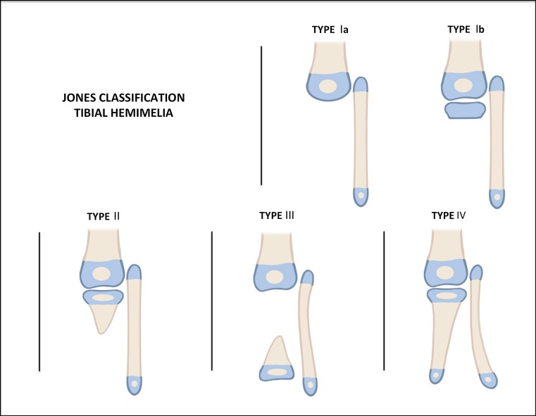
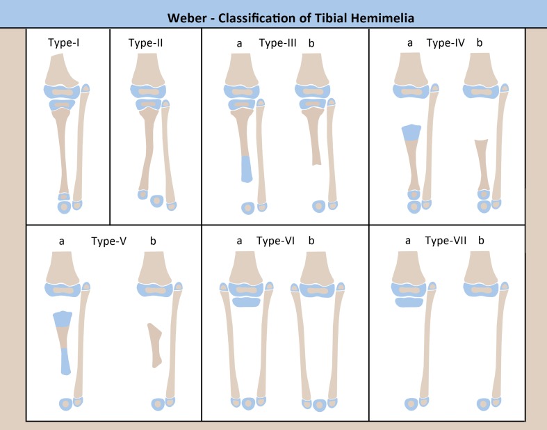
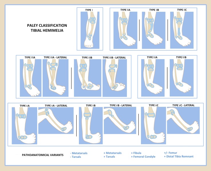
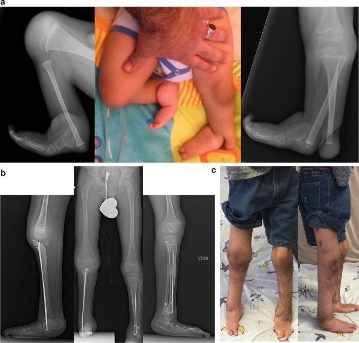
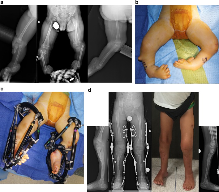
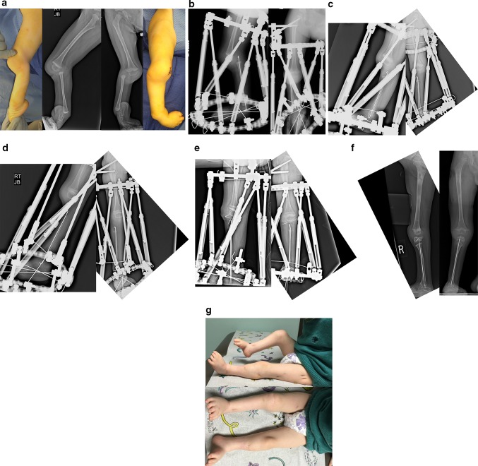
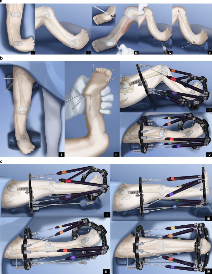
Tibial hemimelia is a rare congenital lower limb deficiency presenting with a wide spectrum of associated congenital anomalies, deficiencies and duplications. Reconstructive options have been limited, and the gold standard for treatment has remained amputation with prosthetic fitting. There is now a better understanding of the genetics, etiology and pathoanatomy of tibial hemimelia. Armed with this knowledge, I present here a new classification to guide treatment and prognosis and then discuss new treatment strategies and techniques for limb reconstruction based on this new classification scheme.

Keywords: Brown centralization of fibula; Clubfoot; Paley classification; Tibial aplasia; Tibial hemimelia; Weber patelloplasty.


Figures

Fig. 1
Fig. 1
Jones classification. Reproduced with permission by the Paley Foundation
Fig. 2
Fig. 2
Weber classification. Reproduced with permission by the Paley Foundation
Fig. 3
Fig. 3
Paley classification. Reproduced with permission by the Paley Foundation
Fig. 4
Fig. 4
a Antero-posterior (AP) and lateral radiographs of 20-year-old woman with bilateral Paley type 1 tibial hemimelia. The tibia is well formed at both the knee and ankle joints. The fibulas are relatively overgrown at their proximal ends and are articulating with the side of the femurs. The knees are both in valgus due to both tibial and femoral deformities. The tibias have mild procurvatum diaphyseal bowing. Since both tibias are relatively short compared to the femurs, the patient has a mesomelic disproportion and short stature. b AP and lateral radiographs after treatment. Both tibias were lengthened with external fixators. The tibial valgus-procurvatum was corrected. The fibulas were not pulled down. Bilateral femur varus osteotomies were performed after completion of the tibial correction. The lengthening restored the proportion of the tibias and femurs to normal
Fig. 5
Fig. 5
a Radiographs of 2-year-old girl with Paley type 2A unilateral tibial hemimelia, with equino-varus deformity of foot and varus of tibial diaphysis. There is no diastasis of the distal tibio-fibular joint. The ankle joint is present. The ossification of the distal tibia shows a regular trumpet shaped metaphyseal line indicating that this is the region of the distal tibial physis. b Lateral radiograph of tibia. Taylor spatial frame (TSF) is in place with proximal tibial and foot rings programmed for gradual correction of foot deformity. There is also an independent mechanism (threaded rod with cube and two half pins connected to the proximal ring) to simultaneously lengthen the tibia through a proximal osteotomy. There is a distal fibular epiphysiodesis screw in place to slow the growth of the faster growing fibula. c Lateral (left) and AP (right) radiographs at the end of the foot correction and 8 cm of lengthening after a second stage surgery to openly reduce the talus to the tibia combined with axial pinning of the ankle joint. Syndesmotic washer-suture device (Ziptite™ Fixation System; Biomet Sports Medicine, LLC, Warsaw, IN) inserted to stabilize the lengthened tibia to the fibula at its new syndesmosis level. The tibial lengthening bone shows poor regenerate formation. d Plate fixation at time of removal of fixator to prevent fracture. The tibia is well aligned. The talus is under the distal tibia. The fibula is now at station at the ankle and the ankle is stable. The foot is plantigrade
Fig. 6
Fig. 6
a Radiographs of a 3-year-old girl with unilateral tibial hemimelia, Paley type 2B tibial hemimelia + metatarsals/toes. The obvious delta tibia is seen with two secondary ossific nuclei. The foot has seven metatarsals and eight toes. The foot is in equino-varus. The ankle and knee joint are maloriented. The proximal fibula is relatively overgrown and sits proximal to the distal femoral physis. b Clinical photograph of same girl. c Arthrogram of knee and ankle showing joint malorientation and dysplasia. d AP (left) and lateral (right) radiographs with TSF in place and osteotomy lengthening and deformity correction of the tibia. The foot deformity correction is partially through the distal tibial reorientation. Note there is no osteotomy of the fibula and that it is only fixed to the distal tibial ring using one distal tibio–fibular wire and half pin. e AP (left) and lateral (right) radiographs following 8 cm of tibial lengthening and deformity correction with distal fibular transport. The frame was modified connecting the distal tibiofibular wire and fibular half pin to the proximal ring. This frees the ankle joint for distraction correction of the remaining foot deformity. Note that the proximal fibula has been pulled down almost to station. f Two years after removal of the external fixator the tibia is well consolidated. The fibula is already showing significant relative proximal and distal overgrowth. The distal fibular epiphysiodesis screw’s head is migrated into the epiphysis and screw shaft is broken rendering it mostly ineffective at this juncture. The foot varus deformity is slowly recurring. This will require another differential lengthening surgery in a year or two. g Clinical photos showing knee and ankle range of motion two years after lengthening. h This girl is very active despite recurrent leg length difference and mild foot deformity. Note how she plays with the other girls while she wears a shoe lift. She is the tall girl in the middle
Fig. 7
Fig. 7
a Magnetic resonance imaging (MRI) scan (left), lateral radiograph (middle), and standing AP radiograph (right) showing Paley type 2C unilateral tibial hemimelia. The foot is in significant varus. The distal tibia has an unossified region contiguous with the ankle. There is no obvious distal tibial physis, but the plafond is present. Note that the ossification of the distal tibia is irregular and sloped, not like a normal metaphysis associated with a distal tibial physis (Fig. 5a). The proximal fibula is relatively overgrown and proximally migrated. b Arthrogram of ankle showing AP (left) and lateral (right) views. c Lateral (left) and AP (right) radiographs after arthrotomy of ankle, and open valgus-extension osteotomy and shortening of distal tibia to realign ankle joint by acute correction. Bone morphogenetic protein 2 (BMP2) was inserted in drill holes of the cartilage remnant of the distal tibia to get it to ossify. A second osteotomy was performed proximally for lengthening. The fibula is being pulled distally with an intramedullary wire hooked over the proximal epiphysis (see AP view). Because the tibial/ankle/foot deformities were corrected acutely, the external fixator is programmed for pure lengthening. The external fixator is extended to the femur with knee hinges to protect the cruciate deficient knee during lengthening while permitting knee flexion and extension motion. d Lateral (left) and AP (right) radiographs, after lengthening with excellent consolidation of the tibia, including ossification of the delayed distal tibia. The foot is plantigrade with forefoot supination deformity present. The proximal fibula is at station
Fig. 8
Fig. 8
a Standing (left), mortis view (middle) and lateral view (right) radiographs of a 2-year-old girl with Paley type 3A unilateral tibial hemimelia. The diastasis of the distal tibia and fibula with the talus in between is very evident. The talus always remains in line with the distal fibula and together they internally rotate around the distal tibia. The talus is shortened relative to the distal tibia but not relative to the distal fibula. There is relative overgrowth with proximal migration of the fibula. There is a mild distal tibial varus diaphyseal bowing. b AP (left) and lateral (right) radiographs showing TSF applied to the tibia and foot. There is no fixation in the fibula or talus. More current constructs would fix to these as described in the text. The foot is in internal rotation and equinus. c AP (left) and lateral (right) radiographs after gradual correction using TSF. The talus is under the distal tibial epiphysis and the foot and fibula have rotated externally relative to the tibia. There is no longer any tibio-fibular diastasis. The equinus deformity is also corrected. This gradual correction took 12 weeks. d AP (left) and lateral (right) radiographs after removal of the external fixator 3 months following open ankle reconstruction. The ankle reconstruction included a biologic arthroplasty to create a concave surface on the distal tibial epiphysis that is congruent to the talar dome convexity. The ankle joint was pinned with an axial wire and the diastasis of the tibia and fibula were fixed with a suture-washer syndesmotic repair (TightRope®, Arthrex, Naples, FL). The axial wire was left in place for another 3 months to prevent recurrent deformity at the ankle. Note the proximal fibula was brought down to station. e AP (left) and lateral (right) radiographs at age 14 years after she underwent two successful lengthenings; one external fixator lengthening at age 6, and one implantable nail lengthening at age 14 (Precice™; NuVasive Inc., San Diego, CA). She also had a supramalleolar and subtalar osteotomy. Note the ankle joint is stable and well preserved. The ankle has about 20° of motion. The proximal fibula is at station. Most of this hardware was removed at a later date. Due to the limited ankle motion, the leg lengths were intentionally corrected to leave a 1-cm difference
Fig. 9
Fig. 9
a Preoperative photograph (center), and radiographs (right on the left, left on the right) of bilateral tibial hemimelia in a 3-year-old boy; Paley type 5A (right) and Paley type 3B tibial hemimelia (left). The patella on the right has not yet ossified. Other than the cleft on the left, this is very similar to type 3A. The cleft makes this a very rare type. It should be noted that the foot is found on the fibular side of the cleft on the left. Both feet are in severe equino-varus and even upside down. There is a greater than 90° flexion contracture and dislocation at the right knee. b Standing (center), right lateral (left) and left lateral (right) radiographs after the staged reconstruction method was completed, 2 years prior on the left and 3 months prior on the right. The Paley–Weber patellar arthroplasty was performed on the right, with insertion of BMP2 to ossify the patella and fuse it to the proximal fibular epiphysis. Note the preservation of the proximal fibular physis. On the left the talus was brought under the tibia, a biologic arthroplasty performed together with plastic closure of the cleft. The distal and proximal tibial and fibular physes are preserved on both sides. The fusion of the talus to the distal fibular epiphysis did not occur and may require revision in the future. Both feet are plantigrade. c Clinical photographs showing the patient standing with both feet plantigrade and both knees straight and well aligned
Fig. 10
Fig. 10
a Preoperative radiographs of a 13-month-old boy with bilateral tibial hemimelia; Paley type 4A tibial hemimelia on the right and Paley type 4B tibial hemimelia on the left. Both sides have severe equino-varus deformities of both feet. The proximal tibial remnant on the right is prominent under the skin. There is proximal migration of the fibula on both sides. There is an unossified proximal tibial cartilagenous anlage on the left side. b Clinical photograph showing the severe equino-varus feet. Note that the tip of the proximal tibial remnant has its own skin pouch. c Bilateral TSF devices were applied to both legs. The thigh rings are perpendicular to the femurs and the foot rings are parallel to the soles of both feet. This computer-dependent external fixator is programmed first to correct the deformities between the femur and fibula at the knee, including transporting the fibula distally. It is then reprogrammed to correct the foot deformity. No additional surgery is required to switch from knee to ankle correction. On the left leg the proximal fibular epiphysis was transported distally and brought under the tibial epiphysis. d Standing AP photograph (center right) and radiograph (center left) of both lower limbs at age 5 years, showing that the legs are well aligned. Lateral radiographs of right leg (left) and left leg (right) showing the feet are fused at the ankle in a plantigrade position. Both distal fibular physes are closed despite best efforts to preserve them. The right foot has some adductus supination deformity. The patient is shown after lengthening of both lower limbs was completed. The left proximal tibial epiphysis is ossified and fused to the proximal fibular epiphysis. The unossified proximal tibial epiphysis was ossified by insertion of BMP2 into drill holes in the cartilage of the tibial anlage. The ossification of the tibial epiphysis and fusion to the fibula occurred within 3 months. Note, there is preservation of the proximal physis of the fibula. On the right the fibula was transferred to the tibia. It has auto-bridged across to the proximal fibula. Initially, knee–ankle–foot–orthotic (KAFO) braces were used. The knee was stable enough to discontinue these after these radiographs
Fig. 11
Fig. 11
a Lateral (left and center left) and AP (right and center right) photographs and radiographs of the right leg of a 2-year-old boy with bilateral tibial hemimelia. Only the right side is shown. The right side has Paley type 5A tibial hemimelia and the left side Paley type 4A. Note the flexion contracture of the right knee and the equino-varus-adductus foot deformity. b Lateral (left) and AP (right) radiographs showing the TSF in place with femoral and foot rings. The fibula is secured to the foot ring with a transverse wire. Note the temporary epiphysiodesis wire in the fibula hooked at both ends. Also note the wire across the neck of the talus. c Lateral (left) and AP (right) radiographs at the end of gradual distraction with TSF. The fibular head is centered under the end of the femur. The foot position has not changed and the transverse fibular wires remains connected to the foot ring. d Lateral (left) and AP (right) radiographs showing that the transverse fibular wire was fixed to the proximal ring using a threaded rod and post. This wire was released from the distal ring to allow gradual correction of the equino-varus foot contracture and to bring the talus under the distal fibula. A second computer planning is carried out to generate a new adjustment schedule for the patient. Note that there is no ossification of the proximal fibular epiphysis. Also note the growth lines related to preoperative infusion of zolidronic acid to prevent disuse osteoporosis during distraction. e Intraoperative lateral (left) and AP (right) radiographs after the surgery to perform a patellar arthroplasty and fibula–talar fusion is completed. Note the hemovac drain can be seen at the knee. The Paley–Weber patellar arthroplasty was performed fixing the proximal fibular epiphysis to the patella with a hooked intramedullary wire secured to the bottom of the frame below the foot. The talus was fused to the distal tibial epiphysis. BMP2 was inserted into a drill hole in the patella and proximal fibula to lead to ossification and fusion of both the patella and fibular epiphysis. A transverse distal fibular wire is arced to the foot ring to apply compression across the fusion site. f Lateral (left) and AP (right) radiographs showing the patella and proximal fibular epiphysis are ossified and fused together with preservation of the proximal fibula physis. The patella now serves as a tibial plateau. The talus has also fused to the distal fibula but the distal fibular physis has closed. g Clinical photographs, frontal view (bottom) and medial view (top) showing that the right knee can bend to 90° (top). There is active motion present. The other leg was also successfully treated for type 4A tibial hemimelia
Fig. 12
Fig. 12
Paley–Weber patellar arthroplasty. a Frontal (i) and lateral (ii) view illustrations of Paley type 5A tibial hemimelia: complete tibial agenesis, patella present, fibula dislocated and proximally migrated, knee flexion contracture and fixed equino-varus foot deformity. First surgery consists of Achilles tenotomy (iii). Two 1.5-mm intramedullary wires are inserted retrograde into the fibula and both are curled and hooked into the proximal fibular epiphysis. One is curled around the distal fibular epiphysis and the other is bent 90° to stick out of the skin (iv, v). Reproduced with permission by the Paley Foundation. b A prophylactic wire is inserted in the femur and hooked over the greater trochanter. Two proximal femoral half pins are inserted—one at the level of the lesser trochanter and one up the femoral neck (i). Three wires are inserted in the calcaneus in the plane of the sole of the foot—one mid posterior to exit near the interspace of the first and second toes; one posterolateral to exit anteromedial; one posteromedial to exit anterolateral (ii). The proximal femoral ring is fixed to the two half pins and an olive wire is added from antero-medial to postero-lateral in the mid femur. A foot ring is fixed to the three wires in the foot and a transverse talar wire added. The 90° bent fibular wire is fixed to the foot ring. Finally, six struts are added between the rings. Computer planning is done to gradually correct the knee deformity (iii, iv). Reproduced with permission by the Paley Foundation. c After several weeks of gradual correction of the knee joint contracture, the fibula is centered on the femur. At this point the fibular wire is connected to a long threaded rod with post and then liberated from the foot ring (i, ii). A new computer planning is carried out to gradually correct the foot deformity. The talus is repositioned under the end of the fibula. The knee and ankle are now ready for the next stage surgery (iii, iv). Reproduced with permission by the Paley Foundation. d Second surgery starts with removal of the foot ring and wires. The leg and proximal ring are prepped and draped, and the proximal ring is covered with a sterile towel. An Esmarch is used as a tourniquet. A transverse concave proximal incision is made at the level of the knee joint (i, ii) and the knee joint capsule exposed. Two “visor” flaps should be outlined with three lines that should converge near the lateral and medial aspects of the knee. The upper line passes across the top end of the patella and the middle line at the lower end of the patella. The most distal line passes as posteriorly as possible (iii, iv). The superior pole of the patella is cut through as part of the upper incision, and a second incision is then made below the lower end of the patella (v, vi). Reproduced with permission by the Paley Foundation. e Before the distal part of the lower visor flap is incised, the biceps tendon is detached laterally and the semitendinosis tendon medially. The lateral border of the medial head of the gastrocnemius is located, and the popliteal vessels are identified (i). Staying clear of the vessels cut the posterior limb of the inferior visor flap (ii). The hook in the fibular wires is unfolded and one wire is removed. The epiphysis of the proximal fibula is cut through to expose its ossific nucleus (iii, iv). Reproduced with permission by the Paley Foundation. f A transverse incision is made at the lateral aspect of the tip of the lateral malleolus and the distal fibula and talus exposed. One fibular wire is removed and the second uncurled. The talus is cut across parallel to the sole of the foot and the distal fibula is cut across perpendicular to the fibula, exposing the bone of both ossific nuclei in preparation for fibula-talar fusion (i, ii). Remove the remaining fibular wire and replace the previous wires with two new wires. These wires are advanced through the fibula and drilled across into the talus and out the plantar aspect of the foot to fuse the fibula to the talus (iii, iv). Reproduced with permission by the Paley Foundation. g Slide the proximal visor flap which contains the patella posteriorly, underneath the distal visor flap, which moves anteriorly (i). The anterior surface of the cartilagenous patella is exposed by reflecting back two perichondral flaps (H flap; ii). A small hole is drilled in the patella from proximal to distal and a second hole is drilled to intersect the first in a T-junction from anterior towards posterior (iii). The articular surface must not be penetrated. A small shallow hole is also drilled in the epiphysis of the fibula but not deep enough to reach the physis. A BMP2 sponge (INFUSE® Bone Graft; Medtronic, Dublin, Ireland) is inserted into the holes to induce ossification of the patella and patella–fibular fusion (iv). The anterior and posterior holes in the patella are plugged with bone wax to prevent leakage of the BMP2 (v). Reproduced with permission by the Paley Foundation. h The retrograde fibular wires are advanced through the patella (i), then bent over themselves and the hook pulled back into the patella (ii). The apex of the bend must be submerged in the cartilage of the patella (iii). The perichondral flaps are sutured to the medial and lateral sides of the fibula (iv). The patella now sits conformed and congruent with the distal femur to act as a tibial plateau (v). The visor flaps are sutured together. The quadriceps muscle and patellar remnant are sutured to the proximal edge of the superior visor flap, and the the biceps and semitendinosis tendons are sutured to the lateral and medial aspects of the fibula, respectively (vi). Reproduced with permission by the Paley Foundation. i After the incision is closed layer by layer, three wires are inserted in the foot, a foot ring applied and six struts connected. One transverse distal fibular wire is arched and tensioned to compress the ankle fusion. The axial wires are fixed below to the under surface of the distal ring. The foot is plantigrade and the knee is well centered and aligned. The frame stays on the leg another 3–4 months (i, ii). After the external fixator is removed the hook wires are left in place and cut short to stay buried in the calcaneus (iii, iv). Reproduced with permission by the Paley Foundation
Fig. 12
Fig. 12
Paley–Weber patellar arthroplasty. a Frontal (i) and lateral (ii) view illustrations of Paley type 5A tibial hemimelia: complete tibial agenesis, patella present, fibula dislocated and proximally migrated, knee flexion contracture and fixed equino-varus foot deformity. First surgery consists of Achilles tenotomy (iii). Two 1.5-mm intramedullary wires are inserted retrograde into the fibula and both are curled and hooked into the proximal fibular epiphysis. One is curled around the distal fibular epiphysis and the other is bent 90° to stick out of the skin (iv, v). Reproduced with permission by the Paley Foundation. b A prophylactic wire is inserted in the femur and hooked over the greater trochanter. Two proximal femoral half pins are inserted—one at the level of the lesser trochanter and one up the femoral neck (i). Three wires are inserted in the calcaneus in the plane of the sole of the foot—one mid posterior to exit near the interspace of the first and second toes; one posterolateral to exit anteromedial; one posteromedial to exit anterolateral (ii). The proximal femoral ring is fixed to the two half pins and an olive wire is added from antero-medial to postero-lateral in the mid femur. A foot ring is fixed to the three wires in the foot and a transverse talar wire added. The 90° bent fibular wire is fixed to the foot ring. Finally, six struts are added between the rings. Computer planning is done to gradually correct the knee deformity (iii, iv). Reproduced with permission by the Paley Foundation. c After several weeks of gradual correction of the knee joint contracture, the fibula is centered on the femur. At this point the fibular wire is connected to a long threaded rod with post and then liberated from the foot ring (i, ii). A new computer planning is carried out to gradually correct the foot deformity. The talus is repositioned under the end of the fibula. The knee and ankle are now ready for the next stage surgery (iii, iv). Reproduced with permission by the Paley Foundation. d Second surgery starts with removal of the foot ring and wires. The leg and proximal ring are prepped and draped, and the proximal ring is covered with a sterile towel. An Esmarch is used as a tourniquet. A transverse concave proximal incision is made at the level of the knee joint (i, ii) and the knee joint capsule exposed. Two “visor” flaps should be outlined with three lines that should converge near the lateral and medial aspects of the knee. The upper line passes across the top end of the patella and the middle line at the lower end of the patella. The most distal line passes as posteriorly as possible (iii, iv). The superior pole of the patella is cut through as part of the upper incision, and a second incision is then made below the lower end of the patella (v, vi). Reproduced with permission by the Paley Foundation. e Before the distal part of the lower visor flap is incised, the biceps tendon is detached laterally and the semitendinosis tendon medially. The lateral border of the medial head of the gastrocnemius is located, and the popliteal vessels are identified (i). Staying clear of the vessels cut the posterior limb of the inferior visor flap (ii). The hook in the fibular wires is unfolded and one wire is removed. The epiphysis of the proximal fibula is cut through to expose its ossific nucleus (iii, iv). Reproduced with permission by the Paley Foundation. f A transverse incision is made at the lateral aspect of the tip of the lateral malleolus and the distal fibula and talus exposed. One fibular wire is removed and the second uncurled. The talus is cut across parallel to the sole of the foot and the distal fibula is cut across perpendicular to the fibula, exposing the bone of both ossific nuclei in preparation for fibula-talar fusion (i, ii). Remove the remaining fibular wire and replace the previous wires with two new wires. These wires are advanced through the fibula and drilled across into the talus and out the plantar aspect of the foot to fuse the fibula to the talus (iii, iv). Reproduced with permission by the Paley Foundation. g Slide the proximal visor flap which contains the patella posteriorly, underneath the distal visor flap, which moves anteriorly (i). The anterior surface of the cartilagenous patella is exposed by reflecting back two perichondral flaps (H flap; ii). A small hole is drilled in the patella from proximal to distal and a second hole is drilled to intersect the first in a T-junction from anterior towards posterior (iii). The articular surface must not be penetrated. A small shallow hole is also drilled in the epiphysis of the fibula but not deep enough to reach the physis. A BMP2 sponge (INFUSE® Bone Graft; Medtronic, Dublin, Ireland) is inserted into the holes to induce ossification of the patella and patella–fibular fusion (iv). The anterior and posterior holes in the patella are plugged with bone wax to prevent leakage of the BMP2 (v). Reproduced with permission by the Paley Foundation. h The retrograde fibular wires are advanced through the patella (i), then bent over themselves and the hook pulled back into the patella (ii). The apex of the bend must be submerged in the cartilage of the patella (iii). The perichondral flaps are sutured to the medial and lateral sides of the fibula (iv). The patella now sits conformed and congruent with the distal femur to act as a tibial plateau (v). The visor flaps are sutured together. The quadriceps muscle and patellar remnant are sutured to the proximal edge of the superior visor flap, and the the biceps and semitendinosis tendons are sutured to the lateral and medial aspects of the fibula, respectively (vi). Reproduced with permission by the Paley Foundation. i After the incision is closed layer by layer, three wires are inserted in the foot, a foot ring applied and six struts connected. One transverse distal fibular wire is arched and tensioned to compress the ankle fusion. The axial wires are fixed below to the under surface of the distal ring. The foot is plantigrade and the knee is well centered and aligned. The frame stays on the leg another 3–4 months (i, ii). After the external fixator is removed the hook wires are left in place and cut short to stay buried in the calcaneus (iii, iv). Reproduced with permission by the Paley Foundation
Fig. 13
Fig. 13
a Antero-posterior (left) and lateral (middle) radiographs of a 12-month-old girl with Paley type 5B unilateral tibial hemimelia. The fibula is hypertrophied and centered on the femur. The foot is in equinovarus. A sagittal section of the magnetic resonance imaging scan (right) shows an absent patella, with a quadriceps muscle extending to the fibula. b Lateral radiograph showing a TSF device with six struts applied between the upper fibula and foot to correct the equino-varus foot deformity and to bring the talus under the end of the fibula. An Ilizarov apparatus on the femur connects to the TSF with hinges at the knee joint. A distraction mechanism is in place to correct the knee contracture while permitting removal of this mechanism to allow physical therapy to move and exercise the knee through the hinges. c Standing AP (left) and lateral (right) radiographs showing the fibula is centralized, hypertrophied and lengthened. A KAFO brace is used to protect the stability of the knee and promote hypertrophy. The talus is fused to the fibula with the foot in a plantigrade position. The distal and proximal fibular physes are both patent
Fig. 14
Fig. 14
a Preoperative AP (left) and lateral (right) radiographs of the right leg of a 15-month-old boy born with unilateral tibial hemimelia, Paley type 5C. There is no patella or quadriceps muscle. The knee and ankle have severe contractures. b Lateral (left) and AP (right) radiographs showing TSF in place with one ring on femur and one on the foot. There is a temporary epiphysiodesis wire in the fibula hooked at both ends. There is a transverse wire through the fibula to transport it distally. c Intraoperative (hemovac seen) lateral (left) and AP (right) radiographs after complete correction of the knee and ankle and after the second staged surgery to fuse the ankle and to temporarily arthrodese the knee using an axial wire. Tendon transfers were also done to replace the absent quadriceps muscle. d AP standing radiograph following removal of external fixator 3 months later. The axial wire was left in place to protect the knee for 6 more months. There is excellent alignment with significant leg length difference. e Standing lateral (left) and AP (middle, right) radiographs at age 5 yers, after lengthening of the femur and tibia and after a pelvic osteotomy was done to stabilize the hip. Separate intramedullary wires remain in place to protect the bones from fracture and to guide hypertrophy. A KAFO is used for several years—until the knee becomes stable—to allow knee flexion and extension while protecting varus and valgus bending on the hypertrophying knee joint for several years until the knee becomes stable

Similar articles

Cited by

References

    1. Aitkin GT, Bose K, Brown FW et al (1998) Tibial hemimelia. In: Canale ST (ed) Campbell’s operative orthopaedics. Mosby-Year Book, St Louis, pp 937–938, 967–972, 1001–1003
    1. Spiegel DA, Loder RT, Crandall RC. Congenital longitu-dinal deficiency of the tibia. Int Orthop. 2003;27(6):338–342. doi: 10.1007/s00264-003-0490-5. - DOI - PMC - PubMed
    1. Chinnakkannan S, Das RR, Rughmini K, Ahmed S. A case of bilateral tibial hemimelia type VIIa. Indian J Hum Genet. 2013;19(1):108–110. doi: 10.4103/0971-6866.112924. - DOI - PMC - PubMed
    1. Jose RM, Kamath AK, Vijayaraghavan S, Varghese S, Nair SR, Nandakumar UR. Tibial hemimelia with ‘mirror foot’. Eur J Plast Surg. 2004;27(1):39–41. doi: 10.1007/s00238-004-0609-y. - DOI
    1. Yetkin H, Cila E, Bilgin Guzel V, Kanatli U. Femoral bifurcation associated with tibial hemimelia. Orthopedics. 2001;24:389. - PubMed