. 2016 Dec 2:6:38171.
doi: 10.1038/srep38171.

Paper-based CRP Monitoring Devices

Affiliations

Paper-based CRP Monitoring Devices

Shang-Chi Lin et al. Sci Rep. .

Erratum in

  • Corrigendum: Paper-based CRP Monitoring Devices.
    Lin SC, Tzeng CY, Lai PL, Hsu MY, Chu HY, Tseng FG, Cheng CM. Lin SC, et al. Sci Rep. 2017 Mar 16;7:44721. doi: 10.1038/srep44721. Sci Rep. 2017. PMID: 28300187 Free PMC article. No abstract available.

Abstract

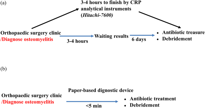
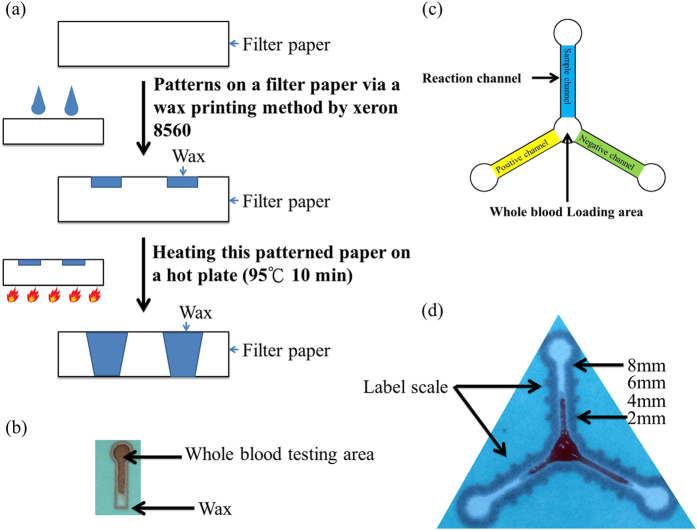
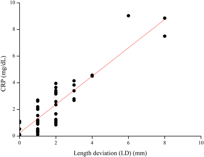
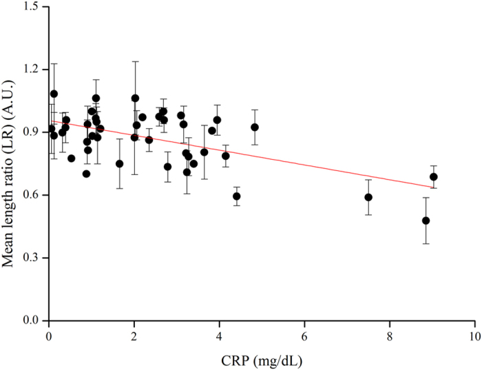
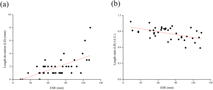
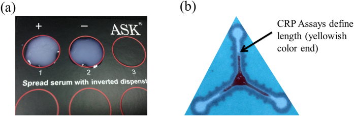
Here, we discuss the development of a paper-based diagnostic device that is inexpensive, portable, easy-to-use, robust, and capable of running simultaneous tests to monitor a relevant inflammatory protein for clinical diagnoses i.e. C-reactive protein (CRP). In this study, we first attempted to make a paper-based diagnostic device via the wax printing method, a process that was used in previous studies. This device has two distinct advantages: 1) reduced manufacturing and assay costs and operation duration via using wax printing method to define hydrophobic boundaries (for fluidic devices or general POC devices); and, 2) the hydrophilicity of filter paper, which is used to purify and chromatographically correct interference caused by whole blood components with a tiny amount of blood sample (only 5 μL). Diagnosis was based on serum stain length retained inside the paper channels of our device. This is a balanced function between surface tension and chromatographic force following immune reactions (CRP assays) with a paper-embedded biomarker.


Figures

Figure 1
Figure 1. A comparison between standard clinical analysis and paper-based CRP assay device analysis in osteomyelitis patients.
(a) The traditional process (Hitachi 7600) required approximately 3–4 hours to obtain results. (b) The paper-based process required approximately 5 minutes.
Figure 2
Figure 2. The fabrication and design of our paper-based CRP monitoring devices.
(a) Fabrication of the paper-based diagnostic device via wax printing. (b) An optical image of the paper-based diagnostic device with one channel for whole blood testing. (c) Schematic drawing of our paper-based diagnostic device designed for CRP assays (negative channel, sample channel, positive channel – a gradient for reagent concentration). Latex was loaded from the 3 outer circles for immobilization. Blood sample was loaded into the center circle for testing. (d) Actual picture of experiment result shows the profiles of one CRP assay on our paper-based diagnostic device (paper chromatography).
Figure 3
Figure 3. The length deviation (LD) results of paper-based device CRP analysis using diluted blood.
Visual determination (naked eye) of blood flow length for serum of blood samples spotted onto our paper-based device using a measuring stick (smallest unit of measure 1 mm) and CRP concentration as measured via conventional means (Hitachi 7600). The curve fitting to the data using the equation display Adj. R-Square: 0.775 and Pearson’s r: 0.883.
Figure 4
Figure 4. The length ratio (LR) results of paper-based device CRP analysis using diluted blood.
Visual examination of spotted serum length on our paper-based device. CRP concentration as measured via conventional means (Hitachi 7600). Samples number n = 3. The curve fitting to the data using the equation displays R-Square: 0.330, and Pearson’s r: −0.588.
Figure 5
Figure 5. The results of paper-based device analysis of ESR showing LD and LR in diluted blood.
We obtained sensitivity using a linear equation to generate an Orginpro 8.5 curve fitting. This figure shows the calibration plot for the length ratio of the results for the reagent compound (latex) reaction in our paper-based device compared to medical mainframe examinations of ESR and CRP data. (a) Patient samples indicating osteomyelitis inflammation by LD. (b) Patient samples indicating osteomyelitis inflammation by LR. The curve fitting to the data using the equation displays R-Square LD: 0.301 LR: 0.264, Pearson’s r LD: 0.563 LR: 0.533 and Residual Sum of Squares LD: 32109.263 LR: 0.351.
Figure 6
Figure 6. A comparison between commercial CRP assays and paper-based CRP devices.
(a) A display of positive and negative space when observing aggregated latex, (b) Each CRP assay channel defines stain length from serum (yellowish color) to calculate LD or LR.

References

    1. Gubala V., Harris L. F., Ricco A. J., Tan M. X. & Williams D. E. Point of care diagnostics: status and future. Anal. Chem. 84, 487–515 (2011). - PubMed
    1. Chin C. D., Linder V. & Sia S. K. Lab-on-a-chip devices for global health: past studies and future opportunities. Lab Chip 7, 41–57 (2007). - PubMed
    1. Sia S. K., Linder V., Parviz B. A., Siegel A. & Whitesides G. M. An integrated approach to a portable and low-cost immunoassay for resource-poor settings. Angew. Chem. 43, 498–502 (2004). - PubMed
    1. Daar A. S. et al.. Top ten biotechnologies for improving health in developing countries. Nat. Genet. 32, 229–232 (2002). - PubMed
    1. Yager P. et al.. Microfluidic diagnostic technologies for global public health. Nature 442, 412–418 (2006). - PubMed

Publication types