. 2016 Dec 2:6:38446.
doi: 10.1038/srep38446.

Disentangling the roles of cholesterol and CD59 in intermedilysin pore formation

Affiliations

Disentangling the roles of cholesterol and CD59 in intermedilysin pore formation

Courtney M Boyd et al. Sci Rep. .

Abstract

The plasma membrane provides an essential barrier, shielding a cell from the pressures of its external environment. Pore-forming proteins, deployed by both hosts and pathogens alike, breach this barrier to lyse target cells. Intermedilysin is a cholesterol-dependent cytolysin that requires the human immune receptor CD59, in addition to cholesterol, to form giant β-barrel pores in host membranes. Here we integrate biochemical assays with electron microscopy and atomic force microscopy to distinguish the roles of these two receptors in mediating structural transitions of pore formation. CD59 is required for the specific coordination of intermedilysin (ILY) monomers and for triggering collapse of an oligomeric prepore. Movement of Domain 2 with respect to Domain 3 of ILY is essential for forming a late prepore intermediate that releases CD59, while the role of cholesterol may be limited to insertion of the transmembrane segments. Together these data define a structural timeline for ILY pore formation and suggest a mechanism that is relevant to understanding other pore-forming toxins that also require CD59.


Figures

Figure 1
Figure 1. CD59 coordinates a specific geometry for ILY oligomeric prepores.
(a) Wildtype ILY was incubated with CD59-decorated liposomes; monomers and oligomers were separated by SDS-AGE and transferred to nitrocellulose. His-tagged toxin was detected using western blot analysis. The + or − denoted at the top of the panel indicates whether the toxin was incubated with or without cholesterol-containing liposomes (Chol) or cytoCD59. ILY only contains neither liposomes nor CD59. ILY and CD59 refer to the incubation of soluble proteins in the absence of membranes. Molecular weight marker is in the far left lane. (bd) Electron micrographs of negatively stained ILY oligomers formed on lipid monolayers. (b) ILY-cytoCD59 complexes on cholesterol containing monolayers. (c) ILY-cytoCD59 complexes formed on cholesterol-lacking monolayers. (d) ILY oligomers on cholesterol-rich monolayers formed in the absence of CD59. Two types of oligomers are shown in left and right panels. Scale bar, 50 nm.
Figure 2
Figure 2. CD59 triggers a collapse of the ILY prepore.
(a) Schematic of ILY structural transitions. ILY colored by domains (D1, blue; D2, yellow; D3, green; D4, red) and CD59 in purple. Top panel denotes the ILYTI-CD59 crystal structure (PDB ID: 4BIK). Bottom panel illustrates a model for the complex after collapse. (be) AFM images (top panel) and corresponding height data (bottom panel) for ILY-CD59 complexes on supported lipid bilayers. Height data is plotted as a histogram of the z measurements for each pixel in the image, with the inset showing a cross-section of the area highlighted by the blue dotted line. All bilayers contain cholesterol unless otherwise indicated by –Chol. Color scale, 15 nm. (b) cytoCD59 on a supported lipid bilayer. (c) ILYTI bound to a cytoCD59-decorated bilayer. (d) wildtype ILY bound a to cytoCD59-decorated bilayer. (e) Wildtype ILY bound to a cytoCD59-decorated bilayer lacking cholesterol.
Figure 3
Figure 3. CD59 releases an ILY late-prepore independent of cholesterol.
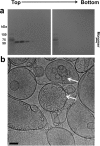
(a) Schematic illustrating the liposome flotation assay. Liposomes decorated with cytoCD59 were incubated with ILY and subjected to density centrifugation. Monomeric and oligomeric forms of the toxin in each fraction were separated by SDS-AGE and detected by western blot analysis. Samples containing cholesterol-rich liposomes and the ILYTI variant are shown in (b), while those in (c) contain wildtype ILY prepores formed on liposomes lacking cholesterol. Fractions from a single gradient span two gels (left and right). Directionality of fractionation is indicated by Top and Bottom above gels. The first lane in each gel contains molecular weight markers. (d) Cryo-electron micrograph of the sample analyzed in panel c showing ILY oligomeric prepores on liposomes (white arrows) and those released from the membrane (red arrows). Scale bar, 50 nm.
Figure 4
Figure 4. ILYIG forms an SDS-sensitive early prepore that remains membrane-bound.
(a) ILYIG was incubated with cytoCD59-decorated, cholesterol-containing liposomes and subjected to flotation through ficoll. Toxin within gradient fractions was analyzed by SDS-AGE and western blot. Fractions span two gels with direction of fractionation indicated by Top and Bottom. Molecular weight markers are in the first lane of each gel. (b) Cryo-electron micrograph of the sample analyzed in panel a showing SDS-sensitive oligomeric prepores on liposomes (white arrows). Scale bar, 50 nm.
Figure 5
Figure 5. Lytic activity of ILY variants.
(a) ILY crystal structure (PDB ID: 1S3R) with domains colored as in Fig. 2. Residues mutated in ILY variants shown as spheres. (b) ILY variants were incubated with calcein-containing liposomes decorated with cytoCD59, unless otherwise indicated by cytoCD59. All liposomes contain cholesterol unless otherwise indicated by –Cholesterol. Fluorescence measurements, expressed as a percent total lysis, were normalized against background and detergent burst vesicles. Error bars indicate standard deviations across three independent experiments. (c) Schematic illustrating the temporal roles of CD59 and cholesterol in ILY pore formation. ILY and CD59 are represented as space-filled models from the crystal structure of the complex (PDB ID: 4BIK). ILY D3 is in lilac; the remainder of the structure is in blue; CD59 is green. Soluble ILY is targeted to cholesterol-containing membranes (red sphere), whereby CD59 sets a specific geometry that defines the diameter of an oligomeric ILY prepore (blue 3D barrel). CD59 triggers the vertical collapse of this prepore, enabling the transmembrane segments of D3 (lilac) to approach the bilayer. CD59 is released from an SDS-resistant late prepore prior to membrane insertion, while cholesterol is required for the final membrane perforation step.

References

    1. Anderluh G. & Lakey J. H. Disparate proteins use similar architectures to damage membranes. Trends Biochem Sci 33, 482–490, doi: 10.1016/j.tibs.2008.07.004 (2008). - DOI - PubMed
    1. Parker M. W. & Feil S. C. Pore-forming protein toxins: from structure to function. Prog Biophys Mol Biol 88, 91–142, doi: 10.1016/j.pbiomolbio.2004.01.009 (2005). - DOI - PubMed
    1. Hotze E. M. et al.. Identification and characterization of the first cholesterol-dependent cytolysins from Gram-negative bacteria. Infect Immun 81, 216–225, doi: 10.1128/IAI.00927-12 (2013). - DOI - PMC - PubMed
    1. Giddings K. S., Johnson A. E. & Tweten R. K. Redefining cholesterol’s role in the mechanism of the cholesterol-dependent cytolysins. Proc Natl Acad Sci USA 100, 11315–11320, doi: 10.1073/pnas.2033520100 (2003). - DOI - PMC - PubMed
    1. Farrand A. J., LaChapelle S., Hotze E. M., Johnson A. E. & Tweten R. K. Only two amino acids are essential for cytolytic toxin recognition of cholesterol at the membrane surface. Proc Natl Acad Sci USA 107, 4341–4346, doi: 10.1073/pnas.0911581107 (2010). - DOI - PMC - PubMed

Publication types