. 2016 Dec 2:6:38199.
doi: 10.1038/srep38199.

Ascl3 transcription factor marks a distinct progenitor lineage for non-neuronal support cells in the olfactory epithelium

Affiliations

Ascl3 transcription factor marks a distinct progenitor lineage for non-neuronal support cells in the olfactory epithelium

Pei-Lun Weng et al. Sci Rep. .

Abstract

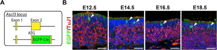
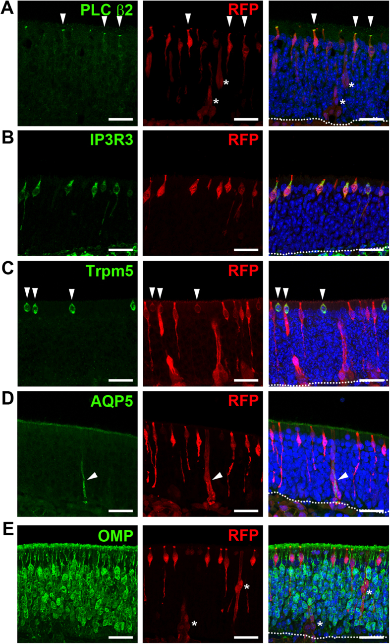
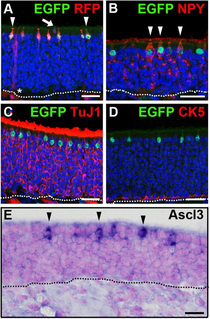
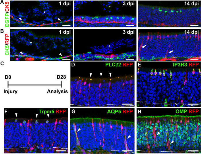
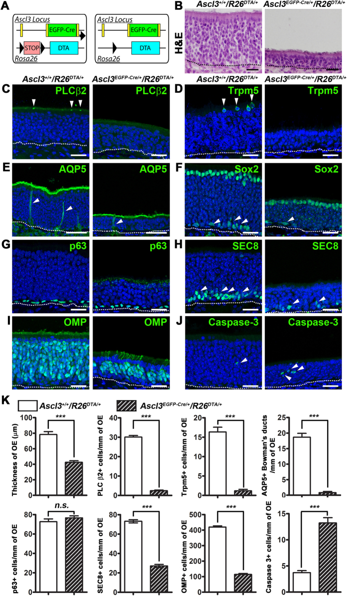
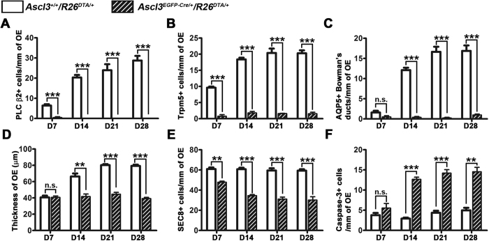
The olfactory epithelium (OE) is composed of olfactory sensory neurons (OSNs), sustentacular supporting cells, and several types of non-neuronal cells. Stem and progenitor cells are located basally, and are the source of all cell types needed to maintain OE homeostasis. Here, we report that Ascl3, a basic helix-loop-helix transcription factor, is expressed in the developing OE. Lineage tracing experiments demonstrate that the non-neuronal microvillar cells and Bowman's glands are exclusively derived from Ascl3+ progenitor cells in the OE during development. Following chemically-induced injury, Ascl3 expression is activated in a subset of horizontal basal cells (HBCs), which repopulate all microvillar cells and Bowman's glands during OE regeneration. After ablation of Ascl3-expressing cells, the OE can regenerate, but lacks the non-neuronal microvillar and Bowman's gland support cells. These results demonstrate that Ascl3 marks progenitors that are lineage-committed strictly to microvillar cells and Bowman's glands, and highlight the requirement for these cell types to support OE homeostasis.


Figures

Figure 1
Figure 1. Ascl3 is expressed in the OE during embryonic development.
(A) The Ascl3 gene locus includes 2 exons. In Ascl3EGFP-Cre/+ mice, the non-coding exon1 was maintained, and a fusion cassette encoding EGFP-Cre replaced the entire Ascl3 coding sequence in exon 2, through homologous recombination. (B) OE was isolated from Ascl3EGFP-Cre/+ mice at E12.5, E14.5, E16.5 and E18.5 of development. Immunohistochemistry was performed using antibodies to EGFP and TuJ1. Ascl3-EGFP+ cells (arrowheads) remain apically localized and are not colocalized with the marker for immature neurons, TuJ1, during embryonic development. Dotted line indicates basal lamina. Nuclei are stained by DAPI (blue). Scale bars: 25 μm.
Figure 2
Figure 2. Ascl3-expressing cells are precursors of microvillar cells and Bowman’s glands.
Immunohistochemistry was performed on OE isolated from Ascl3EGFP-Cre/+/R26tdTomato/+ mice (2 months), using antibodies to tdTomato (RFP) and (A) PLC β2, which marks the apical microvilli of microvillar cells, (B) IP3R3, (C) Trpm5, (D) AQP5 and (E) OMP. RFP expression colocalized with microvillar cell markers: PLC β2 (arrowheads), IP3R3 and Trpm5 (arrowheads) and Bowman’s glands markers: AQP5 (arrowheads). (E) No colocalization was detected between RFP and the mature OSN marker OMP. White asterisks mark Bowman’s gland duct cells. Dotted line indicates basal lamina. Nuclei are stained by DAPI (blue). Scale bars: 25 μm.
Figure 3
Figure 3. Ascl3 is expressed in the adult olfactory epithelium.
(A) Immunohistochemistry was performed on sections of OE isolated from Ascl3EGFP-Cre/+/R26tdTomato/+ mice (2 months) using antibodies to EGFP and RFP. (BD) Immunohistochemistry was performed on sections of OE isolated from Ascl3EGFP-Cre/+ mice (2 months), using antibodies to EGFP and (B) NPY, (C) TuJ1, (D) CK5. Arrowheads indicate co-localization of EGFP and NPY. (E) In situ hybridization for Ascl3 mRNA in the OE of 2-month old mice (arrowheads). Dotted line indicates basal lamina. Nuclei are stained by DAPI (blue). Scale bars: (AD), 25 μm. (E), 20 μm.
Figure 4
Figure 4. Ascl3-expressing cells regenerate microvillar cells and Bowman’s gland/ducts after injury.
(A) Immunohistochemistry with antibodies to EGFP and CK5 on sections of OE isolated from Ascl3EGFP-Cre/+ after methimazole injection. At day one (1 dpi) post injury, Ascl3-expressing cells marked by EGFP co-localize with HBCs marked by CK5 expression (arrowheads). EGFP+ cells gradually migrate away from the basal layer toward the apical OE at 3 and 14 dpi. (B) Lineage tracing in Ascl3EGFP-Cre/+/R26tdTomato/+mice after injury. Antibody to RFP detected Ascl3-expressing cells co-localized with HBCs marked by CK5 antibodies at 1 dpi (arrowheads). RFP-labeled cells become apically localized at 3 and 14 dpi. White asterisk marks delaminated OE tissue. (C) Ascl3EGFP-Cre/+/R26tdTomato/+ mice were treated with methimazole and OE was analyzed after 28 days. (DH) Confocal images of OE sections stained with antibodies to RFP and (D) PLC β2, (E) IP3R3, (F) Trpm5, (G) AQP5 and (H) OMP. RFP expression colocalized with PLC β2, IP3R3, Trpm5 and AQP5 (arrowheads). (H) No colocalization was detected between RFP and OMP. Arrowheads indicate Bowman’s gland ducts within the OE. Dotted line indicates basal lamina. Nuclei are stained by DAPI (blue). Scale bars: 25 μm.
Figure 5
Figure 5. Ablation of Ascl3-expressing cells results in absence of microvillar cells and Bowman’s glands, and decreases GBCs and mature OSNs.
(A) Ascl3EGFP-Cre/+/R26DTA/+ mice, DTA expression is activated only in the Ascl3-expressing cells. OE was isolated from Ascl3+/+/R26DTA/+ and Ascl3EGFP-Cre/+/R26DTA/+ mice at 2 months of age. (B) H&E staining showed a significant decrease in thickness of the OE in the Ascl3EGFP-Cre/+/R26DTA/+ mice compared to Ascl3+/+/R26DTA/+ mice. (C) Staining with antibody to PLC β2 (arrowheads) in OE from Ascl3+/+/R26DTA/+ mice and Ascl3EGFP-Cre/+/R26DTA/+mice. (D) Trpm5-positive microvillar cells are present at the apical surface of the OE (arrowheads) in Ascl3+/+/R26DTA/+ mice, but not detected in OE of Ascl3EGFP-Cre/+/R26DTA/+mice. (E) Antibodies to aquaporin 5 (AQP5) mark the apical surface of the duct cells in the Bowman’s glands extending through the OE in Ascl3+/+/R26DTA/+ mice (arrowheads). Ducts cells of the Bowman’s glands were only rarely observed in OE from Ascl3EGFP-Cre/+/R26DTA/+ mice (arrowhead). (F) Antibodies to Sox2 revealed no difference in number of sustentacular cells at the apical surface of the OE between mice of the two genotypes. The number of Sox2+ GBCs was decreased (arrowheads). (G) p63+ HBC numbers are not changed in Ascl3EGFP-Cre/+/R26DTA/+ mice. (H) SEC8 antibody labels GBCs near the basal layer of the OE in Ascl3+/+/R26DTA/+ mice (arrowheads). Significantly fewer GBCs were detected in OE of Ascl3EGFP-Cre/+/R26DTA/+mice. (I) Labeling with antibody to OMP showed a significant decrease in the number of labeled mature OSNs in Ascl3EGFP-Cre/+/R26DTA/+ mice compared to controls. (J) Antibodies to active caspase-3 showed an increase in number of apoptotic cells in the OE of Ascl3EGFP-Cre/+/R26DTA/+ mice. (K) Quantified results show significant decrease in the thickness of OE and the numbers of PLC β2+ and Trpm5+ microvillar cells, AQP5+ duct cells of the Bowman gland, but no difference in number of p63+ HBCs in the Ascl3EGFP-Cre/+/R26DTA/+mice. Quantification also showed a significant decrease in SEC+ GBCs and OMP+ mature OSNs in the Ascl3EGFP-Cre/+/R26DTA/+mice. In addition, an increase of caspase-3+ cells was observed in Ascl3EGFP-Cre/+/R26DTA/+mice (arrowheads). N ≥ 3 for Ascl3+/+/R26DTA/+ and Ascl3EGFP-Cre/+/R26DTA/+. ***P < 0.001. n.s., No significance. Data are shown with mean ± SEM. Dotted line indicates basal lamina. Nuclei are stained by DAPI (blue). Scale bars: (A), 20 μm. (BH), 25 μm.
Figure 6
Figure 6. Time course of OE regeneration in the absence of non-neuronal support cells.
Quantification of PLC β2+, Trpm5+ microvillar cells, duct cells of AQP5+ Bowman’s glands, OE thickness, SEC8+ GBCs and caspase-3+ apoptotic cells from Ascl3+/+/R26DTA/+ and Ascl3EGFP-Cre/+/R26DTA/+ mice at days 7, 14, 21, 28 post-injury. (AC) Quantified results showed significantly reduced numbers of PLC β2+ and Trpm5+ microvillar cells, AQP5+ Bowman gland ducts in the Ascl3-DTA mice at days 7, 14, 21, 28 post-injury. (D) Decrease in the thickness of OE was detected from day 14 dpi during regeneration. (E) Decrease of SEC8+ GBCs was observed in the Ascl3-DTA mice at days 7, 14, 21, 28 post-injury. (F) More caspase-3+ apoptosis cells were observed in the Ascl3-DTA mice at days 7, 14, 21, 28 post-injury. N ≥ 3 for control and Ascl3-DTA mice. *P < 0.05, **P < 0.01, ***P < 0.001. n.s., No significance. Data are shown with mean ± SEM.

Similar articles

Cited by

References

    1. Whitby-Logan G. K., Weech M. & Walters E. Zonal expression and activity of glutathione S-transferase enzymes in the mouse olfactory mucosa. Brain Res 995, 151–7 (2004). - PubMed
    1. Holbrook E. H., Szumowski K. E. & Schwob J. E. An immunochemical, ultrastructural, and developmental characterization of the horizontal basal cells of rat olfactory epithelium. J Comp Neurol 363, 129–46 (1995). - PubMed
    1. Solbu T. T. & Holen T. Aquaporin Pathways and Mucin Secretion of Bowman’s Glands Might Protect the Olfactory Mucosa. Chemical Senses (2011). - PubMed
    1. Hansen A. & Finger T. E. Is TrpM5 a reliable marker for chemosensory cells? Multiple types of microvillous cells in the main olfactory epithelium of mice. BMC Neuroscience 9, 1–12 (2008). - PMC - PubMed
    1. Lin W., Ezekwe E. A. Jr., Zhao Z., Liman E. R. & Restrepo D. TRPM5-expressing microvillous cells in the main olfactory epithelium. BMC Neurosci 9, 114 (2008). - PMC - PubMed

Publication types

MeSH terms

Substances