Review
. 2017 Jan;10(1):65-79.
doi: 10.1080/17474086.2016.1268047. Epub 2016 Dec 12.

Mitochondria and Iron: current questions

Affiliations
Review

Mitochondria and Iron: current questions

Bibbin T Paul et al. Expert Rev Hematol. 2017 Jan.

Erratum in

  • Erratum.
    [No authors listed] [No authors listed] Expert Rev Hematol. 2017 Mar;10(3):275. doi: 10.1080/17474086.2017.1287270. Epub 2017 Feb 8. Expert Rev Hematol. 2017. PMID: 28245742 No abstract available.

Abstract

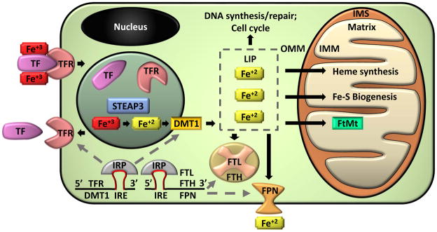
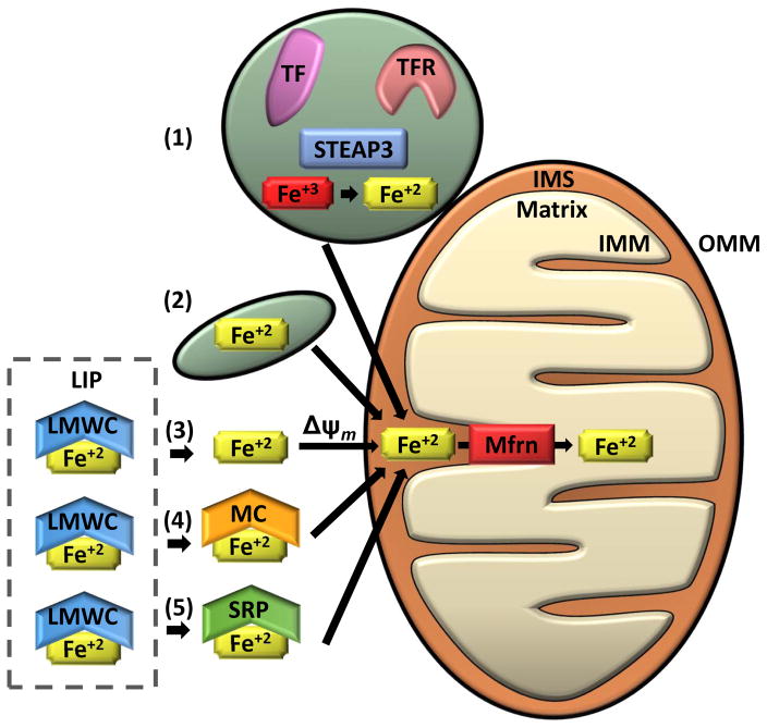
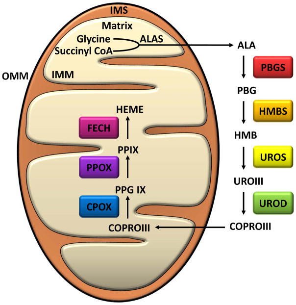
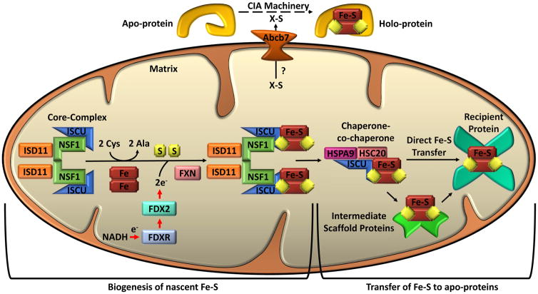
Mitochondria are cellular organelles that perform numerous bioenergetic, biosynthetic, and regulatory functions and play a central role in iron metabolism. Extracellular iron is taken up by cells and transported to the mitochondria, where it is utilized for synthesis of cofactors essential to the function of enzymes involved in oxidation-reduction reactions, DNA synthesis and repair, and a variety of other cellular processes. Areas covered: This article reviews the trafficking of iron to the mitochondria and normal mitochondrial iron metabolism, including heme synthesis and iron-sulfur cluster biogenesis. Much of our understanding of mitochondrial iron metabolism has been revealed by pathologies that disrupt normal iron metabolism. These conditions affect not only iron metabolism but mitochondrial function and systemic health. Therefore, this article also discusses these pathologies, including conditions of systemic and mitochondrial iron dysregulation as well as cancer. Literature covering these areas was identified via PubMed searches using keywords: Iron, mitochondria, Heme Synthesis, Iron-sulfur Cluster, and Cancer. References cited by publications retrieved using this search strategy were also consulted. Expert commentary: While much has been learned about mitochondrial and its iron, key questions remain. Developing a better understanding of mitochondrial iron and its regulation will be paramount in developing therapies for syndromes that affect mitochondrial iron.

Keywords: Iron; cancer; heme synthesis; iron trafficking; iron-sulfur cluster; mitochondria.


Conflict of interest statement

Declaration of Interest

DH Manz received grant T90 DE021989, SV Torti has received grant R01 CA188025, and FM Torti has received grant R01 CA171101, from the US National Institutes of Health. The authors have no other relevant affiliations or financial involvement with any organization or entity with a financial interest in or financial conflict with the subject matter or materials discussed in the manuscript apart from those disclosed.

Figures

Figure 1
Figure 1. Cellular Iron Homeostasis
Ferric iron (Fe+3) is transported through the circulation bound to a carrier protein transferrin (TF). Diferric-TF binds to transferrin receptors (TFR) on the plasma membrane. The complex is endocytosed and a reduction in pH results in dissociation of iron from the complex. TFR and TF are recycled back to the cell surface or into the circulation, respectively. Within the endosome, ferric iron is converted into ferrous iron (Fe+2) by a metalloreductase Six-Transmembrane Epithelial Antigen of Prostate 3 (STEAP3). Divalent metal transporter 1 (DMT1) transports ferrous iron out of the endosome and into the cytoplasm were it enters a transient pool of metabolically active iron known as the labile iron pool (LIP). LIP iron can be utilized for cellular processes such as DNA synthesis, repair, and cell cycling. Alternatively, excess LIP iron can be stored in ferritin, a hetero-polymer of ferritin heavy (FTH) and light (FTL) chains, or can be exported from the cell by ferroportin (FPN). Ferrous iron may also be transported to the mitochondria. Mitochondria are organelles with an outer (OMM) and inner (IMM) membrane that separate an intermembrane space (IMS). Iron is imported through the IMM to the innermost compartment, the mitochondrial matrix, where it can be utilized for heme synthesis, iron-sulfur cluster (Fe-S) biogenesis, or can be stored in mitochondrial ferritin (FtMt). Cellular iron homeostasis is regulated by iron regulatory proteins (IRP). IRPs bind to iron response elements (IRE) in the mRNA of iron regulating proteins. IRP/IRE binding to the 3′ end of mRNA, which occurs in TFR and DMT1, stabilizes the mRNA and enhances translation. IRP/IRE binding to the 5′ end of mRNA, which occurs in FTH, FTL, and FPN, blocks the binding of translational machinery and reduces mRNA translation.
Figure 2
Figure 2. Mechanisms of Iron transport to the Mitochondria
Several mechanisms for transporting iron into the mitochondria have been suggested. (1) “Kiss and Run” mechanism: Ferric iron (Fe+3) taken up by endocytosis of the transferrin receptor (TFR)-transferrin (TF) complex is likely reduced to ferrous iron (Fe+2) by a metalloreductase Six-Transmembrane Epithelial Antigen of Prostate 3 (STEAP3) and directly transported from the endosome to the mitochondria without entering the cytosol. The “Kiss and Run” mechanism has only been demonstrated in erythroid cells. (2) Non-transferrin bound iron may be taken up via fluid-phase endocytosis by a partially solvent occluded mechanism. This method of iron transport was proposed from data from murine cardiac cells. (3–6) Cytosolic labile iron (LIP) is thought to be bound by low molecular weight complexes (LMWC) and is presumed to be the source of iron delivered to the mitochondria through the cytosol. (3) Iron has been shown to be transported into the mitochondria via a mitochondrial membrane potential-dependent manner (Δψm) in yeast and rat hepatocytes. (4) Alternatively, iron may be trafficked to the mitochondria by high molecular weight complexes such as metallochaperones (MC) or (5) siderophores (SRP). Once iron is transported through the outer mitochondrial membrane (OMM) and into the intermembrane space (IMS), it is transferred through the inner mitochondrial membrane (IMM) and into the mitochondrial matrix by mitoferrins (Mfrn) and associated regulatory proteins (i.e. ATP Binding Cassette Subfamily B Member 10 [Abcb10] in erythroid cells).
Figure 3
Figure 3. Heme Biosynthesis in Mammals
Enzymatic steps and intermediates formed during the biosynthesis of Heme from 5-aminolevelunic acid. Heme synthesis in the mitochondria begins in the mitochondrial matrix. Glycine and succinyl CoA are condensed by 5-aminolevulinate synthase to 5-aminolevelunic acid (ALA). ALA is exported to the cytosol through the inner mitochondrial membrane (IMM), the intermembrane space (IMS), and the outer mitochondrial membrane (OMM). In the cytosol, ALA undergoes four steps of enzymatic modification to generate coproporphyrinogen III (COPRO III). COPRO III is transported back to mitochondrial matrix where the last three enzymatic reactions occur. Abbreviations: ALA, 5-aminolevelunic acid; ALAS, 5-Aminolevulinate synthase; PBG, Porphobilinogen; PBGS, Porphobilinogen synthase; HMB, Hydroxymethylbilane; HMBS, Hydroxymethylbilane synthase; UROIII, Uroporphyrinogen III; UROS, Uroporphyrinogen III synthase; COPRO III, Coproporphyrinogen III; UROD, Uroporphyrinogen decarboxylase; PPG IX, Protoporphyrinogen IX; CPOX, Coproporphyrinogen oxidase; PP IX, Protoporphyrin IX; PPOX, Protoporphyrinogen oxidase; FECH, Ferochelatase.
Figure 4
Figure 4. Iron-Sulfur Cluster Biogenesis in Mammals
Separate pathways are involved in mitochondrial iron-sulfur cluster biogenesis and cytosolic iron-sulfur cluster biogenesis, but both depend on mitochondria. Mitochondrial iron-sulfur cluster biogenesis begins in the mitochondrial matrix with the nascent iron-sulfur cluster (Fe-S) assembly core-complex. This core-complex consists of a cysteine desulfurase (NFS1) dimer, two monomers of the scaffold protein ISCU, two regulatory proteins ISD11, and frataxin (FXN). NFS1 abstracts sulfur from cysteine (Cys), converting it to alanine (Ala), for nascent Fe-S cluster assembly. The stability of NFS1 depends on its binding partner ISD11. Iron entry and cysteine desulfurase activity are possibly regulated by FXN. Iron and electrons (e-) provided by NADH, ferredoxin reductase (FDXR), and ferredoxin (FDX2) facilitate the assembly of the nascent ISC on the ISCU. The ISCU-bound nascent Fe-S is transferred to recipient apo-proteins or intermediate carrier proteins by a dedicated chaperone-co-chaperone (HSPA9/HSC20) system. For cytosolic iron-sulfur cluster biogenesis, an undetermined sulfur-containing compound (X-S) is exported from the mitochondrial to the cytosol by ATP-binding cassette sub-family B member 7 (ABCB7). Cytoplasmic Fe-S assembly (CIA) machinery then facilitates the loading of Fe-S onto apo-proteins to form holo-proteins. Numerous proteins are involved in the transfer of Fe-S to specific proteins; see text for discussion.

References

    1. Lane N, Martin W. The energetics of genome complexity. Nature. 2010;467(7318):929–934. - PubMed
    1. Bayrhuber M, Meins T, Habeck M, et al. Structure of the human voltage-dependent anion channel. Proceedings of the National Academy of Sciences of the United States of America. 2008;105(40):15370–15375. - PMC - PubMed
    1. Kuhlbrandt W. Structure and function of mitochondrial membrane protein complexes. BMC biology. 2015;13:89. - PMC - PubMed
    1. Friedman JR, Nunnari J. Mitochondrial form and function. Nature. 2014;505(7483):335–343. - PMC - PubMed
    1. Johnson LV, Walsh ML, Chen LB. Localization of mitochondria in living cells with rhodamine 123. Proceedings of the National Academy of Sciences of the United States of America. 1980;77(2):990–994. - PMC - PubMed