

Validation of antibody reagents for mucin analysis in chronic inflammatory airway diseases
- PMID: 27911216
- PMCID: PMC5297535
- DOI: 10.1080/19420862.2016.1264551
Validation of antibody reagents for mucin analysis in chronic inflammatory airway diseases
Abstract
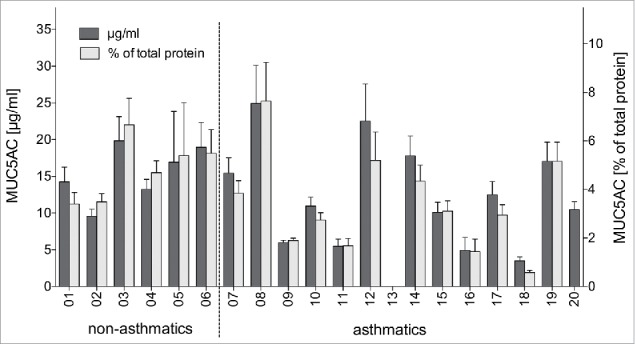
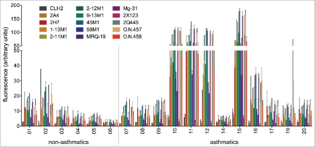
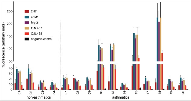
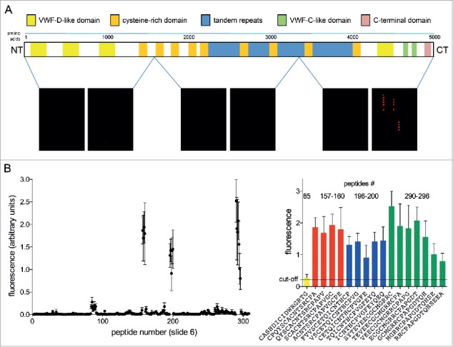
In chronic inflammatory airway diseases, mucins display disease-related alterations in quantity, composition and glycosylation. This opens the possibility to diagnose and monitor inflammatory airway disorders and their exacerbation based on mucin properties. For such an approach to be reasonably versatile and diagnostically meaningful, the mucin of interest must be captured in a reliable, patient-independent way. To identify appropriate mucin-specific reagents, we tested anti-mucin antibodies on mucin-content-standardized, human bronchoalveolar lavage fluid samples in immunoblot assays. All commercially available monoclonal antibodies against the major airway mucin MUC5AC were screened, except for those with known specificity for carbohydrates, as glycosylation patterns are not mucin-specific. Our results indicated considerable inter-patient and inter-antibody variability in mucin recognition for all antibodies and samples tested. The best results in terms of signal strength and reproducibility were obtained with antibodies Mg-31, O.N.457 and 45M1. Additional epitope mapping experiments revealed that only one of the antibodies with superior binding to MUC5AC recognized linear peptide epitopes on the protein backbone.
Keywords: Asthma; COPD; MUC5AC; chronic inflammatory airway diseases; mucin capturing; mucin quantification; mucus.
Figures
References
-
- Rose MC, Voynow JA. Respiratory tract mucin genes and mucin glycoproteins in health and disease. Physiol Rev 2006; 86:245-78; PMID:16371599; http://dx.doi.org/10.1152/physrev.00010.2005 - DOI - PubMed
-
- Thornton DJ, Rousseau K, McGuckin MA. Structure and function of the polymeric mucins in airways mucus. Annu Rev Physiol 2008; 70:459-86; PMID:17850213; http://dx.doi.org/10.1146/annurev.physiol.70.113006.100702 - DOI - PubMed
-
- Rogan MP, Geraghty P, Greene CM, O'Neill SJ, Taggart CC, McElvaney NG. Antimicrobial proteins and polypeptides in pulmonary innate defence. Respir Res 2006; 7:29; PMID:16503962; http://dx.doi.org/10.1186/1465-9921-7-29 - DOI - PMC - PubMed
-
- Rose MC. Mucins: structure, function, and role in pulmonary diseases. Am J Physiol 1992; 263:L413-29; PMID:1415719. - PubMed
-
- Rogers DF. Airway mucus hypersecretion in asthma: an undervalued pathology? Curr Opin Pharmacol 2004; 4:241-50; PMID:15140415; http://dx.doi.org/10.1016/j.coph.2004.01.011 - DOI - PubMed
Publication types
MeSH terms
Substances
LinkOut - more resources
Full Text Sources
Other Literature Sources