Review
. 2017 Jan;6(1):10.1002/wdev.216.
doi: 10.1002/wdev.216. Epub 2016 Dec 1.

Neural plasticity across the lifespan

Affiliations
Review

Neural plasticity across the lifespan

Jonathan D Power et al. Wiley Interdiscip Rev Dev Biol. 2017 Jan.

Abstract

An essential feature of the brain is its capacity to change. Neuroscientists use the term 'plasticity' to describe the malleability of neuronal connectivity and circuitry. How does plasticity work? A review of current data suggests that plasticity encompasses many distinct phenomena, some of which operate across most or all of the lifespan, and others that operate exclusively in early development. This essay surveys some of the key concepts related to neural plasticity, beginning with how current patterns of neural activity (e.g., as you read this essay) come to impact future patterns of activity (e.g., your memory of this essay), and then extending this framework backward into more development-specific mechanisms of plasticity. WIREs Dev Biol 2017, 6:e216. doi: 10.1002/wdev.216 For further resources related to this article, please visit the WIREs website.


Figures

Figure 1
Figure 1
Neurons, their parts, their actual complexity. Top, a schematic view of several neurons, illustrating the major parts of neurons and the direction of information flow. Bottom, microscopic images of a single neuron. This neuron has extensive dendrites with thousands of spines representing possible inputs from other neurons. The neuron also has a prominent cell body, and a smooth axonal process that leads down and left from the cell body. Each dendritic section may receive inhibitory and excitatory signals, and the neuron only fires if excitatory signals, within a small time window, sufficiently outweigh inhibitory signals. Images of neurons courtesy of Austen Milnerwood and the Centre for Applied Neurogenetics at the University of British Columbia. With permission from Austen Milnerwood of the Centre for Applied Neurogenetics, University of British Columbia.
Figure 2
Figure 2
Spike-timing-dependent tuning of synaptic strength. At left, a 3-neuron system in an initial state. If the purple neuron fires milliseconds before the black neuron, the synapse is selectively strengthened. If the purple neuron fires independently of the black neuron, the synapse is unaltered. If the purple neuron fires milliseconds after the black neuron, the synapse is selectively weakened.
Figure 3
Figure 3
Plasticity in adult cortical finger representations in the monkey. At top, a monkey brain is shown where the strip indicates the primary somatosensory area. This area has a map of the body, part of which is a map of the hand. The representation of the tips of the second digit before and after intense and prolonged stimulation is shown in gray shading. Stimulation selectively enlarges the representation of the manipulated skin. Modified from Jenkins et al.
Figure 4
Figure 4
Ocular dominance columns plasticity in macaques. Sections through the visual cortex of macaques. Radiolabeled tracer (white) was injected into one eye prior to sectioning, labeling neurons receiving input from that eye. At left, a normal monkey exhibits ocular dominance columns of equal size. At right, a monkey with the right eye occluded since 2 weeks of age has much larger left eye representations (white, unoccluded) than right eye representations (black, occluded). Modified from Hubel et al..
Figure 5
Figure 5
Signal gradients influence expression of transcription factors that regulate cortical area patterning. At top, 4 transcription factors present in gradients that function in establishing cortical patterning in the mouse brain. At bottom left, a diagrammatic illustration of several cortical areas in a normal mouse. At bottom right, the patterning in “knock-out” (KO) mice in which various transcription factors are no longer expressed. Loss of these factors greatly alters the layout of cortical areas in the knock-out mice. V1 denotes visual cortex, A1 auditory cortex, S1 somatosensory cortex, and F/M motor cortex. A denotes anterior, P posterior, L lateral, M medial. Modified from O'Leary et al..

References

    1. Kandel ER, Schwartz JH, Jessell TM, Siegelbaum SA, Hudspeth AJ. Principles of Neural Science. 5th. New York: McGraw-Hill Professional; 2012.
    1. Hebb DO. The Organization of Behavior: A Neuropsychological Theory. New York: John Wiley and Sons, Inc; 1949.
    1. Feldman DE. The spike-timing dependence of plasticity. Neuron. 2012;75:556–571. - PMC - PubMed
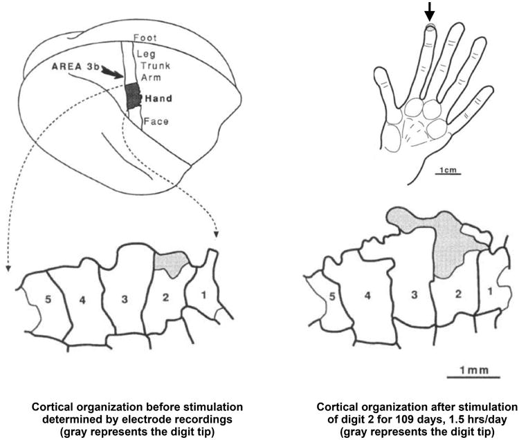
    1. Merzenich MM, Kaas JH, Wall J, Nelson RJ, Sur M, Felleman D. Topographic reorganization of somatosensory cortical areas 3b and 1 in adult monkeys following restricted deafferentation. Neuroscience. 1983;8:33–55. - PubMed
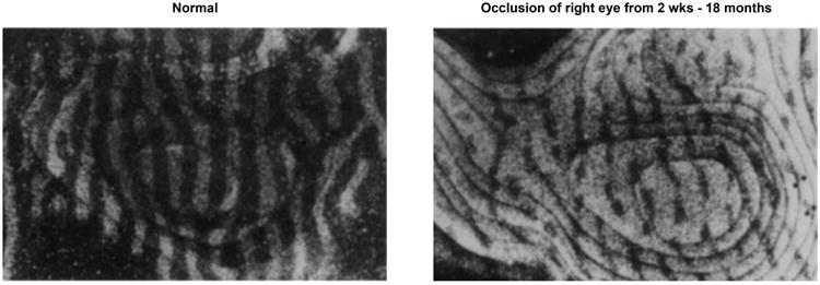
    1. Clark SA, Allard T, Jenkins WM, Merzenich MM. Receptive fields in the body-surface map in adult cortex defined by temporally correlated inputs. Nature. 1988;332:444–445. - PubMed

Further Reading

    1. Sammons RP, Keck T. Adult plasticity and cortical organization after peripheral lesions. Curr Opin Neurobiol. 2015;35:136–141. - PubMed
    1. Sengpiel F. Plasticity of the visual cortex and treatment of amblyopia. Curr Biol. 2014;24:R936–940. - PubMed
    1. Sur M, Nagakura I, Chen N, Sugihara H. Mechanisms of plasticity in the developing and adult visual cortex. Prog Brain Res. 2013;207:243–254. - PubMed
    1. Kerschensteiner D. Spontaneous network activity and synaptic development. Neuroscientist. 2013;20:272–290. - PMC - PubMed

Publication types

LinkOut - more resources