Review
. 2016 Oct 15;44(5):1253-1263.
doi: 10.1042/BST20160116.

The PLK4-STIL-SAS-6 module at the core of centriole duplication

Affiliations
Review

The PLK4-STIL-SAS-6 module at the core of centriole duplication

Christian Arquint et al. Biochem Soc Trans. .

Abstract

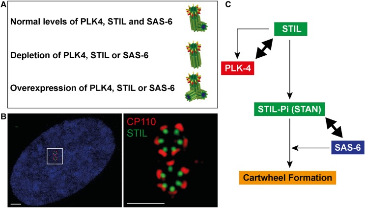
Centrioles are microtubule-based core components of centrosomes and cilia. They are duplicated exactly once during S-phase progression. Central to formation of each new (daughter) centriole is the formation of a nine-fold symmetrical cartwheel structure onto which microtubule triplets are deposited. In recent years, a module comprising the protein kinase polo-like kinase 4 (PLK4) and the two proteins STIL and SAS-6 have been shown to stay at the core of centriole duplication. Depletion of any one of these three proteins blocks centriole duplication and, conversely, overexpression causes centriole amplification. In this short review article, we summarize recent insights into how PLK4, STIL and SAS-6 co-operate in space and time to form a new centriole. These advances begin to shed light on the very first steps of centriole biogenesis.

Keywords: PLK4; SAS-6; cartwheel formation; centriole duplication; centrosomes.


Figures

Figure 1.
Figure 1.. The PLK4-STIL-SAS-6 centriole duplication module.
(A) Abundance of PLK4, STIL or SAS-6 controls centriole numbers. Under physiological conditions, normal levels of PLK4, STIL and SAS-6 ensure the formation of only one procentriole per mother centriole (upper row). In contrast, depletion of either protein inhibits centriole formation (middle row) and, conversely, overexpression triggers the formation of several procentrioles per mother centriole (lower row). (B) Super-resolution immunofluorescence images illustrate the (near-) simultaneous formation of several procentrioles around each of the two mother centrioles of a U2OS cell conditionally overexpressing PLK4 [43]. Centrioles are stained for STIL (green) and CP110 (a marker for the distal ends; red). The right panel shows a ×10 magnification of the area marked in the left panel (DNA is shown in blue). Error bars denote 2 μM (left image) and 1 μM (right image). (C) Highly simplified scheme summarizing key interactions between components of the PLK4–STIL–SAS-6 module during cartwheel formation (for more detailed information see text and Figures 2 and 3).
Figure 2.
Figure 2.. Precise interplay of PLK4, STIL and SAS-6 at the core of centriole duplication.
(A) Illustration of the co-operation between PLK4, STIL and SAS-6. The schematic emphasizes the interaction between the STIL CC domain and both the PLK4 PB3 and the L1 linker region. This interaction then triggers phosphorylation of key residues within the STIL STAN domain. Phosphorylation of the STIL STAN domain in turn enables recruitment of SAS-6. HD, head domain; CC, coiled coil; CAT, catalytic domain; L, linker; PB, polo-box; STAN, STIL/Ana2 domain. (B) Summary of PLK4 phosphorylation sites within STIL, as mapped by in vitro assays. Sites that have also been observed in vivo by mass spectrometry (www.phosphosite.org) [101] are marked in red. A sequence alignment of the STAN domain compiles several vertebrate STIL and Drosophila Ana2 sequences. Two PLK4 phosphorylation sites within this region (S1108 and S1116) are highlighted in bold, because they were shown to be phosphorylated by PLK4 in vivo by the use of site-specific phospho-antibodies [93]. PRXXPXP, CPAP-binding motif; CC, coiled coil; STAN, STIL/Ana2 domain; KEN, destruction motif (recognized by APC/CCdh [1]) important for STIL degradation.
Figure 3.
Figure 3.. How PLK4, STIL and SAS-6 initiate centriole duplication.
(A) Hypothetical model summarizing the first steps in centriole duplication. For further explanation, see text. (B) Detail of the above model, with focus on the release of PLK4 autoinhibition upon binding of STIL CC to both PB3 and L1 linker domains of PLK4, and the phosphorylation of the STIL STAN domain. Note that binding of STIL CC to L1 may mask the DSG motif, thereby protecting PLK4 from degradation.

References

    1. Lüders J. and Stearns T. (2007) Microtubule-organizing centres: a re-evaluation. Nat. Rev. Mol. Cell Biol. 8, 161–167 doi:10.1038/nrm2100 - DOI - PubMed
    1. Bettencourt-Dias M. and Glover D.M. (2007) Centrosome biogenesis and function: centrosomics brings new understanding. Nat. Rev. Mol. Cell Biol. 8, 451–463 doi:10.1038/nrm2180 - DOI - PubMed
    1. Bornens M. (2012) The centrosome in cells and organisms. Science 335, 422–426 doi:10.1126/science.1209037 - DOI - PubMed
    1. Arquint C., Gabryjonczyk A.-M. and Nigg E.A. (2014) Centrosomes as signalling centres. Philos. Trans. R Soc B Biol.l Sci. 369, 20130464 doi:10.1098/rstb.2013.0464 - DOI - PMC - PubMed
    1. Gönczy P. (2012) Towards a molecular architecture of centriole assembly. Nat. Rev. Mol. Cell Biol. 13, 425–435 doi:10.1038/nrm3373 - DOI - PubMed

MeSH terms

Substances

LinkOut - more resources