. 2016 Dec 13;113(50):14432-14437.
doi: 10.1073/pnas.1604099113. Epub 2016 Nov 28.

Phosphatidylinositol 3-kinase inhibition restores Ca2+ release defects and prolongs survival in myotubularin-deficient mice

Affiliations

Phosphatidylinositol 3-kinase inhibition restores Ca2+ release defects and prolongs survival in myotubularin-deficient mice

Candice Kutchukian et al. Proc Natl Acad Sci U S A. .

Abstract

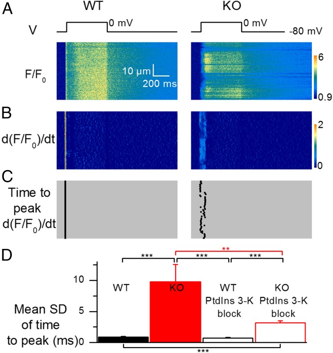
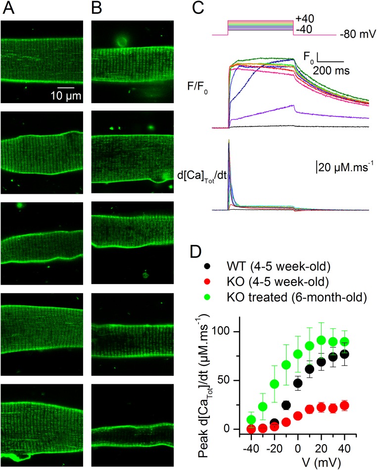
Mutations in the gene encoding the phosphoinositide 3-phosphatase myotubularin (MTM1) are responsible for a pediatric disease of skeletal muscle named myotubular myopathy (XLMTM). Muscle fibers from MTM1-deficient mice present defects in excitation-contraction (EC) coupling likely responsible for the disease-associated fatal muscle weakness. However, the mechanism leading to EC coupling failure remains unclear. During normal skeletal muscle EC coupling, transverse (t) tubule depolarization triggers sarcoplasmic reticulum (SR) Ca2+ release through ryanodine receptor channels gated by conformational coupling with the t-tubule voltage-sensing dihydropyridine receptors. We report that MTM1 deficiency is associated with a 60% depression of global SR Ca2+ release over the full range of voltage sensitivity of EC coupling. SR Ca2+ release in the diseased fibers is also slower than in normal fibers, or delayed following voltage activation, consistent with the contribution of Ca2+-gated ryanodine receptors to EC coupling. In addition, we found that SR Ca2+ release is spatially heterogeneous within myotubularin-deficient muscle fibers, with focally defective areas recapitulating the global alterations. Importantly, we found that pharmacological inhibition of phosphatidylinositol 3-kinase (PtdIns 3-kinase) activity rescues the Ca2+ release defects in isolated muscle fibers and increases the lifespan and mobility of XLMTM mice, providing proof of concept for the use of PtdIns 3-kinase inhibitors in myotubular myopathy and suggesting that unbalanced PtdIns 3-kinase activity plays a critical role in the pathological process.

Keywords: excitation–contraction coupling; myotubularin; ryanodine receptor; sarcoplasmic reticulum Ca2+ release; skeletal muscle.


Conflict of interest statement

M.W.L. receives research funding support from Audentes Therapeutics, Solid GT, and Demeter Therapeutics; is a member of the Scientific Advisory Board of Audentes Therapeutics; and was recently a consultant for Sarepta Therapeutics. A.B.-B. is a scientific advisor of Audentes Therapeutics.

Figures

Fig. 1.
Fig. 1.
Defects in voltage-activated global Ca2+ release in MTM1-deficient muscle fibers are alleviated by PtdIns 3-kinase inhibition. (A) Changes in rhod-2 fluorescence (F/F0) and corresponding calculated rate of Ca2+ release (d[Ca]Tot/dt) in response to 500 ms-long voltage-clamp depolarizing pulses of various amplitude (top traces) in a WT (Left) and in a MTM1-deficient muscle fiber (KO, Right). F/F0 traces correspond to the average change in fluorescence over the entire scanned line. The raw rhod-2 fluorescence image collected while applying the pulse to –10 mV is shown above each series of F/F0 traces. (B) Voltage dependence of the peak rate of Ca2+ release in WT (black symbols) and MTM1-deficient muscle fibers (red symbols) under control conditions (filled symbols) and following a 1-h exposure to the PtdIns 3-kinase inhibitors wortmannin and LY294002 (PtdIns 3-K block, open symbols). The Inset shows the mean values for the maximum rate of Ca2+ release in the different conditions: For this, a Boltzmann function was fitted to the individual sets of values in each fiber. Corresponding mean values for the other Boltzmann parameters are presented in Table S1. (C) Voltage dependence of the mean time to peak rate of Ca2+ release in the different conditions. Black and red asterisks report a significant difference between WT and KO fibers and between KO fibers not treated and treated with the PtdIns 3-kinase blockers, respectively. In C and D, mean values are from 20 and 13 WT and KO fibers under control conditions and 17 and 12 WT and KO fibers treated with the PtdIns 3-kinase inhibitors, respectively. All data are expressed as mean ± SEM. *P < 0.05; **P < 0.01; ***P < 0.001 with unpaired t test.
Fig. S1.
Fig. S1.
Evidence for Ca2+-induced Ca2+ release in MTM1-deficient muscle fibers. (A) Change in cytosolic [Ca2+] (Δ[Ca2+]) and corresponding rate of Ca2+ release (d[Ca]Tot/dt) calculated from line-averaged rhod-2 fluorescence signals collected while applying a 500 ms-long depolarizing pulse to –10 mV in a WT fiber (Left) and in a MTM1-deficient fiber (Right). The MTM1-deficient fiber yields a delayed onset phase of Ca2+ release during the pulse: Its amplitude and the corresponding threshold [Ca2+] level were determined as indicated. (B) Peak amplitude of the delayed onset phase of Ca2+ release as a function of the threshold [Ca2+] level. Each data point is from a separate MTM1-deficient muscle fiber.
Fig. S2.
Fig. S2.
CAV1.1 Ca2+ current in WT and MTM1-deficient muscle fibers untreated and treated with PtdIns 3-kinase blockers. (A) Ca2+ current traces in response to depolarizing steps to values ranging between –30 and +30 mV in a WT (Left) and in a MTM1-deficient fiber (Right). (B) Mean voltage dependence of peak Ca2+ current in WT (black symbols) and MTM1-deficient muscle fibers (red symbols) under control conditions (filled symbols, n = 19 WT fibers and 12 KO fibers, respectively) and following a 1-h exposure to the PtdIns 3-kinase inhibitors wortmannin and LY294002 (PtdIns 3-K block, open symbols; n = 16 and 12 fibers, respectively). (C) Mean values for the maximal conductance (Gmax), reversal potential (Vrev), midactivation voltage (V0.5), and steepness (k) obtained from fitting the appropriate function to the individual series of peak Ca2+ current values versus voltage in each fiber under the different conditions. All data are expressed as mean ± SEM. *P < 0.05; **P < 0.01; ***P < 0.001 with unpaired t test.
Fig. S3.
Fig. S3.
Defects in voltage-activated global Ca2+ release in MTM1-deficient muscle fibers are alleviated by a prolonged exposure to PtdIns 3-kinase inhibitors. (A) Voltage dependence of the peak rate of Ca2+ release in WT (black symbols) and MTM1-deficient muscle fibers (red symbols) under control conditions (filled symbols, n = 7 and 8 WT and KO fibers, respectively) and following overnight exposure to the PtdIns 3-kinase inhibitors wortmannin and LY294002 (PtdIns 3-K block, open symbols, n = 8 and 9 WT and KO fibers, respectively). The Inset shows the mean values for the maximum rate of Ca2+ release in the different conditions: For this, a Boltzmann function was fitted to the individual sets of values in each fiber. (B) Voltage dependence of the mean time to peak rate of Ca2+ release in the different conditions. Black and red asterisks report a significant difference between WT and KO fibers and between KO fibers untreated and treated with the PtdIns 3-kinase blockers, respectively. All data are expressed as mean ± SEM. *P < 0.05; **P < 0.01; ***P < 0.001 with unpaired t test.
Fig. S4.
Fig. S4.
3-MA does not rescue the defects in voltage-activated global Ca2+ release in MTM1-deficient muscle fibers. (A) Voltage dependence of the peak rate of Ca2+ release in WT (black symbols) and MTM1-deficient muscle fibers (red symbols) under control conditions (filled symbols, n = 7 and 7 WT and KO fibers, respectively) and following 1 h exposure to 1 mM of the PtdIns 3-kinase inhibitor 3-MA (open symbols, n = 9 and 6 WT and KO fibers, respectively). The Inset shows the mean values for the maximum rate of Ca2+ release in the different conditions: For this, a Boltzmann function was fitted to the individual sets of values in each fiber. (B) Voltage dependence of the mean time to peak rate of Ca2+ release in the different conditions. Asterisks report a significant difference between WT and KO fibers. All data are expressed as mean ± SEM. *P < 0.05; **P < 0.01; ***P < 0.001 with unpaired t test.
Fig. 2.
Fig. 2.
Spatial heterogeneity of Ca2+ release in MTM1-deficient muscle fibers. (A) Line-scan rhod-2 F/F0 images collected in a WT fiber (Left) and in a MTM1-deficient fiber (Right) while a 500 ms-long depolarizing pulse to 0 mV was applied. (B) Corresponding images of the rate of change in F/F0 (dF/F0/dt); these images were resampled from 512 to 64 pixels (0.1–0.8 µm per pixel) after linear averaging in the space domain, and the time derivative was calculated with a dt of 4 pixels (4.6 ms). (C) Corresponding distribution of the time to peak rate of change in F/F0 along the scanned line. (D) Mean SD of the time to peak rate of change in F/F0 in WT and KO fibers (n = 30 and 26, respectively) and in WT and KO fibers treated with the PtdIns 3-kinase blockers (n = 26 and 24, respectively). All data are expressed as mean ± SEM. **P < 0.01; ***P < 0.001 with unpaired t test.
Fig. 3.
Fig. 3.
Local alterations in voltage-activated SR Ca2+ release in MTM1-deficient muscle fibers. (A–C) Rhod-2 F/F0 line-scan images collected in MTM1-deficient fibers while a 500 ms-long depolarizing pulse to the indicated value was applied. In each panel, bottom traces show the changes in rhod-2 fluorescence (F/F0) and the corresponding calculated rate of Ca2+ release (d[Ca]Tot/dt) at three different positions of the line (colored arrows).
Fig. S5.
Fig. S5.
Line-scan rhod-2 fluorescence images taken from a WT (Left) and from a MTM1-deficient muscle fiber (Right) while a 500 ms-long pulse at –10 or +10 mV was applied.
Fig. S6.
Fig. S6.
T-tubule defects and local alterations of voltage-activated Ca2+ release in MTM1-deficient fibers. The t-tubule network was stained with di-8-anepps. (A–C) Each panel shows a line-scan rhod-2 F/F0 Ca2+ transient (Right) triggered by a voltage-clamp depolarization to the indicated value. Traces below correspond to the rate of Ca2+ release at three positions of the scanned line (indicated by colored arrows). The green image on Left is an x,y frame of the di-8-anepps fluorescence in the same fiber. The longitudinal profile of di-8-anepps fluorescence measured within a rectangular region (white rectangle) encompassing five pixels on each side of the position of the rhod-2 scanned line is reported next to the x,y di-8-anepps image (green trace) to better view the spatial correlation or absence of spatial correlation between defects in t-tubule pattern and in Ca2+ transient. (A) Example of clear correlations between localized defects in t-tubule pattern and in Ca2+ release. (B) Example showing a large fiber region with depressed Ca2+ release but no apparent sign of related alteration in t-tubule pattern. (C) Example of slow and delayed Ca2+ release in a fiber yielding an overall very much altered t-tubule pattern.
Fig. 4.
Fig. 4.
In vivo wortmannin treatment enhances life expectancy of Mtm1-KO mice. (A) Survival curves of WT and Mtm1-KO mice treated with wortmannin (n = 10 and n = 11, respectively) and untreated (n = 9 and n = 10, respectively). The x axis corresponds to the number of days following the beginning of the treatment. An analysis with the log-rank test gave a statistically increased survival rate in treated versus nontreated KO mice (P = 0.001). (B) Absolute body weight of the WT and Mtm1-KO mice treated with wortmannin and untreated. The Inset shows the corresponding relative weight of the animals. One untreated KO mouse unexpectedly survived about 3 wk longer than all other ones; corresponding data points during that period are presented as stars.
Fig. S7.
Fig. S7.
Absence of changes in the expression of genes of the EC coupling machinery and in muscle fiber structure after 3 wk of wortmannin treatment. (A) Relative fold changes in mRNA level between tibialis muscles from WT, WT treated, KO, and KO treated animals for DHPR α1, β1, and γ1 subunits (Cacna1s, Cacnb1, and Cacng1); type 1 ryanodine receptor (Ryr1); type 1 and 2 SERCA pumps (Atp2a1 and Atp2a2); and calsequestrin 1 and 2 (Casq1 and Casq2). Data are mean ± SD. All fold changes were calculated versus the value of the WT group. *P < 0.05; **P < 0.01; ***P < 0.001 with unpaired t test. The major changes in the diseased muscles corresponded to an increased level of DHPR β1 and γ1 subunits and in SERCA2. These changes persisted in muscles from the treated KO animals, whereas no other substantial modification was detected in muscles from both treated WT and KO animals. (B) Immunoblot analysis showing no change in the protein level of RYR1 between treated and untreated KO muscles (n = 5 in each group). GAPDH was used as the internal control. Data are expressed as mean ± SEM. *P < 0.05 with unpaired t test. (C) Ultrastructure evaluation of muscles from wortmannin-treated and untreated animals. Electron microscopy performed on longitudinal sections of skeletal muscle reveals approximately appropriate myofibrillar and mitochondrial morphology, with variable numbers of triad structures (arrows) in the intermyofibrillar space. Images shown are at 30,000× magnification. (Scale bar, 500 nm.) Upon visual inspection, the MTM and WT specimens were easily distinguishable from one another due to the presence of sarcotubular disorganization and differences in myofibril thickness and organization. A distinction between wortmannin-treated and untreated states within each genotype was not possible, and this was supported by our quantitation of t-tubules, l-tubules, and triads (Table S2).
Fig. S8.
Fig. S8.
T-tubule network and voltage-activated Ca2+ release in muscle fibers from a wortmannin-treated, long-term–surviving KO mouse. (A and B) Confocal x,y frames of di-8-anepps fluorescence from 10 muscle fibers: A majority of fibers exhibited either no sign of alteration of the t-tubule network (A) or a few disrupted areas (B, top three frames). Two fibers exhibited a more severely disrupted t-tubule network (B, bottom two frames). (C) Voltage-activated rhod-2 fluorescence transients and corresponding Ca2+ release flux from a muscle fiber from the same mouse. (D) Mean voltage dependence of peak Ca2+ release from six fibers from the same mouse compared with the mean values from untreated young WT and KO animals (same values as in Fig. 1B).

References

    1. Al-Qusairi L, et al. T-tubule disorganization and defective excitation-contraction coupling in muscle fibers lacking myotubularin lipid phosphatase. Proc Natl Acad Sci USA. 2009;106(44):18763–18768. - PMC - PubMed
    1. Ríos E, Pizarro G, Stefani E. Charge movement and the nature of signal transduction in skeletal muscle excitation-contraction coupling. Annu Rev Physiol. 1992;54:109–133. - PubMed
    1. Schneider M-F. Control of calcium release in functioning skeletal muscle fibers. Annu Rev Physiol. 1994;56:463–484. - PubMed
    1. Taylor G-S, Maehama T, Dixon J-E. Myotubularin, a protein tyrosine phosphatase mutated in myotubular myopathy, dephosphorylates the lipid second messenger, phosphatidylinositol 3-phosphate. Proc Natl Acad Sci USA. 2000;97(16):8910–8915. - PMC - PubMed
    1. Tronchère H, et al. Production of phosphatidylinositol 5-phosphate by the phosphoinositide 3-phosphatase myotubularin in mammalian cells. J Biol Chem. 2004;279(8):7304–7312. - PubMed

Publication types

MeSH terms

LinkOut - more resources