. 2016 Dec 13;113(50):E8059-E8068.
doi: 10.1073/pnas.1615392113. Epub 2016 Nov 23.

MyTH4-FERM myosins have an ancient and conserved role in filopod formation

Affiliations

MyTH4-FERM myosins have an ancient and conserved role in filopod formation

Karl J Petersen et al. Proc Natl Acad Sci U S A. .

Abstract

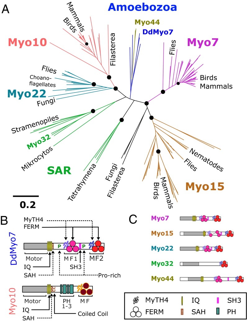
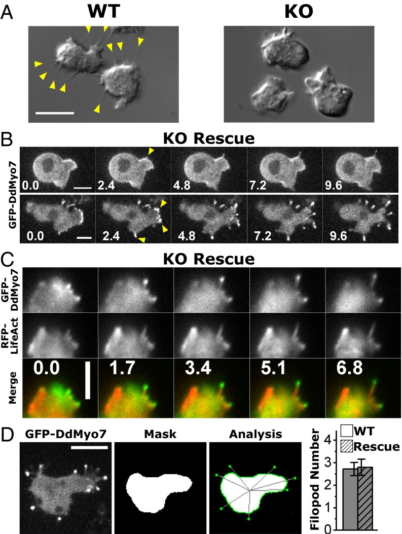
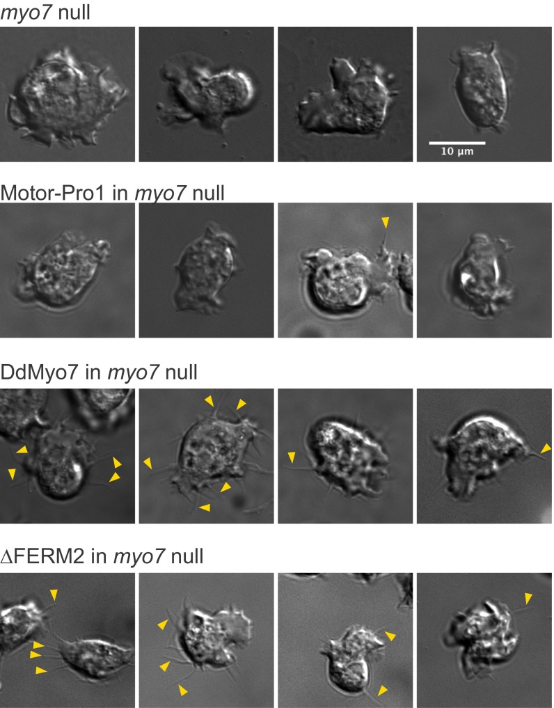
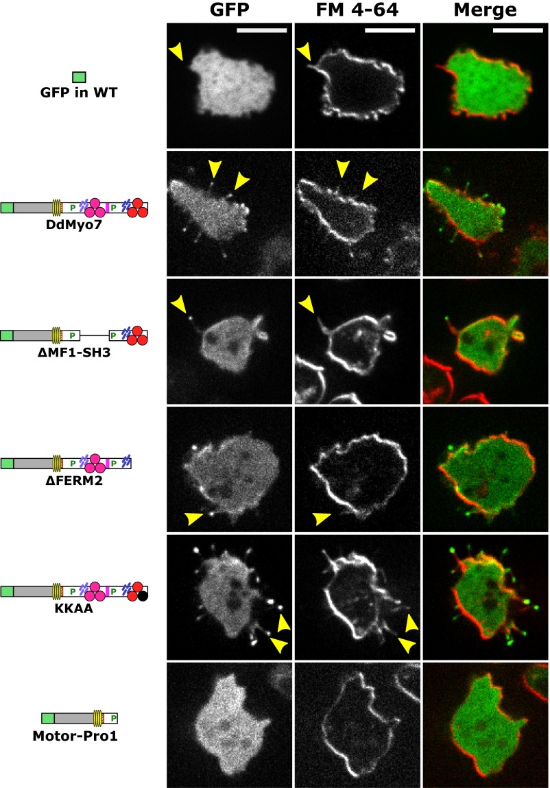
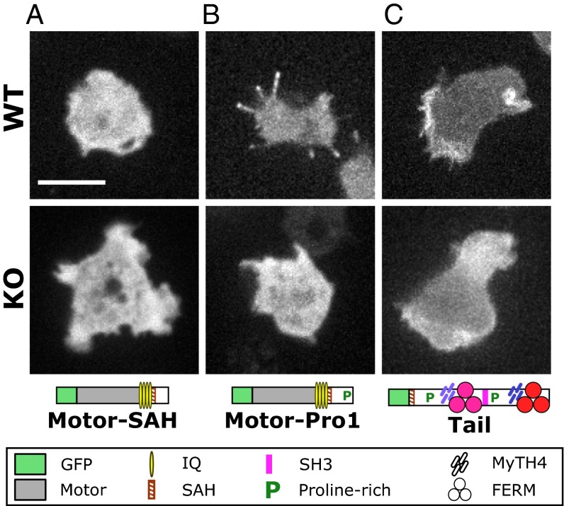
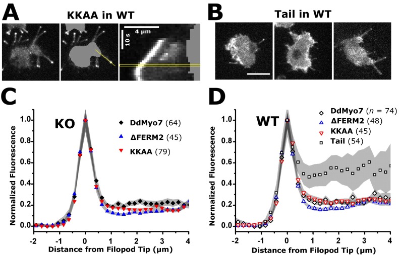
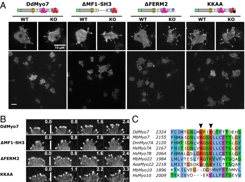
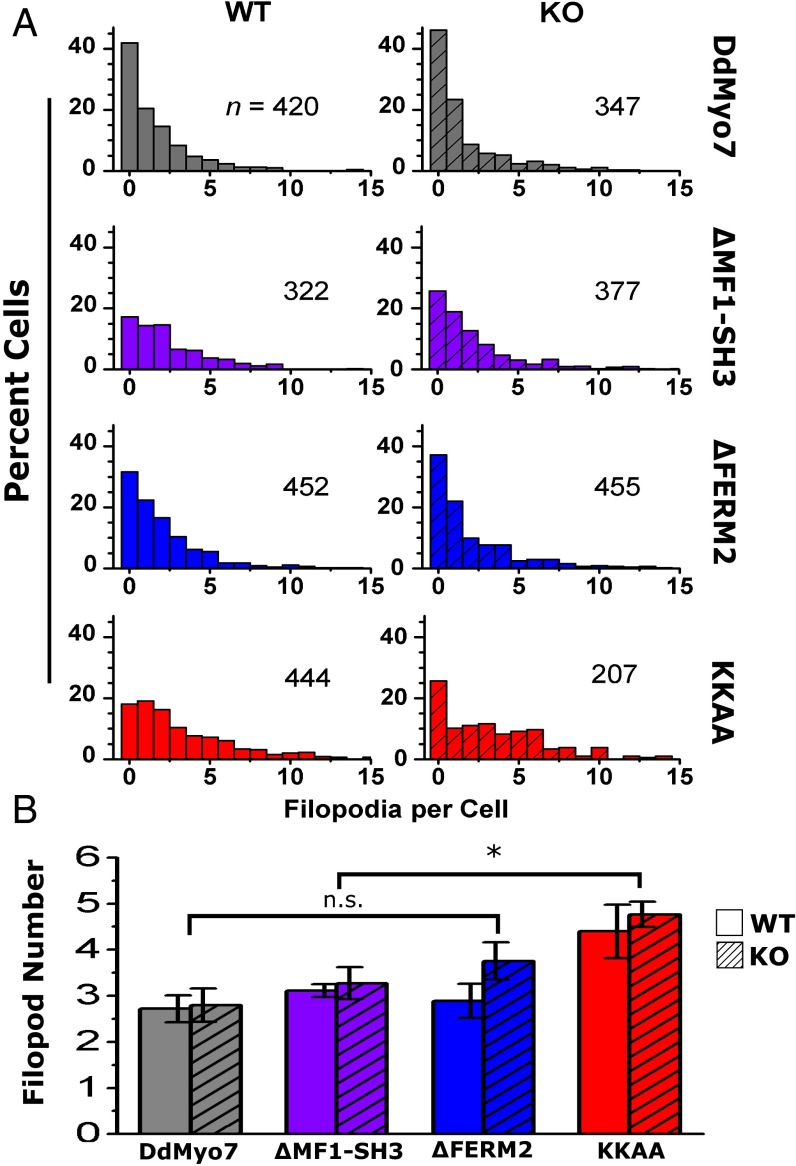
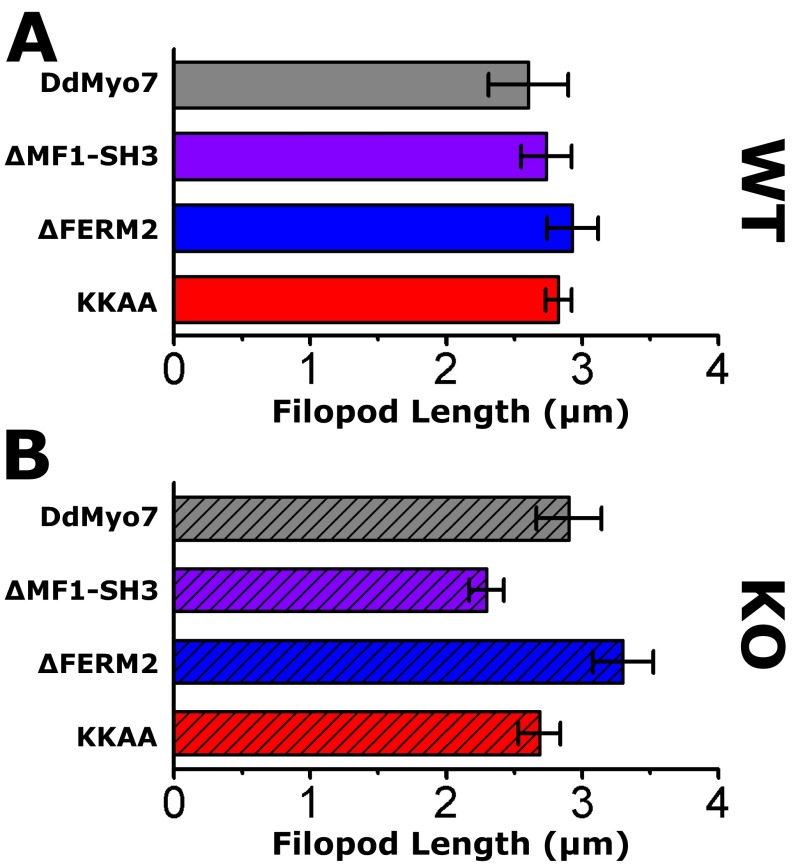
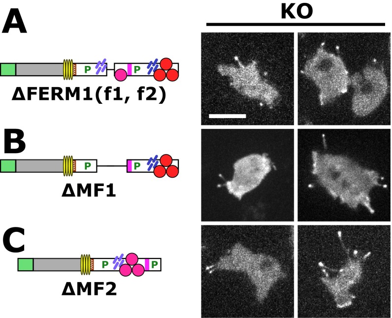
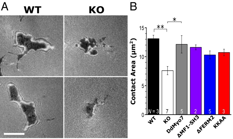
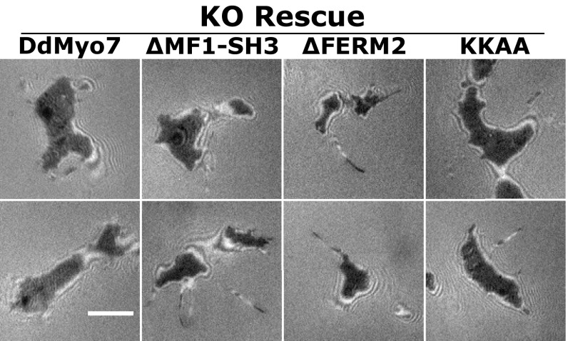
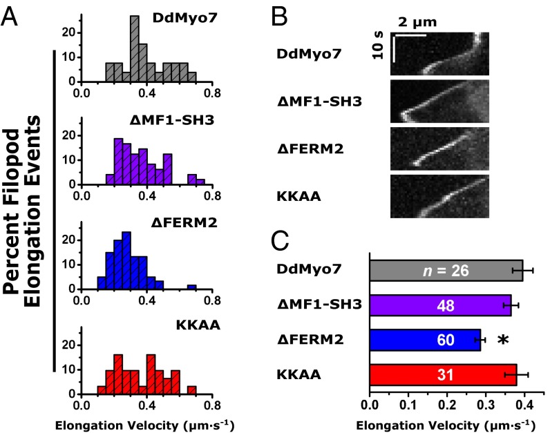
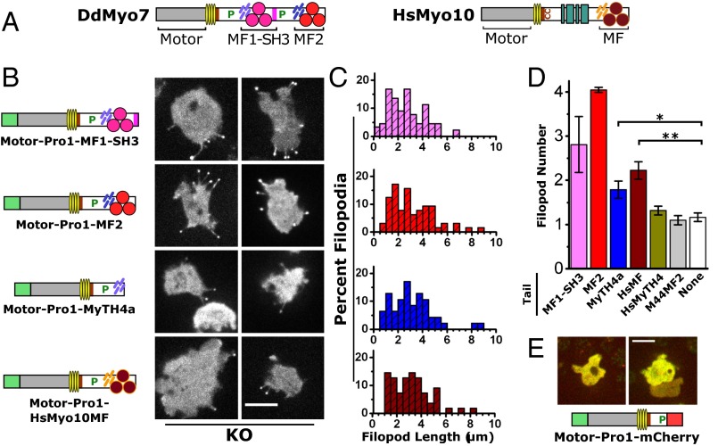
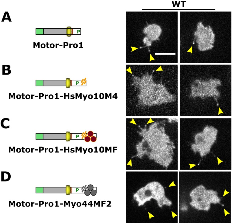
The formation of filopodia in Metazoa and Amoebozoa requires the activity of myosin 10 (Myo10) in mammalian cells and of Dictyostelium unconventional myosin 7 (DdMyo7) in the social amoeba Dictyostelium However, the exact roles of these MyTH4-FERM myosins (myosin tail homology 4-band 4.1, ezrin, radixin, moesin; MF) in the initiation and elongation of filopodia are not well defined and may reflect conserved functions among phylogenetically diverse MF myosins. Phylogenetic analysis of MF myosin domains suggests that a single ancestral MF myosin existed with a structure similar to DdMyo7, which has two MF domains, and that subsequent duplications in the metazoan lineage produced its functional homolog Myo10. The essential functional features of the DdMyo7 myosin were identified using quantitative live-cell imaging to characterize the ability of various mutants to rescue filopod formation in myo7-null cells. The two MF domains were found to function redundantly in filopod formation with the C-terminal FERM domain regulating both the number of filopodia and their elongation velocity. DdMyo7 mutants consisting solely of the motor plus a single MyTH4 domain were found to be capable of rescuing the formation of filopodia, establishing the minimal elements necessary for the function of this myosin. Interestingly, a chimeric myosin with the Myo10 MF domain fused to the DdMyo7 motor also was capable of rescuing filopod formation in the myo7-null mutant, supporting fundamental functional conservation between these two distant myosins. Together, these findings reveal that MF myosins have an ancient and conserved role in filopod formation.

Keywords: MyTH4-FERM; actin; cell motility; filopodia; myosin.


Conflict of interest statement

The authors declare no conflict of interest.

Figures

Fig. 1.
Fig. 1.
Evolutionary relationships of MyTH4-FERM myosins. (A) Phylogenetic analysis of the MF domains of myosins from representative organisms as determined by SATé (30). Likelihood values at major branch points (>95%) are indicated by circles, and the scale is as defined by FastTree (54). (B) Domain organization of DdMyo7 and Myo10, two MF myosins involved in filopod formation. Domains of DdMyo7 include the motor (myosin catalytic domain), IQ (light-chain–binding motifs 1–4); SAH (single α-helix); proline-rich regions; MyTH4 (myosin tail homology 4); FERM (band 4.1, ezrin, radixin, moesin); and SH3 (Src homology 3). Myo10 contains three PH (pleckstrin homology) domains. (C) Domain organization of other major MF myosin families. See the symbol legend at bottom for identification of domains.
Fig. 2.
Fig. 2.
DdMyo7 is present in filopodia from their initiation and at filopod tips during elongation. (A) DIC images of wild-type and myo7-null (KO) cells illustrating the absence of filopodia in cells lacking DdMyo7. (Scale bar: 10 μm.) (B) Filopod initiation in two KO cells expressing GFP-DdMyo7 over 10 s. The arrowheads point to emerging and extending filopodia that show the myosin concentrating near the distal tips. (Scale bars: 5 μm.) (C) Time series showing formation of a filopodium at the edge of an actin-rich pseudopod in a myo7-null cell expressing GFP-DdMyo7 and RFP-LifeAct over time (seconds). (Scale bar: 4 μm.) (D) Automated analysis of filopod number in maximum intensity projection confocal images. Shown is a representative cell expressing GFP-DdMyo7. (Scale bar: 10 μm.) The cell body is masked (Mask), and filopod tips are located by GFP intensity before registering them to the cell (Analysis). Quantification of multiple images from either wild-type or myo7-null cells expressing GFP-DdMyo7 yields an average of 2.8 filopodia per cell, a baseline for further assays (see also Table 1).
Fig. S1.
Fig. S1.
Western blot analysis of GFP-DdMyo7 fusions. Western blotting of total cell lysates to validate the expression of GFP fusions. (A) Wild-type and myo7-null lysates probed with anti-Myo7 antibody showing the presence of the ∼270-kDa heavy chain in wild-type cells (Ax2) that is missing from the myo7-null cells. (B and C) Blots of wild-type (B) or myo7-null (C) cells expressing the DdMyo7 fusions probed with anti-GFP to detect the expressed heavy chain. The asterisk indicates the position of the full-length GFP fusion. All samples shown were also blotted for the 125-kDa MyoB heavy chain that is used as a loading control. Note that lanes from several independent blots are shown.
Fig. S2.
Fig. S2.
DIC images of Dictyostelium cells under optimal conditions for filopodia formation. Randomly selected images of cells imaged by DIC microscopy illustrating the scoring of filopodia for comparison with scoring by filopodial tip GFP fluorescence. Shown are the myo7-null cell line that does not extend filopodia, a nonrescued cell line (motor-Pro1), and two rescued lines (DdMyo7 and ∆FERM2). Examples of filopodial extensions are indicated by arrowheads. Nonrescued cell lines show very rare protrusions compared with rescued cell lines (also see Table S1). (Scale bar: 10 µm.)
Fig. S3.
Fig. S3.
Filopodia visualized by GFP-DdMyo7 and the fluorescent membrane marker FM 4-64. Dictyostelium myo7-null cells expressing full-length or mutant DdMyo7 proteins or GFP alone were starved for 1 h and incubated with 0.25 µg/mL FM 4-64 membrane dye for 1–5 min before confocal imaging. A single image plane is shown for the GFP (Left) and red FM 4-64 (Center) channels; the merge of the two channels is shown on the right. DdMyo7 is enriched in filopod tips, and the filopodia are visible with FM 4-64 as well as GFP staining along their length (yellow arrows). Note the cytosolic localization of GFP expressed in wild-type cells (Top Row) contrasted with the localization of GFP-DdMyo7 at the filopod tip in the myo7-null cells.
Fig. 3.
Fig. 3.
The head and tail of DdMyo7 are required for filopod formation. (A) The DdMyo7 motor-SAH domains do not localize to filopodia when expressed in wild-type cells and do not rescue filopod formation in the myo7-null (KO) cells. (Scale bar: 10 μm.) (B) The motor-Pro1 region localizes to filopodia in wild-type cells but does not rescue filopod formation in myo7-null cells. (C) The DdMyo7 tail localizes to the cell leading edge in both wild-type and myo7-null cells and weakly localizes to filopodia in wild-type cells but does not rescue filopod formation in myo7-null cells.
Fig. S4.
Fig. S4.
Deletion or mutation of the C-terminal FERM2 domain does not alter the distribution of GFP-DdMyo7 along the length of the filopodium. The fluorescence intensity of GFP-DdMyo7 was measured using the peak intensity in filopodia tips to align filopodia on the x axis (positive values are toward the cell body) and to localize DdMyo7 mutants in filopodia using the mean intensity along the length. (A, Left) A representative wild-type (WT) cell expressing the KKAA mutant protein. (Center) Images were processed by segmentation of the cell body followed by an intensity line scan when the filopod was near its maximum length. (Right) Kymograph with the time point used for the line scan outlined in yellow. (Scale bars: 4 μm × 10 s.) (B) The tail expressed in wild-type cells is occasionally localized to filopodia tips and along the length of the filopodium. (Scale bar: 10 μm.) (C) Mean GFP intensity along the length of filopodia extended by myo-7 null (KO) cells expressing full-length DdMyo7, the C-terminal FERM2 deletion (∆FERM2), or the double point mutant (KKAA). (D) Mean GFP intensity along the length of filopodia extended by wild-type cells expressing full-length or mutant DdMyo7 or the DdMyo7 tail. Note that a small portion of DdMyo7 is located uniformly along the actin-rich shaft in cells expressing the wild-type or deletion mutants. In contrast, higher relative levels of the tail are observed along the shaft. The tail does not rescue filopod formation and therefore is not shown in KO cells. The peak value (x = 0) is the normalized mean intensity of n filopodia (indicated for each condition). Shading indicates SEM of the number of filopodia averaged at each x value.
Fig. 4.
Fig. 4.
Rescue of filopod formation by mutant DdMyo7. (A) Domain organization of DdMyo7 mutants, with GFP fused to the N termini, is shown (see Fig. 1 for symbol key) with representative confocal fluorescence images of full-length DdMyo7, deletion mutants lacking the MF1-SH3 or C-terminal FERM2 domain, and the KKAA mutant with changes in conserved basic residues in the FERM2 domain. The large panels show representative fields used in analysis of each DdMyo7 expressed in myo7-null (KO) cells. (Scale bar: 10 μm.) (B) Detail of filopodia in extending pseudopods of KO cells expressing wild-type and mutant DdMyo7 proteins. Note the localization of DdMyo7 to the leading edge and subsequently to filopod tips during elongation. The time lapse is indicated in seconds. (Scale bar: 4 μm.) (C) Sequence alignment showing a motif (K/RxxK/R) that is conserved in the FERM2 domain of MF myosins including DdMyo7, animal and choanoflagellate Myo7, and Myo22 but excluding Myo10. Arrowheads indicate conserved basic residues. Aea, Aedes aegyptii; Dd, Dictyostelium discoideum; Dm, Drosophila melanogaster; Hs, Homo sapiens; Mb, Monosiga brevicollis. Sequence positions are indicated on the left, and conserved residues are highlighted: blue, hydrophobic; red, basic; magenta, acidic; green, Asn/Thr/Ser; cyan, His/Tyr; orange, Gly; yellow, Pro.
Fig. 5.
Fig. 5.
Filopod formation by DdMyo7 is regulated by the C-terminal FERM2 domain. Filopodia were counted in Dictyostelium cells expressing DdMyo7. (A) Filopod number in cells expressing full-length DdMyo7, in DdMyo7 with MF1-SH3 or FERM2 domain deletions (ΔMF1-SH3, ΔFERM2), and in DdMyo7 with a double point mutation in the FERM2 domain (KKAA). Data are shown for wild-type (solid-colored bars) and myo7 null (KO) (hatched bars). The number of GFP+ cells (n) includes cells with and without filopodia. (B) Average number of filopodia per cell. Filopod number was defined as the total filopodia divided by the number of cells with filopodia. Cells expressing the KKAA mutant significantly increased filopod number by 66% compared with DdMyo7 (*P = 0.00008), by 43% relative to ΔMF1-SH3 (P = 0.002), and by 36% relative to ΔFERM2 (P = 0.003, multiple ANOVA with contrasts). Differences among other mutants were not significant (n.s.); error bars indicate the SEM; see Table 1.
Fig. S7.
Fig. S7.
Filopodia length in myo7-null cells expressing DdMyo7 is decreased with the deletion of the MF1-SH3 but not with the deletion of the FERM2 domain. Filopodia length was measured in cells expressing full-length or mutant (ΔMF1-SH3, ΔFERM2, or KKAA) DdMyo7. Average filopodia length is shown for (A) wild-type (WT) (solid bars) and (B) myo7-null (KO) cells (hatched bars). Error bars indicate the SEM; also see Table 1.
Fig. S5.
Fig. S5.
Deletion of the MyTH4-FERM domains does not impair filopod formation by DdMyo7. DdMyo7 mutants expressed in myo7-null (KO) cells were imaged by confocal microscopy. The domain organization of DdMyo7 mutants is shown on the left, and fluorescence images are shown on the right. (A) The ΔFERM1(f1, f2) mutant lacks the f1 and f2 subdomains of the internal FERM domain but not f3 or the associated MyTH4 domain. (B and C) Additional deletions remove the MF1 domain (located in the proximal tail region) (B) or the MF2 domain (the common MF domain at the C terminus) (C). All mutants shown here localize to tips of filopodia and rescue filopod formation. (Scale bar, 10 μm.)
Fig. 6.
Fig. 6.
Rescue of the myo7-null substrate adhesion defect in polarized cells. The cell–substrate contact area was measured by IRM. (A) IRM images of the cell contact area for polarized myo7-null (KO) and wild-type control cells. (Scale bar: 5 μm.) (B) Average contact area in myo7-null Dictyostelium is decreased significantly relative to wild-type control cells (**P = 0.008) and DdMyo7 KO rescue cells (*P = 0.01). Error bars indicate the SEM of the number of independent assays shown in each bar. See also Fig. S6.
Fig. S6.
Fig. S6.
Substrate adhesion in polarized myo7-null (KO) cells expressing DdMyo7 mutants. The cell–substrate contact area was imaged using IRM. Shown are IRM images of the cell contact area for two different myo7-null cells expressing full-length DdMyo7 and the ∆MF1-SH3, ∆FERM2, and KKAA mutant proteins in each column. (Scale bar: 5 μm.) See also Fig. 6.
Fig. 7.
Fig. 7.
Filopod elongation velocity is reduced by deletion of the FERM2 domain. (A) Histograms of the elongation velocity during filopod formation events are shown for full-length DdMyo7 and the ∆MF1-SH3, ΔFERM2, and KKAA mutants. The y axis is the percent of total. (B) Representative kymographs of filopod elongation in myo7-null cells expressing DdMyo7 mutant proteins. (Scale bars: 2 μm × 10 s.) (C) Filopod elongation velocity is significantly decreased (by 28%) in the ∆FERM2 mutant (*P = 0.002) compared with full-length DdMyo7. The filopod elongation velocities in the ΔMF1-SH3 and KKAA mutants do not differ significantly from that of full-length DdMyo7. Error bars show the SEM of the number of events (n) indicated in each bar. (Also see Table 1.)
Fig. 8.
Fig. 8.
Conservation of the requirement for a single MyTH4-FERM domain for MF myosin filopod-forming activity. (A) Domain structure of DdMyo7 and human Myo10. (B) Representative images of cells with filopodia marked by the indicated fusion proteins. All mutants shown rescued filopod formation and localized strongly to filopod tips when expressed in myo7-null (KO) cells. (Scale bar: 10 µm.) (C) Histograms of filopod length in measured in myo7-null cells expressing the indicated fusion proteins. The y axis is the percent of total (also see Table 2). (D) Filopod number in myo7-null cells expressing the motor-Pro1 fused to the indicated tail domains. Filopod number was defined as total filopodia during a 10-s time-lapse observation divided by the number of cells with filopodia. The weighted mean filopod number is shown with the SEM of three to nine independent experiments. Multiple ANOVA analysis (n = 10–50 cells) confirms significant increases in filopod formation for the MyTH4a (*P < 0.05) and HsMyo10MF (**P < 0.01) chimeras (see Table 2). MF1-SH3 and MF2 data were excluded from ANOVA because of their much larger variance. (E) Domain structure of the motor-Pro1-mCherry fusion protein and confocal fluorescence images showing merged GFP and mCherry channels (Scale bar: 10 μm.) The expressed protein is cytosolic, lacks membrane enrichment, and does not support filopod formation.
Fig. S8.
Fig. S8.
Localization of DdMyo7 mutants with chimeric tail domains in wild-type cells. DdMyo7 mutants expressed in wild-type cells were imaged by confocal microscopy. The domain organization of the DdMyo7 mutants is shown on the left, and fluorescence images are shown on the right. (A) GFP-tagged motor-Pro1 is shown. Note localization at filopodia tips (arrowheads). (BD) The equivalent proteins fused at the C terminus with human Myo10 MyTH4 domain (HsMyo10M4) (B), the human Myo10 MF domain (HsMyo10MF) (C), or the Dictyostelium Myo44 MF2 domain (D). (Scale bar: 10 µm.) Also see Fig. 8 and Table 2.

References

    1. Mattila PK, Lappalainen P. Filopodia: Molecular architecture and cellular functions. Nat Rev Mol Cell Biol. 2008;9(6):446–454. - PubMed
    1. Tuxworth RI, et al. A role for myosin VII in dynamic cell adhesion. Curr Biol. 2001;11(5):318–329. - PubMed
    1. Ostap EM, et al. Dynamic localization of myosin-I to endocytic structures in Acanthamoeba. Cell Motil Cytoskeleton. 2003;54(1):29–40. - PubMed
    1. Eilken HM, Adams RH. Dynamics of endothelial cell behavior in sprouting angiogenesis. Curr Opin Cell Biol. 2010;22(5):617–625. - PubMed
    1. Mortimer D, Fothergill T, Pujic Z, Richards LJ, Goodhill GJ. Growth cone chemotaxis. Trends Neurosci. 2008;31(2):90–98. - PubMed

Publication types

MeSH terms