. 2016 Dec 13;113(50):14300-14305.
doi: 10.1073/pnas.1617316113. Epub 2016 Nov 28.

Two crystal structures reveal design for repurposing the C-Ala domain of human AlaRS

Affiliations

Two crystal structures reveal design for repurposing the C-Ala domain of human AlaRS

Litao Sun et al. Proc Natl Acad Sci U S A. .

Abstract

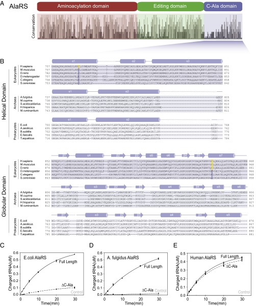
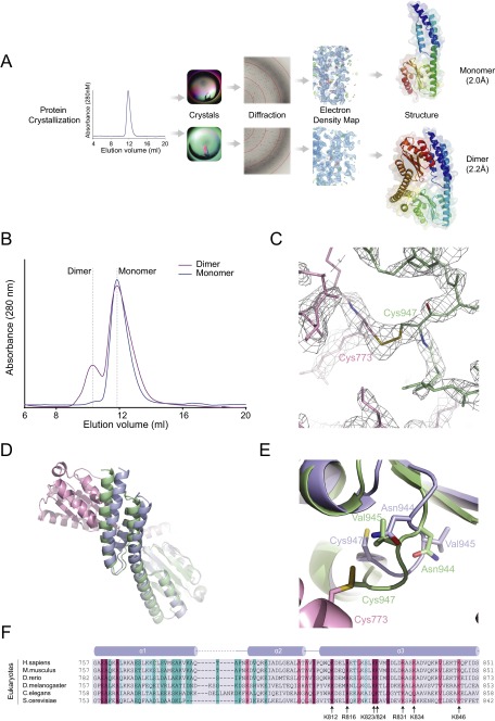
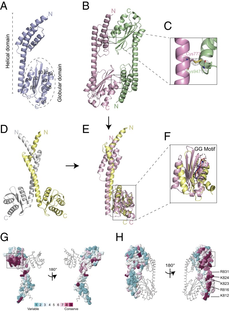
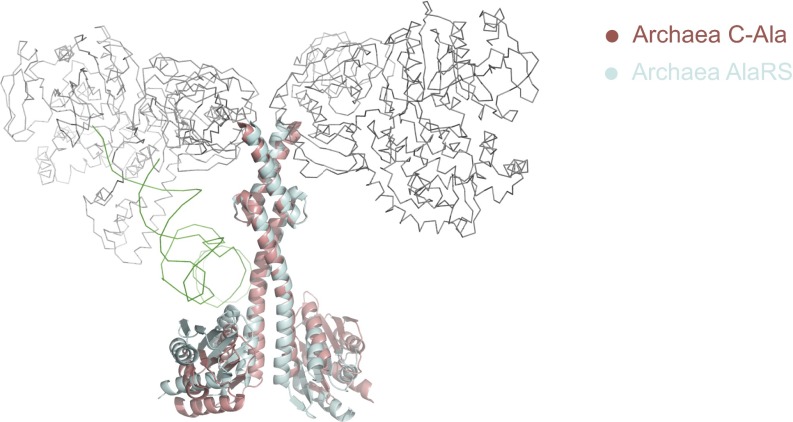
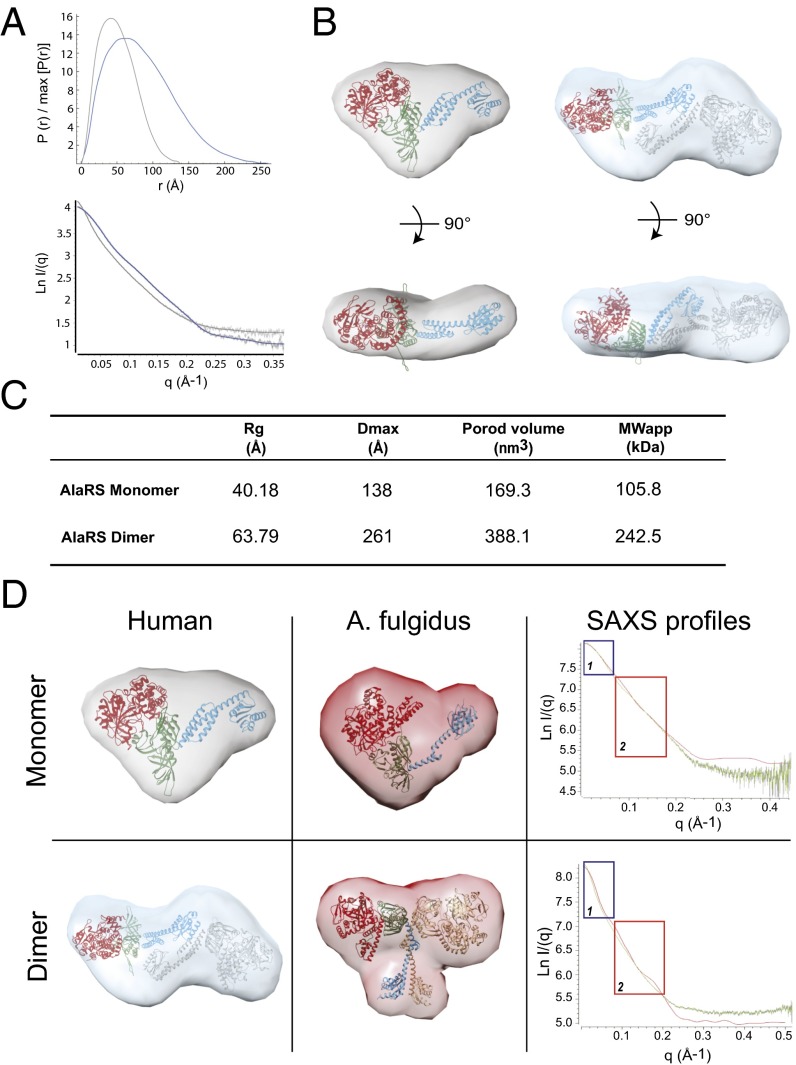
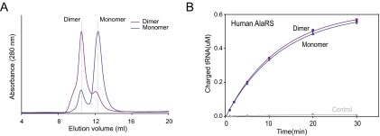
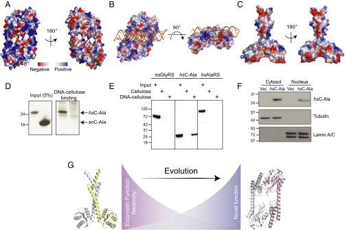
The 20 aminoacyl tRNA synthetases (aaRSs) couple each amino acid to their cognate tRNAs. During evolution, 19 aaRSs expanded by acquiring novel noncatalytic appended domains, which are absent from bacteria and many lower eukaryotes but confer extracellular and nuclear functions in higher organisms. AlaRS is the single exception, with an appended C-terminal domain (C-Ala) that is conserved from prokaryotes to humans but with a wide sequence divergence. In human cells, C-Ala is also a splice variant of AlaRS. Crystal structures of two forms of human C-Ala, and small-angle X-ray scattering of AlaRS, showed that the large sequence divergence of human C-Ala reshaped C-Ala in a way that changed the global architecture of AlaRS. This reshaping removes the role of C-Ala in prokaryotes for docking tRNA and instead repurposes it to form a dimer interface presenting a DNA-binding groove. This groove cannot form with the bacterial ortholog. Direct DNA binding by human C-Ala, but not by bacterial C-Ala, was demonstrated. Thus, instead of acquiring a novel appended domain like other human aaRSs, which engendered novel functions, a new AlaRS architecture was created by diversifying a preexisting appended domain.

Keywords: DNA binding; appended domain; evolution; splice variant; structural plasticity.


Conflict of interest statement

The authors declare no conflict of interest.

Figures

Fig. 1.
Fig. 1.
Human C-Ala has no effect on the charging activity. (A) Conservation analysis of AlaRS sequences across bacteria, archaea, and eukaryotes showing the relative sequence identity of the 410 aligned AlaRS sequences (16). (B) Alignment generated using the online Clustal Omega server (28). Secondary structural elements of C-Ala are indicated above the sequences. The two cysteines (disulfide bond) are colored in yellow. (C, D, and E) In vitro aminoacylation assay showing that human AlaRS-ΔC-Ala has similar activity relative to human full-length AlaRS (E), whereas E. coli (C) or A. fulgidus (D) AlaRS-ΔC-Ala reduces the charging activity toward tRNAAla compared with the corresponding full-length AlaRS. Error bars indicate SDs.
Fig. S1.
Fig. S1.
The monomer and dimer of human C-Ala. (A) The flowchart for crystal structure determination. The resolution for human monomer C-Ala is 2.0Å, while for the dimer is 2.2Å. (B) Gel filtration analysis (Superdex 75 10/300 GL, GE Healthcare) of bacterially purified human C-Ala. Traces are shown of human C-Ala in buffer containing 1mM DTT (blue) and buffer containing 1mM GSSG (purple). The elution positions are indicated by dashed lines. (C) The 2Fo - Fc electron density map at the disulfide bond region (contoured at 1.5 σ), which was formed between Cys773 of one molecule and Cys947 of the other. (D) Superimposition of the monomer and dimer structures of human C-Ala. The structures are colored as indicated above. (E) Asn944 and Val945 in the dimer form pushed Cys947 out of the globular subdomain to contact Cys773 from the helical domain of the other molecule. (F) Sequence alignment colored by Consurf score according to the color key shown. The secondary structure for the helical domain is shown along the top of the alignment. The positively charged residues (lysine or arginine), which is highly conserved from the helical domain, is labeled.
Fig. 2.
Fig. 2.
Crystal structure of human C-Ala. (A) The crystal structure of monomeric human C-Ala. (B) The crystal structure of dimeric human C-Ala. Two disulfide bonds are shown in the black boxes. One molecule is shown in light purple, and the other is in pale green. (C) A 2Fo-Fc electron density map contoured at 1.5 σ. A disulfide bond was formed between Cys773 of one molecule and Cys947 of the other. (D) A dimeric form of A. fulgidus C-Ala. One molecule is shown in light yellow, and the other is in gray. (E) Superimposition of the monomers of human and A. fulgidus C-Ala. (F) A zoom-in view showing the superimposition of the globular domains of human and A. fulgidus C-Ala. Human C-Ala GG motif is colored in red, and A. fulgidus C-Ala GG motif is shown in blue. (G) Structure of A. fulgidus C-Ala with surface residues colored in accordance with evolutionary conservation (high, magenta; low, cyan) among amino acid sequences from different 150 C-Ala sequences. The boxed area shows the highly conserved GG motif. These figures were prepared using ConSurf server. (H) Structure of human C-Ala with surface residues colored in accordance with evolutionary conservation among amino acid sequences from different 150 C-Ala sequences. The positively charged residues (lysine or arginine), which is highly conserved from the helical domain, is labeled.
Fig. S2.
Fig. S2.
Superimposition of the full-length and dimerization domain (C-Ala) of A. fulgidus AlaRS. The aminoacylation domain (gray), editing domain (gray), and tRNA (green) are shown in ribbon, and the C-Ala domain is shown in cyan. The C-Ala domain alone is shown in red.
Fig. 3.
Fig. 3.
Comparison of human AlaRS with A. fulgidus AlaRS. (A) The pairwise distance distribution function, P(r), of AlaRS-monomer (gray) and AlaRS-dimer (blue) (Top), and the theoretical scattering calculated from the average of 20 ab initio reconstructions (continuous lines, with AlaRS-monomer in gray and AlaRS-dimer in blue), plotted with the experimental scattering intensity curves (Bottom). The data are presented as the natural logarithm of the intensity. (B) The human full-length AlaRS model docked into the average ab initio SAXS envelope of the monomeric AlaRS (Left) and the dimeric full-length AlaRS model docked into the average ab initio SAXS envelope of the dimeric AlaRS (Right). The dimerization interface is based on the crystal structure of human dimer C-Ala. The aminoacylation domain is in red (PDB ID code 4XEM), the editing domain is in green, and C-Ala is in blue. (C) Summary of SAXS parameters. The Rg value was determined from the Guinier plot using AutoRg, and the maximum particle dimension (Dmax) and the Porod volume were calculated using GNOM. An estimate of the molecular weight was obtained by multiplying the Porod volume by 0.625. (D) Comparison of the human (Left) and A. fulgidus (Middle) envelopes for monomeric (Top) and dimeric (Bottom) full-length AlaRS. (Right) Alignment of the experimental SAXS profile for the human AlaRS (green) with the SAXS profile of the A. fulgidus AlaRS extracted from the crystal structure (red).
Fig. S3.
Fig. S3.
The monomer and dimer of human AlaRS. (A) Gel filtration analysis (Superdex 200 Increase 10/300 GL; GE Healthcare) of bacterially purified human AlaRS. Traces are shown of human AlaRS in buffer containing 1 mM DTT (blue) and buffer without DTT (purple). The elution positions are indicated by dashed lines. (B) In vitro aminoacylation assay showing that the monomer and dimer human AlaRS has the similar activity toward tRNAAla.
Fig. 4.
Fig. 4.
Human C-Ala binds DNA. (A) Electrostatic surface views of human C-Ala dimer structure. (B) A DNA-binding model created by ZDOCK program showing that DNA binds to the human C-Ala dimer through the positive charges on surface of the dimer interface. (C) Electrostatic surface views of A. fulgidus C-Ala dimer structure. (D) DNA cellulose-binding assay of Hs C-Ala and Ec C-Ala. (E) DNA cellulose-binding assay of Hs GlyRS, C-Ala and AlaRS. (F) The nuclear distribution of Hs C-Ala. Lamin A/C and tubulin were used as nuclear (N) and cytoplasmic (C) markers, respectively. (G) The distinct dimerization mode for C-Ala during evolution.

References

    1. Guo M, Yang XL, Schimmel P. New functions of aminoacyl-tRNA synthetases beyond translation. Nat Rev Mol Cell Biol. 2010;11(9):668–674. - PMC - PubMed
    1. Kim S, You S, Hwang D. Aminoacyl-tRNA synthetases and tumorigenesis: More than housekeeping. Nat Rev Cancer. 2011;11(10):708–718. - PubMed
    1. Park SG, Schimmel P, Kim S. Aminoacyl tRNA synthetases and their connections to disease. Proc Natl Acad Sci USA. 2008;105(32):11043–11049. - PMC - PubMed
    1. He W, et al. CMT2D neuropathy is linked to the neomorphic binding activity of glycyl-tRNA synthetase. Nature. 2015;526(7575):710–714. - PMC - PubMed
    1. Sampath P, et al. Noncanonical function of glutamyl-prolyl-tRNA synthetase: Gene-specific silencing of translation. Cell. 2004;119(2):195–208. - PubMed

Publication types

MeSH terms

LinkOut - more resources