. 2016 Dec 1;167(6):1610-1622.e15.
doi: 10.1016/j.cell.2016.11.020.

Modular Assembly of the Bacterial Large Ribosomal Subunit

Affiliations

Modular Assembly of the Bacterial Large Ribosomal Subunit

Joseph H Davis et al. Cell. .

Abstract

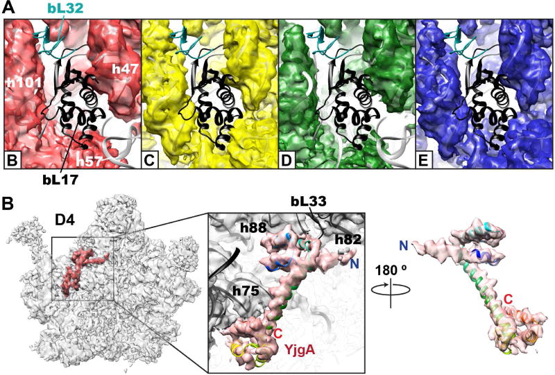
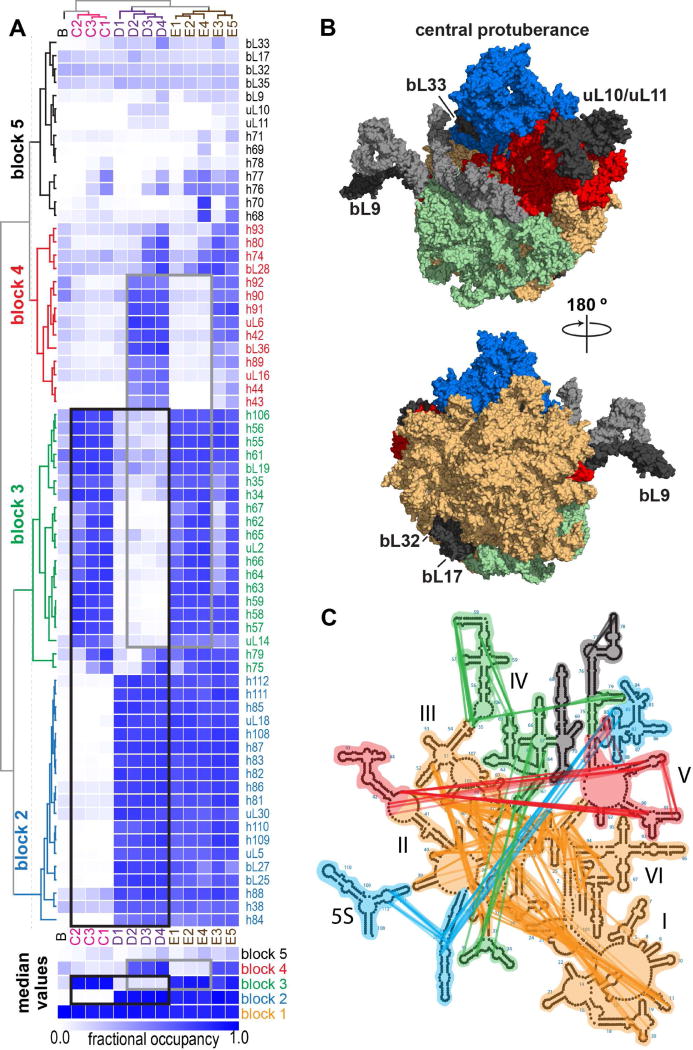
The ribosome is a complex macromolecular machine and serves as an ideal system for understanding biological macromolecular assembly. Direct observation of ribosome assembly in vivo is difficult, as few intermediates have been isolated and thoroughly characterized. Herein, we deploy a genetic system to starve cells of an essential ribosomal protein, which results in the accumulation of assembly intermediates that are competent for maturation. Quantitative mass spectrometry and single-particle cryo-electron microscopy reveal 13 distinct intermediates, which were each resolved to ∼4-5 Å resolution and could be placed in an assembly pathway. We find that ribosome biogenesis is a parallel process, that blocks of structured rRNA and proteins assemble cooperatively, and that the entire process is dynamic and can be "re-routed" through different pathways as needed. This work reveals the complex landscape of ribosome assembly in vivo and provides the requisite tools to characterize additional assembly pathways for ribosomes and other macromolecular machines.

Keywords: 50S subunit; RNA folding; Ribosome assembly; macromolecular assembly; quantitative mass spectrometry; single-particle cryo-electron microscopy.


Figures

Figure 1
Figure 1
Cellular response to an r-protein limitation. (A) Schematic for the genetic system to regulate the production of bL17. The rplQ gene encoding bL17 (dotted) was replaced with a chloramphenicol resistance gene (camR, bold) using site-specific recombineering. The strain was complemented with a plasmid-borne HSL-inducible copy of rplQ (top). Proteins, coding sequences, and promoters are marked with ovals, rectangles, and arrows respectively. Ribosomal genes and proteins are colored red. (B) Growth rate of bL17-titrated (JD321, black circles) and GFP-titrated control (JD270, gray triangles) strains as a function of the HSL inducer concentration. Restrictive (0.1 nM) and permissive (2.0 nM) HSL concentrations used subsequently are noted with vertical red and green dotted lines, respectively. (C) Ribosomal protein and (D) assembly factor or chaperone protein levels as measured by qMS as a function of HSL inducer concentration in bL17-limitation (JD321, dark circles) and control (JD270, light triangles) strains. Traces for ribosomal proteins, assembly factors, and chaperones are colored red, blue, and green, respectively.
Figure 2
Figure 2
LSUbL17dep particles are maturation-competent assembly intermediates. (A) Sucrose density gradient profiles of JD321 cell lysates grown under restrictive (black) or permissive (gray) conditions. Absorbance at 260 nm, which largely corresponds to rRNA is plotted, and LSUbL17dep (red), 50S (purple), 30S, and 70S/polysome peaks are marked with vertical dashed lines. (B) Time-course of sucrose density gradient profiles for JD321 cells grown under restrictive conditions after pulsing to permissive conditions. The center of mass for the LSU is noted by the colored dashed lines for each timepoint harvested pre-, and post-pulse. 30S, 70S/polysome, and the bottom of the gradient are marked with black dashed lines. (C) Representative mass spectra for a bL17 peptide (VVEPLITLAK [47–56]; left) or a bL20 peptide (ILADIAVFDK [94–103]; right) isolated from the 70S fraction post-pulse/bL17-induction as a function of time. Intensities normalized to that of the 14N monoisotopic peak. Colors correspond to timepoints in (B). (D) Quantitation of pulsed-isotope label as a function of time post-pulse/bL17 induction. Median peptide value for bL17 (red), and the median LSU (black) or SSU (gray) proteins isolated from 70S particles are plotted at each timepoint. (E) Representative 70S particle pulse labeling kinetics from cells grown under steady-state bL17-limited conditions (0.3 nM HSL). Maximum labeling rate at this cellular growth rate of λ = 0.007 min−1 noted as max lab (dotted gray), and highlighted by bS21 (red), which is known to exchange and will thus label at approximately the growth rate. Kinetic traces were fit as described previously (Chen et al., 2012) resulting in a relative precursor pool size P, which was large for most LSU proteins and consistent with maturation-competent intermediates, as demonstrated by the delayed labeling of representative LSU protein, bL20 (blue).
Figure 3
Figure 3
R-protein binding is re-routed upon bL17 depletion. (A) Heat map of LSU protein abundance relative to a purified 70S particle across a sucrose gradient purified from bL17-limited cells (JD321, 0.1 nM HSL). Occupancy patterns were hierarchically clustered to reveal 4 binding groups, which are marked with proteins labels from earliest (green, I) to latest binding (bold, red, IV). The trace of the sucrose density gradient analyzed is depicted above. (B) Re-routed Nierhaus assembly map. Proteins are located on the map according to the binding order defined by Chen et al. from earliest (top) to latest (bottom), and these binding groups are colored at the right side of the map (blue to red). In vitro protein binding cooperativities measured by the Nierhaus group are depicted with thick black (strong cooperativity), or thin gray (weak cooperativity) lines. Interactions that are dispensable for binding in vivo are highlighted purple, whereas those confirmed are colored red. Thick dashed orange lines mark newly measured binding dependencies that are absent from the in vitro map. The assembly map is colored according to the protein binding groups in (A), and bL17 is marked with italics.
Figure 4
Figure 4
Cryo-EM 3D classification and refinement of LSUbL17dep particles. (A) Particles were classified and refined using a hierarchical scheme (Methods). Annotations indicate number of particles used at each stage of refinement and global map resolution (Res) of the resulting class. Super-classes (A–F), and sub-classes (C1–E5), which resulted from a second round of classification and refinement are depicted. (B) Side, front, and top views highlighting EM density missing from each super-class relative to a native 50S subunit. The latter is depicted as EM density using the LSU model from a mature ribosome (PDB: 4ybb). Structures are aligned relative to one another, allowing for direct comparison within each view, as guided by dotted lines.
Figure 5
Figure 5
Reconstructed maps of bL17-limited assembly intermediates. (A) A detailed view of the bL17 binding site. Each superclass (B,C,D,E) is shown as a colored semi-transparent surface using the color scheme in Figure 4. The docked 4ybb structure is shown as a gray cartoon model with bL17 and bL32 highlighted in black and cyan, respectively. (B) YjgA bound to the D4 intermediate. Two views of a homology model for the ribosomal cofactor YjgA (rainbow) docked into segmented density (red). Model was calculated using PDB model 2p0t as the seed on the SWISS-MODEL server. YjgA bears an N-terminal extension relative to the seed model. Proximal LSU elements noted.
Figure 6
Figure 6
rRNA and r-protein cooperative folding blocks. (A) Hierarchically clustered heat map of calculated r-protein and rRNA helix occupancy in different intermediate structures. Values calculated as described in Methods. Blocks 1–5 with coordinated occupancy across different intermediate structures colored orange, blue, green, red, and black, respectively. This color scheme is unrelated to that in Figures 3, 4, or 5. Mutually exclusive blocks highlighted with black and gray rectangles. Occupancy heatmap for block 1 is omitted, as elements are largely present in all structures. For each block, the median occupancy value in each structure is displayed as a heat map below. (B) LSU model structure (PDB: 4ybb) with blocks colored according to (A). Block 5 rRNA helices are colored light gray, whereas block 5 proteins missing from all structures are labeled and colored dark gray. (C) 23S rRNA secondary structure map with rRNA helices colored according to blocks in (A). Colored edges represent tertiary contacts linking helices within blocks.
Figure 7
Figure 7
Parallel bL17-independent ribosome assembly pathways. Putative assembly paths from the least mature B class (top) to the most mature E5 class (bottom). Arrows mark allowed transitions, red lines highlight unfavorable transitions. Unresolved structure is highlighted yellow. Resolved elements are colored by block number as in Figure 6, and element formation is noted with *. Protein binding is shown at each transition. YjgA (black) occupancy percentage is indicated, as calculated by sub-classification in classes D3, D4, E3.

References

    1. Ban N, Nissen P, Hansen J, Moore PB, Steitz TA. The complete atomic structure of the large ribosomal subunit at 2.4 A resolution. Science. 2000;289:905–920. - PubMed
    1. Bokov K, Steinberg SV. A hierarchical model for evolution of 23S ribosomal RNA. Nature. 2009;457:977–980. - PubMed
    1. Canton B, Labno A, Endy D. Refinement and standardization of synthetic biological parts and devices. Nat Biotechnol. 2008;26:787–793. - PubMed
    1. Chen SS, Sperling E, Silverman JM, Davis JH, Williamson JR. Measuring the dynamics of E. coli ribosome biogenesis using pulse-labeling and quantitative mass spectrometry. Molecular Biosystems. 2012;8:3325–3334. - PMC - PubMed
    1. Chen SS, Williamson JR. Characterization of the ribosome biogenesis landscape in E. coli using quantitative mass spectrometry. J Mol Biol. 2013;425:767–779. - PMC - PubMed

MeSH terms