. 2016 Dec 1;64(5):993-1008.
doi: 10.1016/j.molcel.2016.10.034.

Characterization of Hippo Pathway Components by Gene Inactivation

Affiliations

Characterization of Hippo Pathway Components by Gene Inactivation

Steven W Plouffe et al. Mol Cell. .

Abstract

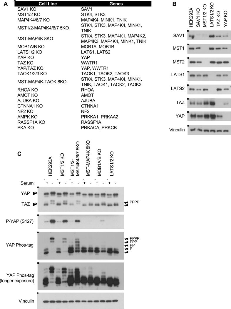
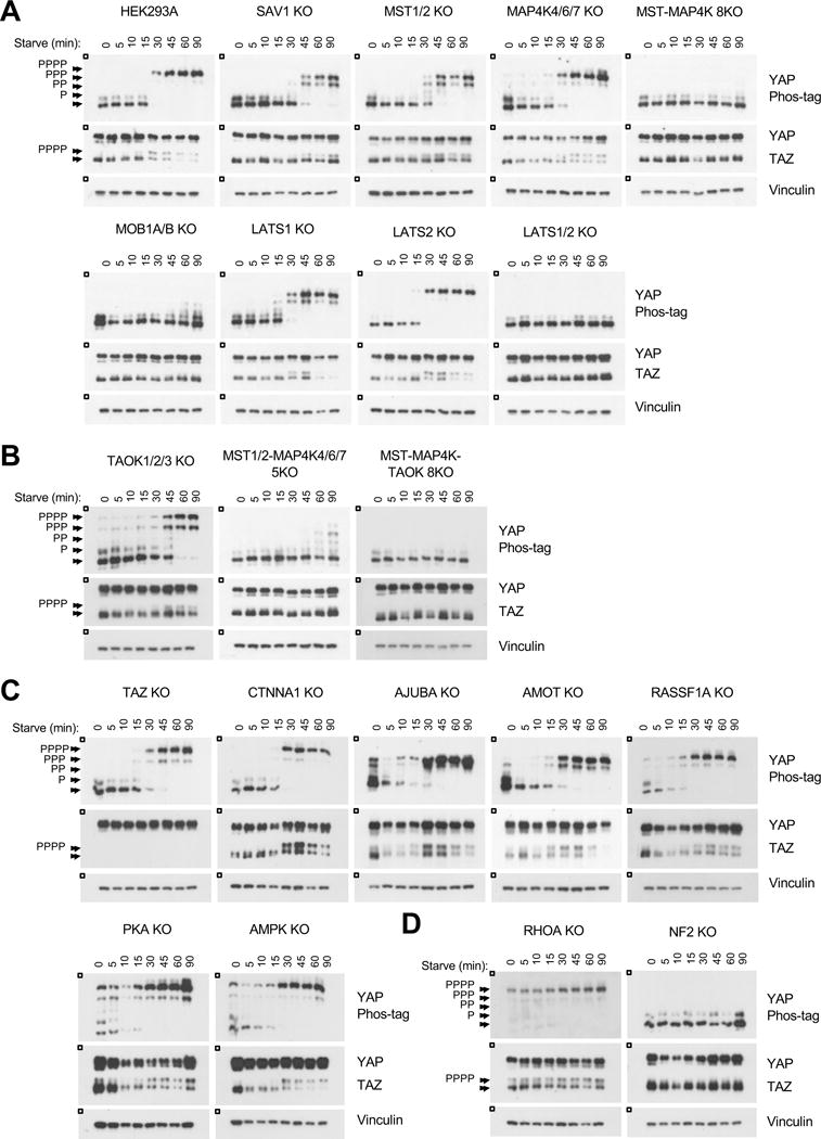
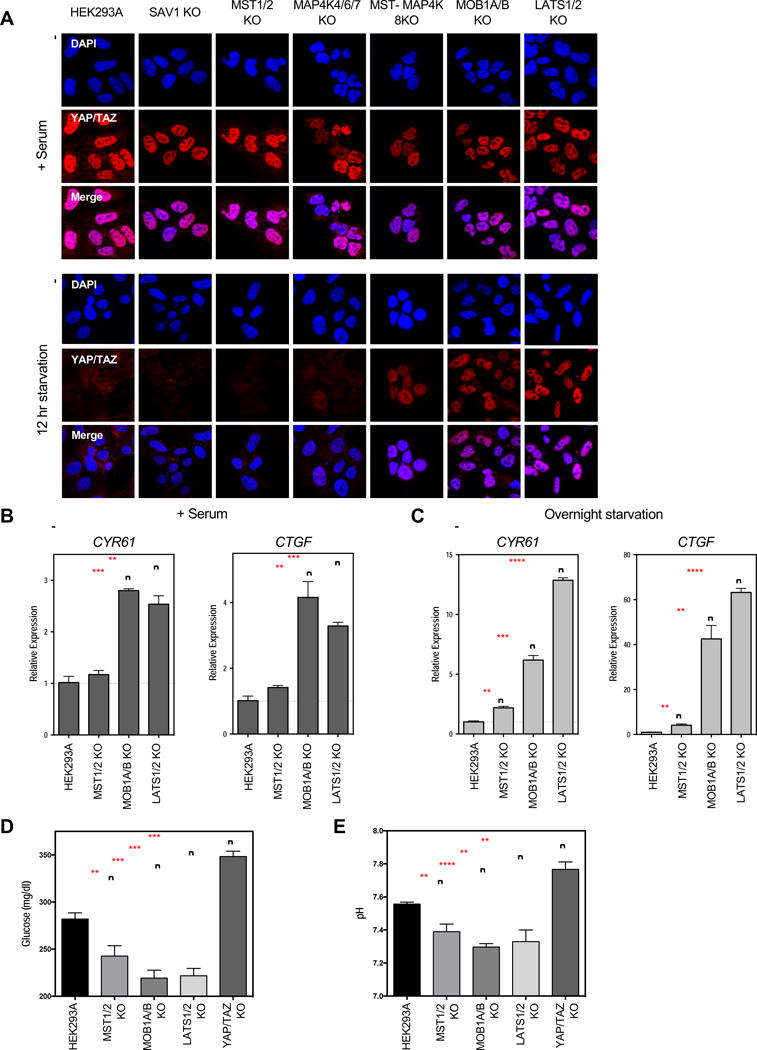
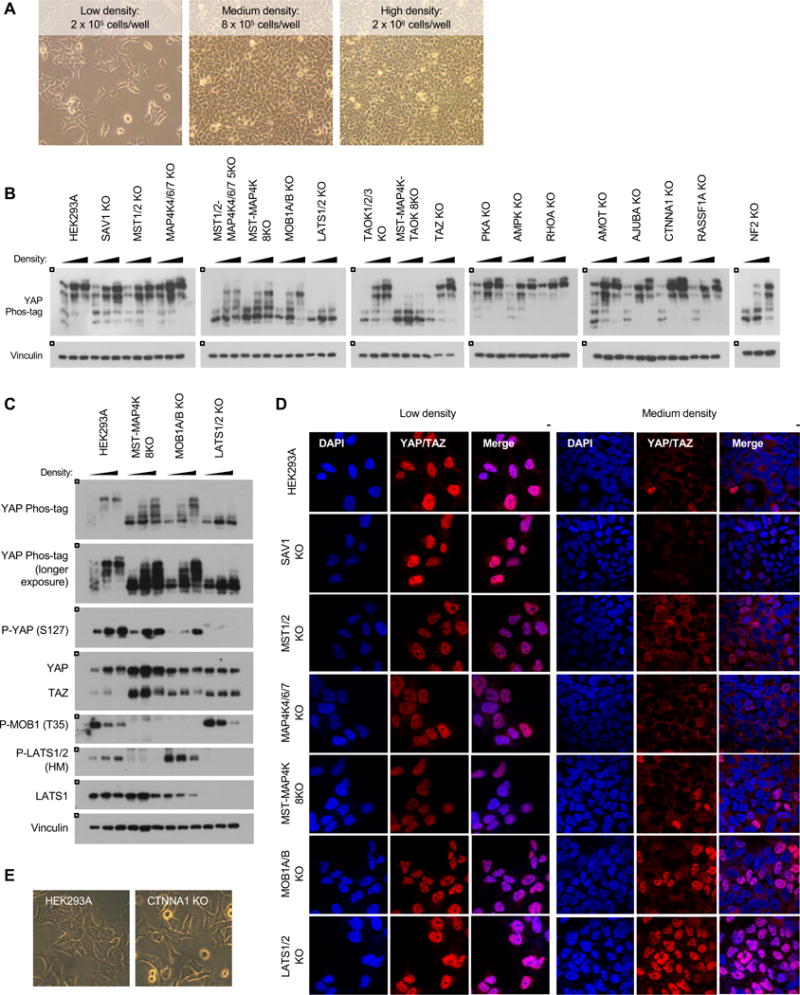
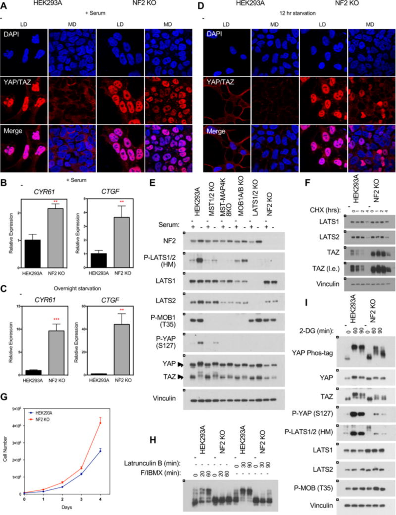
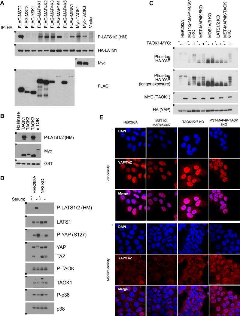
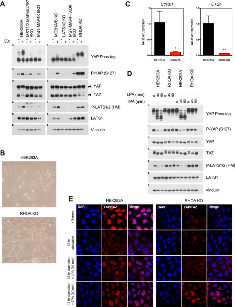
The Hippo pathway is important for regulating tissue homeostasis, and its dysregulation has been implicated in human cancer. However, it is not well understood how the Hippo pathway becomes dysregulated because few mutations in core Hippo pathway components have been identified. Therefore, much work in the Hippo field has focused on identifying upstream regulators, and a complex Hippo interactome has been identified. Nevertheless, it is not always clear which components are the most physiologically relevant in regulating YAP/TAZ. To provide an overview of important Hippo pathway components, we created knockout cell lines for many of these components and compared their relative contributions to YAP/TAZ regulation in response to a wide range of physiological signals. By this approach, we provide an overview of the functional importance of many Hippo pathway components and demonstrate NF2 and RHOA as important regulators of YAP/TAZ and TAOK1/3 as direct kinases for LATS1/2.

Keywords: CRISPR; Hippo pathway; NF2; RHOA; TAOK1; TAOK3; TAZ; YAP.


Figures

Figure 1
Figure 1. Using CRISPR to target the Hippo pathway
A. List of cell lines created and genes deleted in this study. See Table S1 and Figure S1 for sequences and immunoblots. B. Immunoblots showing CRISPR-mediated deletion of core Hippo pathway components. C. Overnight serum starvation induces YAP/TAZ phosphorylation and degradation in wild-type HEK293A cells. See Figure S2 for quantification. See Figure S7 for a schematic of the Hippo pathway.
Figure 2
Figure 2. YAP/TAZ phosphorylation in response to serum starvation
A–D. Immunoblots showing YAP/TAZ phosphorylation status following serum starvation. The HEK293A cells in A are the relevant control for all panels; the figure is subdivided for readability. See Figure S3 for quantification.
Figure 3
Figure 3. Dysregulation of YAP/TAZ phosphorylation results in aberrant YAP/TAZ localization and transcriptional activity
A. Immunofluorescence staining for YAP/TAZ (red) and DAPI (blue) in the presence of serum or following 12 hours of serum starvation. See Figure S4A for quantification. B and C. Relative expression of YAP/TAZ downstream target genes CYR61 and CTGF in the presence of serum (B), or following overnight serum starvation (C), as quantified by qPCR. Data represented as mean +/−S.D. D and E. Glucose levels (D) and pH (E) of the culture media following a 6 hour incubation under normal culture conditions. Data represented as mean +/− S.D.
Figure 4
Figure 4. Inactivation of YAP/TAZ in response to cell-cell contact
A. Cells were plated at low, medium, and high densities in a 6-well plate. Images show wild-type cells at each of the respective densities. B. Cell-cell contact induces YAP phosphorylation. Cells were plated at each of the respective densities and harvested 24 hours later. C. YAP/TAZ phosphorylation in the wild-type, MST-MAP4K 8KO, MOB1A/B KO, and LATS1/2 KO cells in response to cell-cell contact. D. Immunofluorescence staining for YAP/TAZ (red) and DAPI (blue) at low and medium densities. See Figure S4B for quantification. E. Images show cellular morphologies of wild-type and CTNNA1 KO cells. Cells were plated at low density and images were taken 24 hours later.
Figure 5
Figure 5. Deletion of NF2 results in hyper-activated YAP/TAZ
A. Immunofluorescence staining for YAP/TAZ (red) and DAPI (blue) in wild-type and NF2 KO cells at low (LD) and medium (MD) densities in the presence of serum. See Figure S4C for quantification. B and C. Relative expression of CYR61 and CTGF in the presence of serum (B) or following overnight serum starvation (C), as quantified by qPCR. Data represented as mean +/− S.D. D. Immunofluorescence staining for YAP/TAZ (red) and DAPI (blue) in wild-type and NF2 KO cells at low and medium densities following 12 hour serum starvation. See Figure S4C for quantification. E. Deletion of NF2 prevents LATS1/2 (HM) phosphorylation in response to overnight serum starvation. F. Protein stability of LATS1/2 is not affected in NF2 KO cells following treatment with 100 ug/ml cycloheximide. G. Deletion of NF2 confers a growth advantage in HEK293A cells. Cells were plated at 7 × 10⁴ cells/well in a 6-well plate with fresh media and counted after 0, 24, 48, 72, and 96 hours. Data represented as mean +/− S.D. H. NF2 KO cells are sensitive to actin disruption by Latrunculin B. See Figure S5 for YAP/TAZ phosphorylation response of other cell lines. I. NF2 KO cells are sensitive to cellular energy stress.
Figure 6
Figure 6. TAOK1/3 are direct kinases for LATS1/2
A. Overexpression of TAOK1/3 induces LATS1/2 (HM) phosphorylation in wild-type cells. Cells were transfected with HA-LATS1 and various kinases including MST2, TAOK1, and TAOK3, and HA-LATS1 was immunoprecipitated and immunoblotted for HM phosphorylation. B. TAOK1/3 can directly phosphorylate LATS1/2 (HM) in an in vitro kinase assay. TAOK1, TAOK2, and TAOK3 were transfected into wild-type cells and immunoprecipitated, and an in vitro kinase assay was performed with a truncated form of LATS1 (AA 638–1,130). C. Overexpression of TAOK1 induces YAP phosphorylation in the MST-MAP4K 8KO but not the LATS1/2 KO cells. Cells were seeded at 0.7 × 10⁵ cells/well in a 6-well plate, transfected with TAOK1-MYC, and harvested 24 hours later. D. TAOK kinase activity is unaffected in the NF2 KO cells. P38 is a downstream target of TAOK. E. The MST1/2-MAP4K-TAOK 8KO cells are resistant to cell-cell contact. Immunofluorescence staining for YAP/TAZ (red) and DAPI (blue) at low and medium densities. See Figure S4D for quantification.
Figure 7
Figure 7. RHOA is an important mediator of growth signals to activate YAP/TAZ
A. Treatment with the Rho inhibitor C3 exoenzyme (C3) induces YAP/TAZ phosphorylation. Cells were treated with C3 (1 ug/ml) for 4 hours. B. The RHOA KO cells showed altered morphology compared to the wild-type cells. C. Relative expression of CYR61 and CTGF in the presence of serum, as quantified by qPCR. Data represented as mean +/− S.D. D. LPA fails to induce YAP/TAZ dephosphorylation while TPA can induce TAZ dephosphorylation in the RHOA KO cells. Cells were starved overnight before treatment with LPA (0.5 uM) or TPA (5 nM). See Figure S6 for YAP/TAZ phosphorylation response of other cell lines. E. Immunofluorescence staining for YAP/TAZ (red) and DAPI (blue) in the HEK293A wild-type and RHOA KO cells. See Figure S4E for quantification.

References

    1. Boggiano JC, Vanderzalm PJ, Fehon RG. Tao-1 phosphorylates Hippo/MST kinases to regulate the Hippo-Salvador-Warts tumor suppressor pathway. Dev Cell. 2011;21:888–895. - PMC - PubMed
    1. Callus BA, Verhagen AM, Vaux DL. Association of mammalian sterile twenty kinases, Mst1 and Mst2, with hSalvador via C-terminal coiled-coil domains, leads to its stabilization and phosphorylation. FEBS J. 2006;273:4264–4276. - PubMed
    1. Chan EH, Nousiainen M, Chalamalasetty RB, Schafer A, Nigg EA, Sillje HH. The Ste20-like kinase Mst2 activates the human large tumor suppressor kinase Lats1. Oncogene. 2005;24:2076–2086. - PubMed
    1. Chan SW, Lim CJ, Chong YF, Pobbati AV, Huang C, Hong W. Hippo pathway-independent restriction of TAZ and YAP by angiomotin. The Journal of biological chemistry. 2011;286:7018–7026. - PMC - PubMed
    1. Chen Q, Zhang N, Xie R, Wang W, Cai J, Choi KS, David KK, Huang B, Yabuta N, Nojima H, et al. Homeostatic control of Hippo signaling activity revealed by an endogenous activating mutation in YAP. Genes & development. 2015;29:1285–1297. - PMC - PubMed