Review
. 2016 Dec 3;23(1):87.
doi: 10.1186/s12929-016-0303-y.

Mechanisms of inflammatory responses and development of insulin resistance: how are they interlinked?

Affiliations
Review

Mechanisms of inflammatory responses and development of insulin resistance: how are they interlinked?

Kanwal Rehman et al. J Biomed Sci. .

Abstract

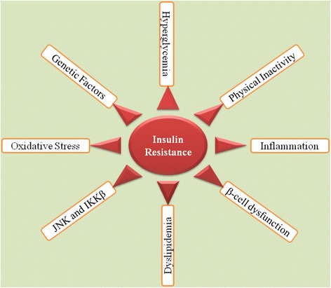
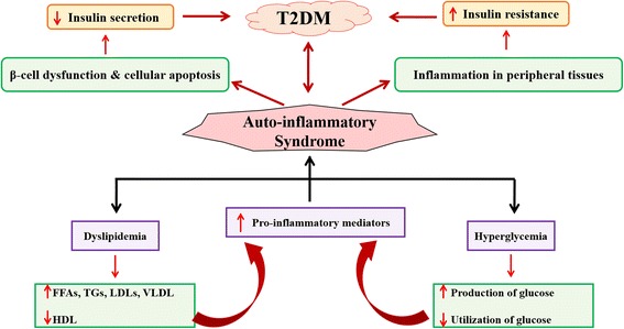
Background: Insulin resistance (IR) is one of the major hallmark for pathogenesis and etiology of type 2 diabetes mellitus (T2DM). IR is directly interlinked with various inflammatory responses which play crucial role in the development of IR. Inflammatory responses play a crucial role in the pathogenesis and development of IR which is one of the main causative factor for the etiology of T2DM.

Methods: A comprehensive online English literature was searched using various electronic search databases. Different search terms for pathogenesis of IR, role of various inflammatory responses were used and an advanced search was conducted by combining all the search fields in abstracts, keywords, and titles.

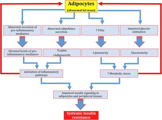
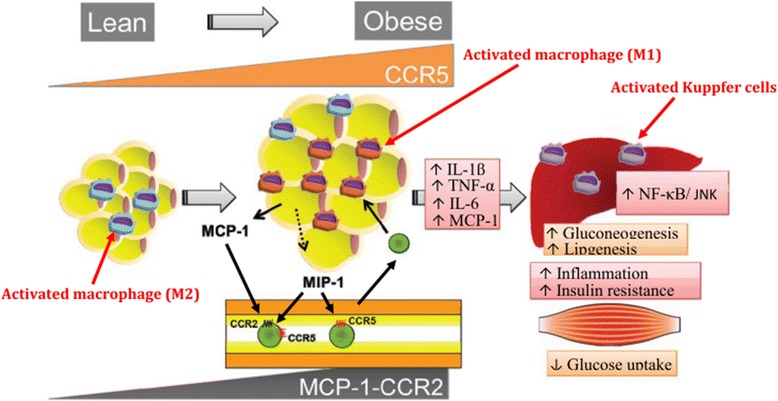
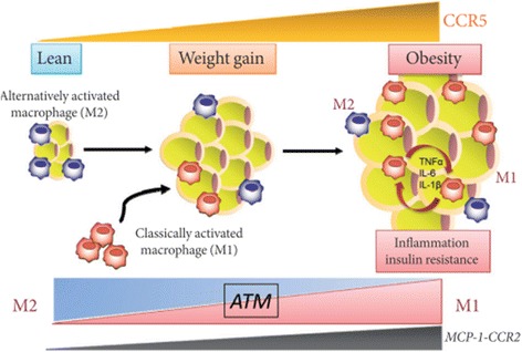
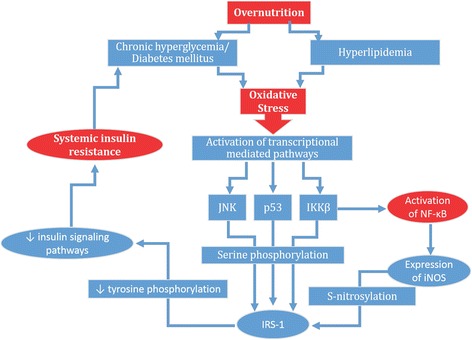
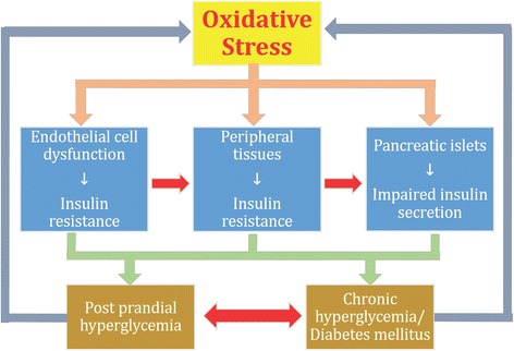
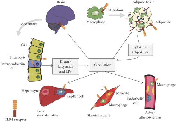
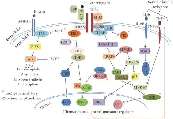
Results: We summarized the data from the searched articles and found that inflammatory responses activate the production of various pro-inflammatory mediators notably cytokines, chemokines and adipocytokines through the involvement of various transcriptional mediated molecular pathways, oxidative and metabolic stress. Overnutrition is one of the major causative factor that contributes to induce the state of low-grade inflammation due to which accumulation of elevated levels of glucose and/or lipids in blood stream occur that leads to the activation of various transcriptional mediated molecular and metabolic pathways. This results in the induction of various pro-inflammatory mediators that are decisively involved to provoke the pathogenesis of tissue-specific IR by interfering with insulin signaling pathways. Once IR is developed, it increases oxidative stress in β-cells of pancreatic islets and peripheral tissues which impairs insulin secretion, and insulin sensitivity in β-cells of pancreatic islets and peripheral tissues, respectively. Moreover, we also summarized the data regarding various treatment strategies of inflammatory responses-induced IR.

Conclusions: In this article, we have briefly described that how pro-inflammatory mediators, oxidative stress, transcriptional mediated molecular and metabolic pathways are involved in the pathogenesis of tissues-specific IR. Moreover, based on recent investigations, we have also described that to counterfeit these inflammatory responses is one of the best treatment strategy to prevent the pathogenesis of IR through ameliorating the incidences of inflammatory responses.

Keywords: Diabetes mellitus; Insulin resistance; Insulin sensitivity; Pro-inflammatory mediators; Transcriptional pathways.


Figures

Fig. 1
Fig. 1
Schematic representation of development of IR. Adopted from Rehman and Akash [5]
Fig. 2
Fig. 2
Overnutrition is responsible to elevate the levels of glucose and FFAs in blood which are responsible to induce metabolic stress in β-cells of pancreatic islets and insulin sensitive tissues notably adipocytes (especially in case of obesity). The metabolic stress induced in these tissues activates the release of various pro-inflammatory cytokines notably IL-1β and IL-1β-dependent various other cytokines and chemokines. As a result, immune cells are recruited which contribute the tissue-specific inflammation. Adapted from Donath and Shoelson [12]
Fig. 3
Fig. 3
Production of IL-1β-induced inflammation in β-cells of pancreatic islets. Prolonged exposure of FFAs and glucose induce the activation of IL-1β from β-cells of pancreatic islets through the involvement of various transcriptional mediated molecular pathways notably TXNIP, MYD88, NF-κB, TLRs, caspases and inflammasomes. Once IL-1β is activated, it recruits various other pro-inflammatory mediators after binding with its receptor IL-1RI and through the involvement of MYD88 and NF-κB. Adopted from Donath and Shoelson [12]
Fig. 4
Fig. 4
Overnutrition is responsible for elevated levels of glucose and FFAs in blood which entered into the β-cells of pancreatic islets. Initially, these augmented levels of glucose and FFAs induce the expression and release of IL-1β from the β-cells of pancreatic islets (Stimulation). Prolonged and/or chronic exposure of glucose and FFAs (also known as metabolic stress) may lead to the activation of IL-1β by activating NF-κB and auto-stimulatory process (Amplification). Once, IL-1β is activated and produced, it leads to the recruitment of various other pro-inflammatory cytokines, chemokines, and macrophages (Precipitation) which further induces apoptosis, amyloidosis and fibrosis in β-cells of pancreatic islets, and hence impaired insulin secretion occurs whereas, in peripheral tissues, IR is developed due to systemic inflammation. Adopted from Donath et al. [17]
Fig. 5
Fig. 5
Schematic representation of adipocytokines-induced IR. Glucolipotoxicity and induction of inflammation in adipocytes are responsible to make the adipocytes abnormal. Once adipocytes are injured, glucose utilization is decreased in adipocytes and levels of FFAs are abnormally increased due to which metabolic stress in adipocytes is increased which ultimately leads to the abnormal secretion of various pro-inflammatory mediators and adipocytokines. Abnormal secretion of these pro-inflammatory mediators and adipocytokines activate various inflammatory pathways which impairs the phosphorylation of various insulin signaling pathways in adipocytes and/or peripheral tissues due to which systemic IR is developed
Fig. 6
Fig. 6
Chemokines-induced IR. M2 macrophages in lean state, maintain the insulin sensitivity in adipose tissues whereas, due to overnutrition, adipose tissues initiates the secretion of MCP-1 which leads to the recruitment of circulating monocytes in adipocytes. CCR2 macrophages are accumulated in obese adipocytes and presumably maintain the inflammation by recruiting M1 macrophages in obese adipocytes. While on the other side, CCR5-adipose tissue macrophages (ATM) also infiltrate from the obese adipocytes and promote the inflammatory responses by involving ATM recruitment and producing various pro-inflammatory mediators notably TNF-α, IL-6, and IL-1β in conjunction with other infiltrated immune cells and adipokines. After production, these pro-inflammatory mediators induce IR in adipocytes and peripheral tissues through activation of several transcriptional pathways such as JNK and NF-κB. Adapted from Xu et al. 2015
Fig. 7
Fig. 7
C-C motif chemokine receptor 5 (CCR5) promotes obesity-induced inflammation and IR. Recently, it has been found that the expression of CCR5 and its ligand MCP-1 is significantly increased in white adipose tissues (WAT) and its accumulation is increased in adipose tissue macrophages (ATM) in WAT of obese mice and provides a novel link between inflammation and IR in adipocytes by stimulating the production of various pro-inflammatory cytokines and chemokines. Adopted from Ota [51]
Fig. 8
Fig. 8
Mechanism of oxidative stress-induced IR: Chronic exposure of hyperglycemia and hyperlipidemia due to over nutrition leads to the production of oxidative stress via activation of reactive oxygen species. Once, oxidative stress is produced within the body, it leads to the activation of various transcriptional mediated pathways such as p38, JNK, IKKβ and/or NF-κB. IKKβ also induces the activation of NF-κB. p38, JNK and IKKβ, further activates the serine phosphorylation of insulin receptor substrate-1 (IRS-1). While on the other side, NF-κB also activates the expression of iNOS which also induces the S-nitrosylation of IRS-1. Both S-nitrosylation and serine phosphorylation of IRS-1 suppress the tyrosine phosphorylation of insulin signaling pathways which ultimately results into the induction of IR in liver, adipocytes and skeletal muscles
Fig. 9
Fig. 9
Impact of oxidative stress on vital organs of the body. β-cells of pancreatic islets, adipocytes and peripheral tissues are more susceptible to the damaging effects of oxidative stress. Oxidative stress independently exhibit its hazardous effects on these organs due to which impaired insulin secretion occurs in β-cells of pancreatic islets and IR develops in adipocytes and peripheral tissues. Impaired insulin secretion and IR lead to the development of post prandial hyperglycemia and overt T2DM both of which also acts as feedback mechanism for the development of oxidative stress
Fig. 10
Fig. 10
Expression TLR4 in integrated tissues and organ systems of the body that regulate the insulin sensitivity. Toll-like receptor 4 (TLR4) present in adipocytes, initiates the inflammatory responses that release various pro-inflammatory mediators. Once, produced, these mediators are entred into the blood stream and thereby promote IR. Liver-resident macrophages known as Kupffer cells are made up of 10% of the cells in liver and 80%–90% of all tissue macrophages within the body. TLR4, expressed on Kupffer cells and other liver cell components, regulates the various inflammatory responses in liver. TLR4, expressed in skeletal muscles, has been shown to regulate the substrate metabolism in muscle, favoring glucose oxidation in the absence of insulin. Hypothalamus and mesolimbic area are important sites that modulate the energy expenditure, pancreatic β-cell function and IR in peripheral tissue. Expression of TLR4 in hypothalamus potentiates various inflammatory responses that contribute to the pathogenesis of IR. Adopted from Kim and Sears 2010
Fig. 11
Fig. 11
Schematic representation of TLR4 signaling cascades. Signal transduction of TLR4 through the activation of MyD88/TIRAP and TRAM/TRIF pathways, leads to potentiate the innate immune responses and inhibit signal transduction of insulin, primarily through serine phosphorylation of IRS. Adopted from Kim and Sears 2010
Fig. 12
Fig. 12
Schematic representation of effect of AMPK activation on various body organs
Fig. 13
Fig. 13
Mechanism of hyperglycemia- and dyslipidemia-induced inflammation for the development of IR and T2DM. Hyperglycemia and dyslipidemia collectively provoke the activation of pro-inflammatory mediators through the involvement of several metabolic pathways. Once, these pro-inflammatory mediators are released, they induce tissue-specific inflammation due to which IR in peripheral tissues and impaired insulin secretion in pancreatic islets occur that ultimately lead to overt T2DM. Adapted from Akash et al. [30]

References

    1. Feve B, Bastard JP. The role of interleukins in insulin resistance and type 2 diabetes mellitus. Nat Rev Endocrinol. 2009;5(6):305–311. doi: 10.1038/nrendo.2009.62. - DOI - PubMed
    1. Hotamisligil GS. Inflammatory pathways and insulin action. Int J Obes Relat Metab Disord. 2003;27(Suppl 3):S53–55. doi: 10.1038/sj.ijo.0802502. - DOI - PubMed
    1. Moller DE. Potential role of TNF-alpha in the pathogenesis of insulin resistance and type 2 diabetes. Trends Endocrinol Metab. 2000;11(6):212–217. doi: 10.1016/S1043-2760(00)00272-1. - DOI - PubMed
    1. Akash MSH, Shen Q, Rehman K, Chen S. Interleukin-1 receptor antagonist: a new therapy for type 2 diabetes mellitus. J Pharm Sci. 2012;101(5):1647–1658. doi: 10.1002/jps.23057. - DOI - PubMed
    1. Rehman K. and Akash M.S.H. Nutrition and Diabetes Mellitus: How are They Interlinked? 2016;26(4):317–332. - PubMed

LinkOut - more resources